Sun, Nov 8, 2026
Volume 16, Issue 4 ( Autumn 2026)
PTJ 2026, 16(4): 409-418 |
Back to browse issues page


Download citation:
BibTeX | RIS | EndNote | Medlars | ProCite | Reference Manager | RefWorks
Send citation to:


BibTeX | RIS | EndNote | Medlars | ProCite | Reference Manager | RefWorks
Send citation to:
Piri E, Sobhani V, Jafarnezhadgero A, Arabzadeh E, Shamsoddini A. Effect of Double Density Insoles on Electromyographic Gait Characteristics in People With Flat Feet. PTJ 2026; 16 (4) :409-418
URL: http://ptj.uswr.ac.ir/article-1-694-en.html
URL: http://ptj.uswr.ac.ir/article-1-694-en.html
Ebrahim Piri1  , Vahid Sobhani *2 , AmirAli Jafarnezhadgero1 , Ehsan Arabzadeh2 , Alireza Shamsoddini2
1- Department of Sports Biomechanics, Faculty of Educational Sciences and Psychology, University of Mohaghegh Ardabili, Ardabil, Iran
2- Exercise Physiology Research Center, Life Style Institute, Baqiyatallah University of Medical Sciences, Tehran, Iran
2- Exercise Physiology Research Center, Life Style Institute, Baqiyatallah University of Medical Sciences, Tehran, Iran
Full-Text [PDF 584 kb]
(1163 Downloads)
| Abstract (HTML) (2629 Views)
Full-Text: (80 Views)
Introduction
Pronated feet (PF) are characterized by excessive rearfoot eversion, forefoot dorsiflexion, and abduction relative to the tibia. In adults aged 18-25 years, the PF prevalence amounts to 11% [1]. Individuals with PF experience a greater rate of overuse injuries in the lower limbs [2], such as tibial stress syndrome [3]. Individuals with PF exhibit abnormal lower-limb mechanics during walking. Individuals exhibit altered foot movement patterns during walking. According to Farahpour et al. [4], individuals with PF demonstrate increased anteroposterior ground reaction forces during the gait stance phase.
According to previous research [5, 6], individuals with PF demonstrate greater peak rearfoot eversion, inversion, and internal rotation during the gait stance phase. Studies have also shown that those with PF exhibit greater activity in ankle invertor muscles during gait [5]. Furthermore, due to the biomechanical link between PF and knee valgus, PF may lead to excessive internal rotation at both the knee and hip joints [7]. The interaction between agonist and antagonist muscle activation is commonly assessed using surface electromyography (EMG) [8]. Muscle co-contraction is defined as the concurrent activation of opposing muscle groups around a joint [9, 10]. While walking is widely accessible and beneficial, it can also lead to injuries if proper foot posture is not maintained [11]. Therefore, rehabilitation or treatment methods should be considered for patients with PF.
EMG serves as a clinical tool for evaluating and recording the electrical signals produced by skeletal muscles during activity [12]. The frequency spectrum of lower-limb muscles pertains to the distribution of electrical activity across different frequencies generated by these muscles during various physical activities [13]. As muscle recruitment patterns change with skill or fatigue, the frequency spectrum can provide insights into the neuromuscular coordination of the lower limbs. Additionally, factors, such as muscle fiber composition, age, and body status, can influence the frequency spectrum. The frequency spectrum of EMG is typically characterized by its power density, revealing how the signal’s power is distributed across different frequency ranges [12]. High-frequency components, specifically above 50 Hz, correlate with the recruitment of fast-twitch muscle fibers and the firing rates of motor units [13]. These higher frequencies are indicative of rapid muscle contractions and vigorous activation [13]. Moreover, the frequency content of an EMG signal can serve as an indicator of muscle fatigue. As muscles fatigue, there is often a notable shift in the frequency spectrum, characterized by a decrease in the power of higher-frequency components and an increase in lower-frequency activities.
Foot orthoses (FOs) are widely utilized as a non-invasive intervention to minimize the likelihood of overuse-related musculoskeletal injuries in individuals exhibiting a PF posture [14]. The primary goal of PF-specific orthotic devices is to enhance dynamic foot function, which can be evaluated by changes in joint movement patterns, force distribution, and muscle activation during gait [15]. Among various types, medial wedge orthoses are most commonly prescribed to reduce excessive PF and modify lower limb biomechanics in this population [16]. Incorporating a rearfoot post within the orthosis facilitates better fit and comfort. In contrast, forefoot posting often presents challenges in terms of shoe accommodation and may irritate [17]. To address these issues, double-density FOs have been introduced, featuring a softer lateral section and a firmer medial portion. However, it is still uncertain whether these orthoses have a unique influence on the frequency characteristics of lower limb muscle activity and co-contraction patterns in adults with PF during walking. To the best of our knowledge, no prior research has specifically examined muscular co-contraction responses during gait in individuals with PF versus healthy controls when using double-density FOs. Therefore, based on existing literature [16, 17], this research aimed to compare muscular co-contraction during the gait stance phase in adults with PF and healthy controls while wearing double-density FOs.
Materials and Methods
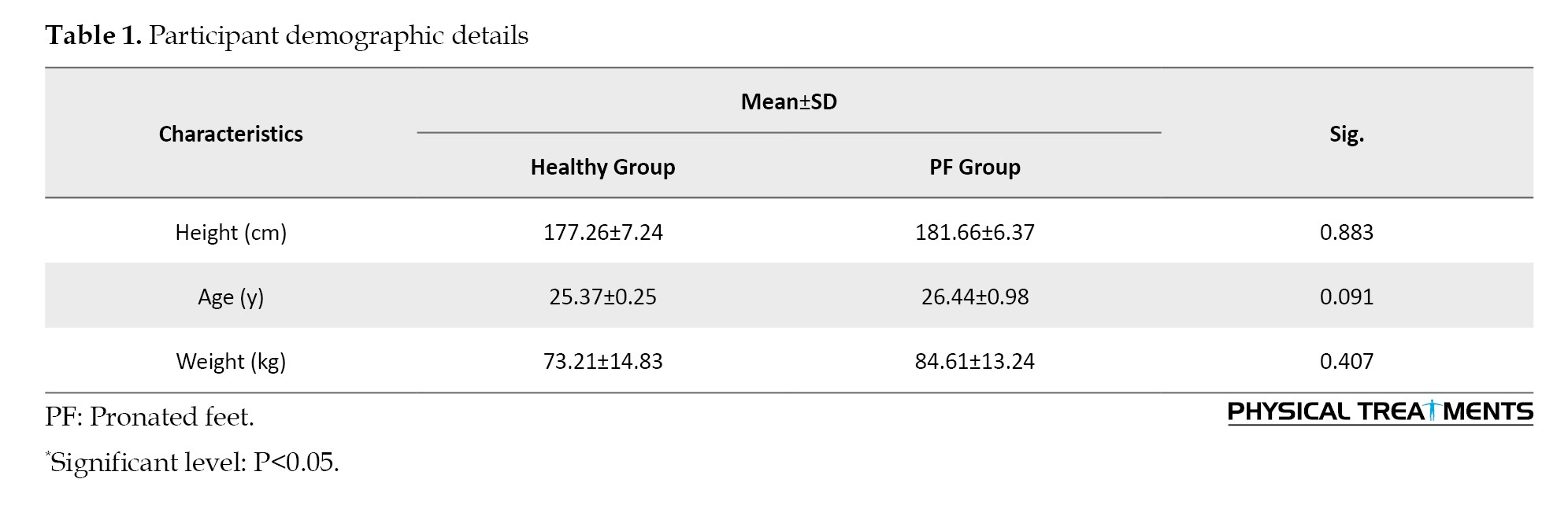
A total of 40 right-handed adult males who exhibited PF participated in the research. The subjects were allocated into PF (n=20) and healthy (n=20) groups. The number of samples was calculated using G*Power software, version 3.1 (University of Kiel, Germany) [18], based on an estimated effect size of 0.65. A priori power analysis was carried out for a two-tailed independent samples t-test, assuming a power (1–β) of 0.80 and a P=0.05. This analysis indicated a minimum required sample of 18 individuals per group. To allow for possible attrition and ensure adequate statistical strength, 20 participants were enrolled in each group. Table 1 summarizes the baseline characteristics of participants.
Right-foot dominance was confirmed in all participants using a ball-kicking test.
The inclusion criteria for the PF group were male gender, body mass index (BMI) <25 kg/m², rearfoot eversion angle >4° [19, 20], navicular drop >10 mm, and (v) a foot posture index (FPI)>10. The validity of this index has been previously supported by literature [21]. The FPI consists of six key indicators used to determine foot alignment [21, 22]. Each item was scored using a 5-point scale ranging from -2 to +2, generating an overall score between -12 and +12. Negative values reflect a supinated PF, whereas positive values correspond to PF posture. Participants were categorized as having PF if their total FPI score ranged from 6 to 10 [21, 22].
The exclusion criteria included a history of musculoskeletal surgery, orthopedic conditions (excluding PF), a lower limb length discrepancy greater than 5 mm [23], and engaging in sports activities within the past two days. Informed consent was obtained from the subjects before participation.
Double density foot orthoses
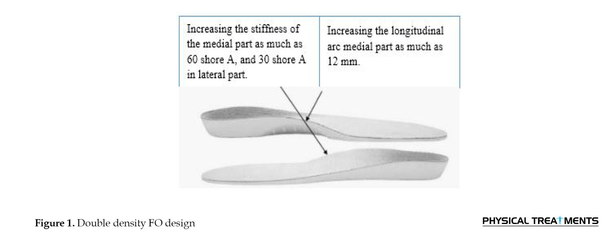
Subjects were introduced to the tests and equipment that would be used. All participants were provided with double-density FOs that were sized to fit their feet. The same model of double-density FOs was used for both the PF and healthy foot groups. These orthoses were made from EVA, with the medial part having a stiffness of Shore 60 and the lateral part having a stiffness of Shore 30. Both parts featured the same height for medial longitudinal arch support.
Over ground walking
All testing sessions were conducted between 10:00 AM and 12:00 PM to ensure consistency in circadian rhythm effects. Before each session, participants performed a warm-up lasting 10 minutes (Figure 1) [18].
 Data collection for lower limb muscle activity, including frequency spectrum analysis and co-contraction assessment, was carried out using an 18-meter walkway fitted with an EMG system (Biometrics Ltd., United Kingdom). Electrodes were placed on relevant muscles of the dominant leg. Participants, who were already familiar with the lab environment and procedures, walked at a controlled speed of 1.2 m/s ±5% throughout the test area. Each experimental condition involved six trials, preceded by three practice trials to help participants adjust to the required pace. A chronometer was used to measure the duration of each trial.
Muscular co-contraction
To assess lower limb muscle activity, an EMG system was employed. This system utilized eight pairs of surface electrodes placed on the dominant limb’s anterior tibialis (TA), medial gastrocnemius (Gas-M), biceps femoris (BF), semitendinosus (ST), vastus lateralis (VL), vastus medialis (VM), rectus femoris (RF), and gluteus medius (Glut-M). The distance between electrode centers was standardized at 25 mm. The median frequency of the EMG signals was determined using Biometric Data LITE software and served as the primary outcome variable for frequency analysis. Prior to electrode placement, the skin over each targeted muscle belly was shaved. Electrodes were then positioned on selected muscles using guidelines from the SENIAM project [5]. EMG amplitudes were assessed using root mean square (RMS) values [5]. For analysis, the stance phase of walking was divided into three phases: Loading, mid-stance, and push-off phases [24]. To normalize EMG values, maximum voluntary isometric contraction (MVIC) was measured. Three valid walking trials were conducted under both orthotic conditions (with and without FOs). MVIC assessments were conducted post-walking trials for each muscle separately. Normalization was achieved by dividing the peak RMS values obtained during walking by the corresponding MVIC peak values, then multiplying by 100 to express results as a percentage of MVIC.
Directed co-contraction (DCCR) was defined for specific muscle groups. These included comparisons between the medial and lateral knee muscles, referred to as MLDCCR, and between knee flexors and extensors, termed FEDCCR [25]. The DCCR was calculated using the Equation 1:
1. DCCR=1-(antagonist/ agonist)
Otherwise:
DCCR=(agonist/ antagonist)–1
In this model, positive DCCR values indicated greater agonist dominance, whereas negative values reflected stronger antagonist activation. A DCCR close to zero represents maximal co-contraction, while values approaching ±1 suggest minimal co-activation between opposing muscles [26].
Statistical Analyses
Normality was assessed using the Shapiro–Wilk test. Two-way analysis of variance (ANOVA) (group [pronated vs healthy]×condition [with vs without FO]) with repeated measures was performed for statistical analysis. Effect sizes, expressed as ƞ2, were obtained from the analysis of variance (ANOVA) and converted into Cohen’s d [27, 28].
Results
The significant impact of “group” was found for general knee co-contraction during loading (P=0.049) and push-off (P=0.027). Pairwise comparisons revealed that the PF group exhibited greater general co-contraction in both loading and push-off phases than the healthy group (Table 2).
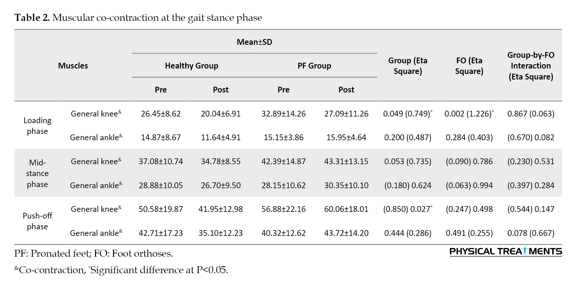
The significant impact of “FO” was observed for general knee co-contraction at loading (P=0.002; d=1.226). Pairwise comparisons showed that general knee co-contraction was lower at loading when walking with FOs than when walking without them (Table 2).
A significant impact of “group” was observed for knee flexor/extensor co-contraction at the loading phase (P=0.008), as well as at midstance (P=0.012) and push-off phase (P=0.015). Additionally, greater knee medio/lateral co-contraction was found in one direction during mid-stance (P=0.043). Results revealed that the PF group had higher values than the healthy group in all these measures. Specifically, the PF group showed increased knee flexor/extensor co-contraction at loading and mid-stance, greater medio/lateral co-contraction at mid-stance, and elevated flexor/extensor co-contraction at push-off (Table 3).
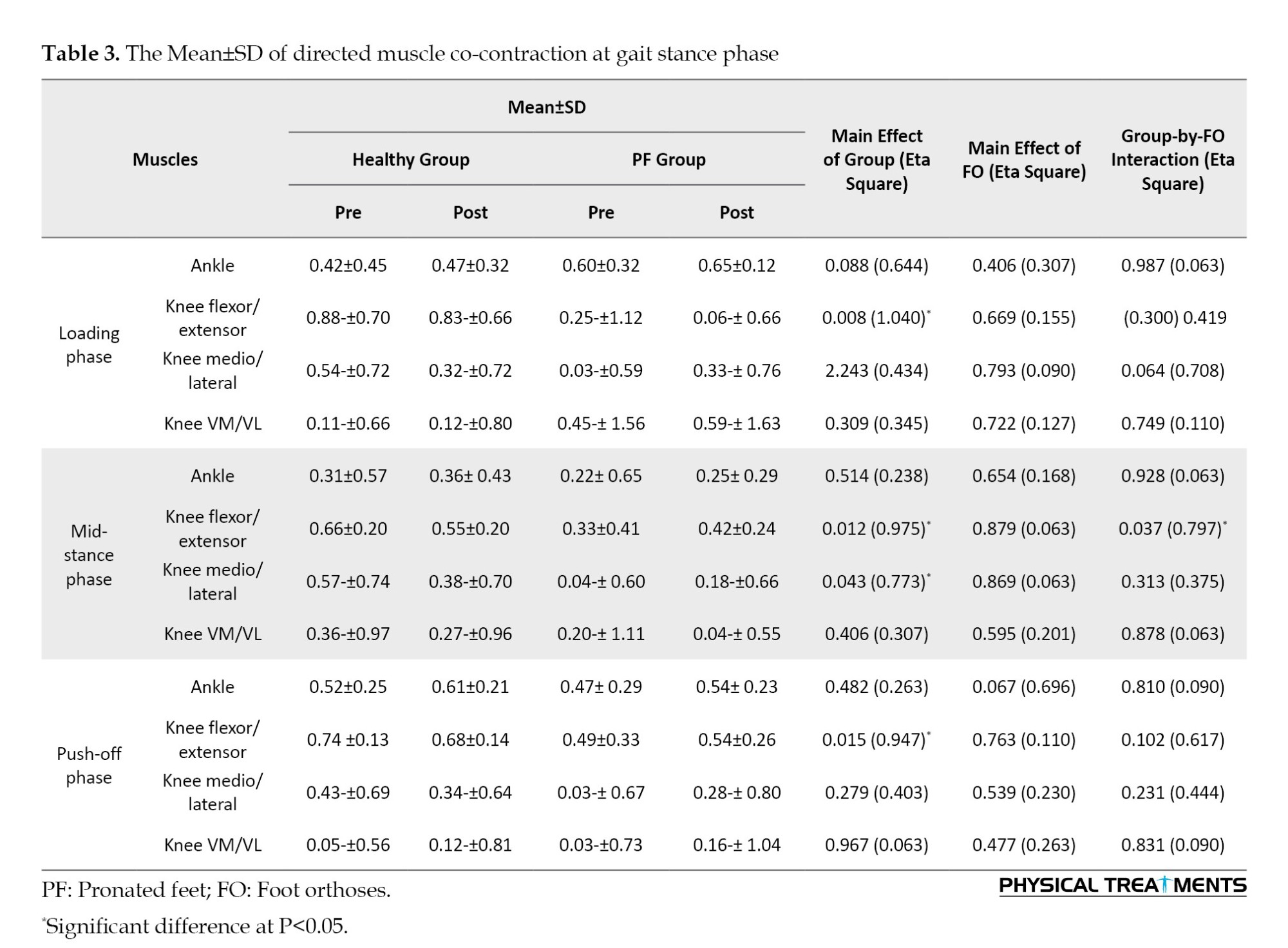
Group-by-condition interaction was significant for directed knee flexor/extensor co-contraction at the mid-stance phase (P=0.037). The results revealed that the healthy group showed greater directed knee flexor/extensor co-contraction at the mid-stance phase while walking with FOs than without them (P=0.030), but no such effect was seen in the PF group.
Significant impact of “group” for Gas-M (P=0.011), VL (P=0.003), VM (P=0.002), RF (P=0.018), BF (P=0.001), ST (P=0.001), and Glut-M (P=0.010) activities at loading phase, and for BF (P=0.002) and ST (P=0.020) frequency content during the midstance phase were found. The results revealed that the PF group had lower frequency content in Gas-M, VL, VM, RF, BF, ST, and Glut-M muscles at the loading phase, and in BF and ST at the mid-stance phase, compared to the healthy group (Table 4).
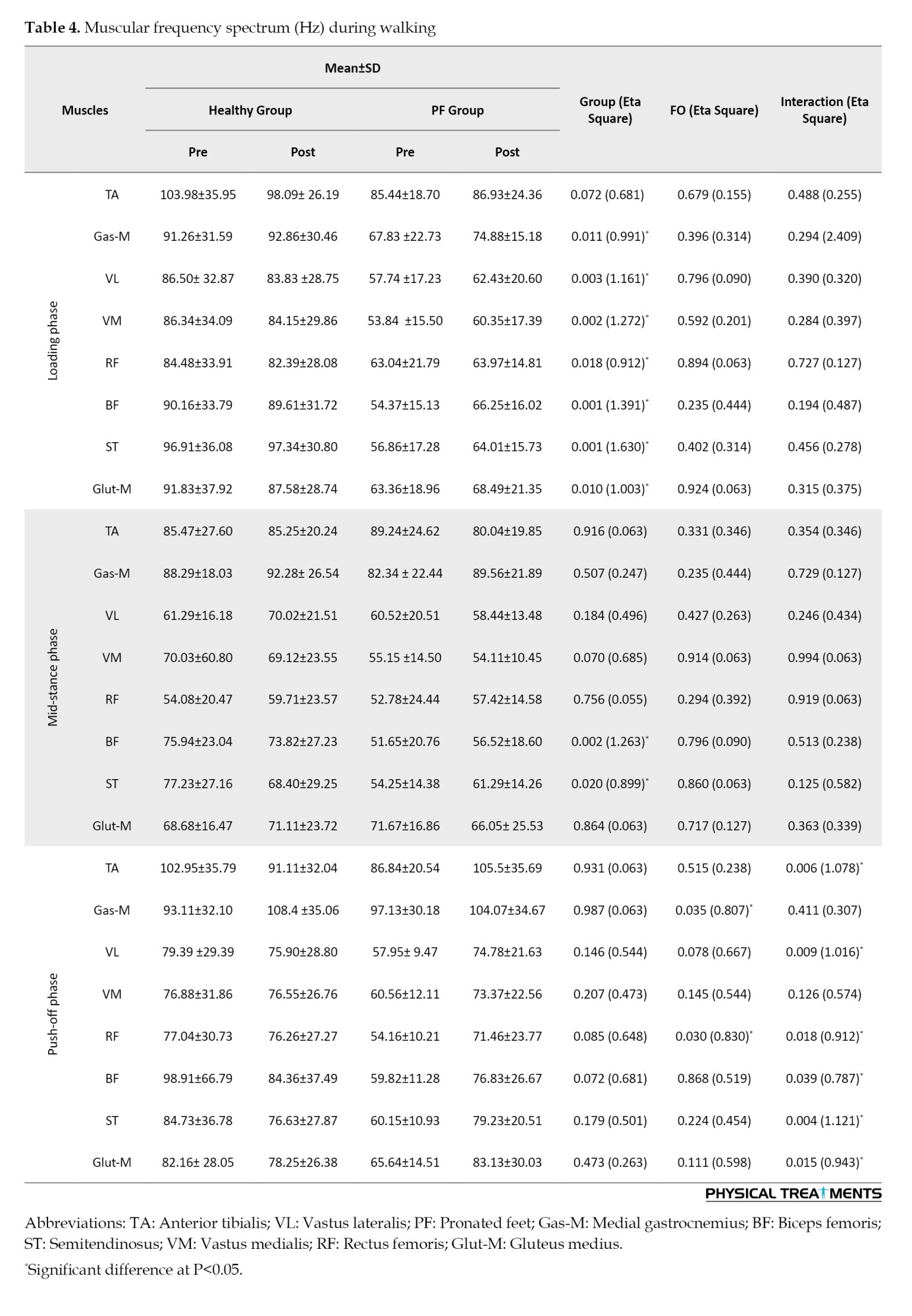
A significant main condition effect was found for Gas-M (P=0.035) and RF (P=0.030) at the push-off phase. The findings showed that Gas-M and RF activities were greater during the push-off phase when walking with FOs than without them (Table 4).
Interactions were significant for TA (P=0.006), VL (P=0.009), RF (P=0.018), BF (P=0.039), ST (P=0.004), and Glut-M (P=0.015) frequency content at the push-off phase. Post-hoc analysis revealed that the PF group, but not the healthy group, exhibited greater TA, VL, RF, BF, ST, and Glut-M activities when walking with FOs than when walking without them (Table 4). These results suggest that FOs may enhance muscle activity at push-off, specifically in individuals with PF, potentially supporting improved propulsion and stability during gait.
Discussion
This study aimed to assess the impact of double-density FOs on lower-limb muscular co-contraction in adults with PF during gait at a constant speed, in comparison to the healthy group. General Muscular Co-contraction.
The results showed higher general knee co-contraction, general ankle co-contraction at the loading phase, and general ankle co-contraction at the push-off phase in the PF group than in the healthy group. In general co-contraction, both the agonist and antagonist muscles of the knee are activated to a similar extent, which may affect joint loading [29, 30]. Importantly, our study found a significant increase in general knee and ankle co-contraction at loading and push-off phases in the PF group compared to the healthy group. Previous studies have indicated that increased general knee muscular co-contraction elevates joint load [31, 32] and has also been associated with greater impact loads. Our findings showed reduced general knee and ankle co-contraction in both groups (especially in the PF group) during the loading phase while running with FO compared with without it. In alignment with our findings, several authors have advocated for the use of FO aimed at reducing co-contraction to minimize joint load, given the potential adverse effects on knee load [33, 34]. This reduction may signify an enhancement in neuromuscular efficiency and a potential alleviation of undue stress on the lower limb joints, enabling individuals with PF to achieve a more biomechanically sound running form. Furthermore, the particularly marked decrease in knee co-contraction within the PF group while using FOs underscores the orthoses’ ability to facilitate improved movement mechanics.
Directed muscular co-contraction
The results showed higher knee flexor/extensor co-contraction at all phases, as well as greater knee medio/lateral co-contraction during the mid-stance phase, in the PF group than in the healthy group. Increased directed knee flexor/extensor and knee medio/lateral co-contraction in the PF group may be linked to a higher risk of anterior cruciate ligament injury [35] and medial knee osteoarthritis [26], respectively. The subjects with PF exhibited greater knee extension angles at the walking stance phase. Consistent with our results, Powell et al. [36] reported that individuals with PF have instability and high mobility during dynamic loading tasks. Our results demonstrated greater knee flexor/extensor co-contraction in the healthy group (but not in the PF group) at mid-stance while walking with FO than without it. Therefore, it could be concluded that double-density FO did not improve knee flexor/extensor co-contraction in individuals with PF.
The current study found no significant main effects of group, FO, or their interaction on directed ankle co-contraction during the gait stance phase. Previous research has suggested that increased directed ankle co-contraction can help reduce the risk of lower limb injuries and ankle instability in individuals with PF [37]. No study has yet examined lower limb muscular co-contraction during walking in adults with PF compared to healthy controls while using double density FOs.
Frequency spectrum of lower limb muscular
The results demonstrated lower frequency content of the Gas-M, VL, VM, RF, BF, ST, Glut-M (loading phase), BF and ST (mid-stance phase) muscles in the PF group than in the healthy group. This finding suggests a compromised neuromuscular system in the PF group, which may impede effective force generation and stability during weight-bearing activities [38]. Such deficits in muscle recruitment could potentially predispose individuals to injuries or further functional impairments. The decreased frequency content observed in the PF group suggests a potential reduction in neuromuscular activation and coordination [5]. Muscles with lower frequency content may exhibit impaired capacity to generate force and stabilize the knee joint, leading to altered gait mechanics and increased susceptibility to injury. For instance, the gastrocnemius and glut-M play critical roles in propulsion and stability; thus, their diminished frequency could adversely impact overall kinetic efficiency and postural control [39].
Results showed higher Gas-M and RF during the push-off phase while walking with FO than without it. This enhancement in activation may be attributed to the supportive nature of the FO, which likely aids in force transmission and propulsion, thereby optimizing gait mechanics [40]. Consequently, this suggests that the application of FOs may serve as a beneficial intervention aimed at ameliorating functional limitations associated with PF dysfunction. The push-off phase of gait is critical because it represents the transition from weight acceptance to propulsion [18]. The observed enhancement in Gas-M and RF activity concomitant with the use of FOs suggests that these devices facilitate more effective muscle engagement. Increased muscular activity in these key muscle groups may enhance the efficiency of locomotion by improving force generation and stability during the push-off, which is paramount for energy conservation and forward propulsion [41]. FOs may also benefit individuals with abnormal gait patterns by improving muscle activity, which can lead to improved walking stability and mobility.
Results showed higher TA, VL, RF, BF, ST, and Glut-M activities while walking with FOs rather than without FOs (push-off phase). This disparity indicates that FOs may not only enhance neuromuscular activation in individuals with PF dysfunction but also play a critical role in re-establishing more typical movement patterns [34, 41]. The reliance on these orthotic supports during push-off suggests a compensatory strategy employed by individuals with PF dysfunction to mitigate the loss of muscle efficiency and stability observed in the absence of the orthoses. The observed increased activation of the TA and VL muscles illuminates the potential of foot orthotics in enhancing lower limb stability and propulsion [42]. The TA muscle plays a vital role in stabilizing the body during walking. It enables dorsiflexion, which enhances stability and conserves energy [43]. Similarly, the elevated activity in the RF and BF, which are instrumental in knee extension and flexion respectively, underscores the compensatory mechanisms engaged during the push-off phase [44]. The interaction between these muscle groups, influenced by FOs, may contribute to improved neuromuscular control during gait in individuals with PF.
We assessed the acute effects of FOs in males with PF; therefore, our findings cannot be generalized to females. Future studies should explore whether FOs are effective as a preventive or therapeutic approach in women with PF. Additionally, the present study did not assess walking kinematics.
Conclusion
Individuals with PF exhibited greater levels of ankle and knee co-contraction during gait, which may reflect compensatory strategies to stabilize the lower limb and could be associated with increased joint loading and a greater risk of musculoskeletal injuries. In this study, we observed reduced overall knee co-contraction during the loading phase when participants with PF walked with FOs, suggesting a potential improvement in neuromuscular efficiency and a decrease in unnecessary joint stress. This change may support a more biomechanically efficient gait pattern in patients with PF.
Ethical Considerations
Compliance with ethical guidelines
This study was approved by the Ethics Committee of Baqiyatallah Medical Sciences University, Tehran, Iran (Code: IR-BMSU-BAQ-REC-1403-066). The study was registered in the Iranian Registry of Clinical Trials (IRCT) (ID: IRCT20220129053865N1).
Funding
This research did not receive any specific grant from funding agencies in the public, commercial, or not-for-profit sectors.
Authors' contributions
Methodology: Vahid Sobhani and Ebrahim Piri; Conceptualization and supervision: Vahid Sobhani; Investigation and writing the original draft: Ehsan Arabzadeh and Alireza Shamsoddini; Methodology, data collection, formal analysis, review and editing: Ebrahim Piri.
Conflict of interest
The authors declared no conflicts of interest.
Acknowledgments
The authors express their sincere appreciation to all participants who voluntarily participated in this study.
References
Pronated feet (PF) are characterized by excessive rearfoot eversion, forefoot dorsiflexion, and abduction relative to the tibia. In adults aged 18-25 years, the PF prevalence amounts to 11% [1]. Individuals with PF experience a greater rate of overuse injuries in the lower limbs [2], such as tibial stress syndrome [3]. Individuals with PF exhibit abnormal lower-limb mechanics during walking. Individuals exhibit altered foot movement patterns during walking. According to Farahpour et al. [4], individuals with PF demonstrate increased anteroposterior ground reaction forces during the gait stance phase.
According to previous research [5, 6], individuals with PF demonstrate greater peak rearfoot eversion, inversion, and internal rotation during the gait stance phase. Studies have also shown that those with PF exhibit greater activity in ankle invertor muscles during gait [5]. Furthermore, due to the biomechanical link between PF and knee valgus, PF may lead to excessive internal rotation at both the knee and hip joints [7]. The interaction between agonist and antagonist muscle activation is commonly assessed using surface electromyography (EMG) [8]. Muscle co-contraction is defined as the concurrent activation of opposing muscle groups around a joint [9, 10]. While walking is widely accessible and beneficial, it can also lead to injuries if proper foot posture is not maintained [11]. Therefore, rehabilitation or treatment methods should be considered for patients with PF.
EMG serves as a clinical tool for evaluating and recording the electrical signals produced by skeletal muscles during activity [12]. The frequency spectrum of lower-limb muscles pertains to the distribution of electrical activity across different frequencies generated by these muscles during various physical activities [13]. As muscle recruitment patterns change with skill or fatigue, the frequency spectrum can provide insights into the neuromuscular coordination of the lower limbs. Additionally, factors, such as muscle fiber composition, age, and body status, can influence the frequency spectrum. The frequency spectrum of EMG is typically characterized by its power density, revealing how the signal’s power is distributed across different frequency ranges [12]. High-frequency components, specifically above 50 Hz, correlate with the recruitment of fast-twitch muscle fibers and the firing rates of motor units [13]. These higher frequencies are indicative of rapid muscle contractions and vigorous activation [13]. Moreover, the frequency content of an EMG signal can serve as an indicator of muscle fatigue. As muscles fatigue, there is often a notable shift in the frequency spectrum, characterized by a decrease in the power of higher-frequency components and an increase in lower-frequency activities.
Foot orthoses (FOs) are widely utilized as a non-invasive intervention to minimize the likelihood of overuse-related musculoskeletal injuries in individuals exhibiting a PF posture [14]. The primary goal of PF-specific orthotic devices is to enhance dynamic foot function, which can be evaluated by changes in joint movement patterns, force distribution, and muscle activation during gait [15]. Among various types, medial wedge orthoses are most commonly prescribed to reduce excessive PF and modify lower limb biomechanics in this population [16]. Incorporating a rearfoot post within the orthosis facilitates better fit and comfort. In contrast, forefoot posting often presents challenges in terms of shoe accommodation and may irritate [17]. To address these issues, double-density FOs have been introduced, featuring a softer lateral section and a firmer medial portion. However, it is still uncertain whether these orthoses have a unique influence on the frequency characteristics of lower limb muscle activity and co-contraction patterns in adults with PF during walking. To the best of our knowledge, no prior research has specifically examined muscular co-contraction responses during gait in individuals with PF versus healthy controls when using double-density FOs. Therefore, based on existing literature [16, 17], this research aimed to compare muscular co-contraction during the gait stance phase in adults with PF and healthy controls while wearing double-density FOs.
Materials and Methods
A total of 40 right-handed adult males who exhibited PF participated in the research. The subjects were allocated into PF (n=20) and healthy (n=20) groups. The number of samples was calculated using G*Power software, version 3.1 (University of Kiel, Germany) [18], based on an estimated effect size of 0.65. A priori power analysis was carried out for a two-tailed independent samples t-test, assuming a power (1–β) of 0.80 and a P=0.05. This analysis indicated a minimum required sample of 18 individuals per group. To allow for possible attrition and ensure adequate statistical strength, 20 participants were enrolled in each group. Table 1 summarizes the baseline characteristics of participants.
Right-foot dominance was confirmed in all participants using a ball-kicking test.
The inclusion criteria for the PF group were male gender, body mass index (BMI) <25 kg/m², rearfoot eversion angle >4° [19, 20], navicular drop >10 mm, and (v) a foot posture index (FPI)>10. The validity of this index has been previously supported by literature [21]. The FPI consists of six key indicators used to determine foot alignment [21, 22]. Each item was scored using a 5-point scale ranging from -2 to +2, generating an overall score between -12 and +12. Negative values reflect a supinated PF, whereas positive values correspond to PF posture. Participants were categorized as having PF if their total FPI score ranged from 6 to 10 [21, 22].
The exclusion criteria included a history of musculoskeletal surgery, orthopedic conditions (excluding PF), a lower limb length discrepancy greater than 5 mm [23], and engaging in sports activities within the past two days. Informed consent was obtained from the subjects before participation.
Double density foot orthoses
Subjects were introduced to the tests and equipment that would be used. All participants were provided with double-density FOs that were sized to fit their feet. The same model of double-density FOs was used for both the PF and healthy foot groups. These orthoses were made from EVA, with the medial part having a stiffness of Shore 60 and the lateral part having a stiffness of Shore 30. Both parts featured the same height for medial longitudinal arch support.
Over ground walking
All testing sessions were conducted between 10:00 AM and 12:00 PM to ensure consistency in circadian rhythm effects. Before each session, participants performed a warm-up lasting 10 minutes (Figure 1) [18].
Muscular co-contraction
To assess lower limb muscle activity, an EMG system was employed. This system utilized eight pairs of surface electrodes placed on the dominant limb’s anterior tibialis (TA), medial gastrocnemius (Gas-M), biceps femoris (BF), semitendinosus (ST), vastus lateralis (VL), vastus medialis (VM), rectus femoris (RF), and gluteus medius (Glut-M). The distance between electrode centers was standardized at 25 mm. The median frequency of the EMG signals was determined using Biometric Data LITE software and served as the primary outcome variable for frequency analysis. Prior to electrode placement, the skin over each targeted muscle belly was shaved. Electrodes were then positioned on selected muscles using guidelines from the SENIAM project [5]. EMG amplitudes were assessed using root mean square (RMS) values [5]. For analysis, the stance phase of walking was divided into three phases: Loading, mid-stance, and push-off phases [24]. To normalize EMG values, maximum voluntary isometric contraction (MVIC) was measured. Three valid walking trials were conducted under both orthotic conditions (with and without FOs). MVIC assessments were conducted post-walking trials for each muscle separately. Normalization was achieved by dividing the peak RMS values obtained during walking by the corresponding MVIC peak values, then multiplying by 100 to express results as a percentage of MVIC.
Directed co-contraction (DCCR) was defined for specific muscle groups. These included comparisons between the medial and lateral knee muscles, referred to as MLDCCR, and between knee flexors and extensors, termed FEDCCR [25]. The DCCR was calculated using the Equation 1:
1. DCCR=1-(antagonist/ agonist)
Otherwise:
DCCR=(agonist/ antagonist)–1
In this model, positive DCCR values indicated greater agonist dominance, whereas negative values reflected stronger antagonist activation. A DCCR close to zero represents maximal co-contraction, while values approaching ±1 suggest minimal co-activation between opposing muscles [26].
Statistical Analyses
Normality was assessed using the Shapiro–Wilk test. Two-way analysis of variance (ANOVA) (group [pronated vs healthy]×condition [with vs without FO]) with repeated measures was performed for statistical analysis. Effect sizes, expressed as ƞ2, were obtained from the analysis of variance (ANOVA) and converted into Cohen’s d [27, 28].
Results
The significant impact of “group” was found for general knee co-contraction during loading (P=0.049) and push-off (P=0.027). Pairwise comparisons revealed that the PF group exhibited greater general co-contraction in both loading and push-off phases than the healthy group (Table 2).
The significant impact of “FO” was observed for general knee co-contraction at loading (P=0.002; d=1.226). Pairwise comparisons showed that general knee co-contraction was lower at loading when walking with FOs than when walking without them (Table 2).
A significant impact of “group” was observed for knee flexor/extensor co-contraction at the loading phase (P=0.008), as well as at midstance (P=0.012) and push-off phase (P=0.015). Additionally, greater knee medio/lateral co-contraction was found in one direction during mid-stance (P=0.043). Results revealed that the PF group had higher values than the healthy group in all these measures. Specifically, the PF group showed increased knee flexor/extensor co-contraction at loading and mid-stance, greater medio/lateral co-contraction at mid-stance, and elevated flexor/extensor co-contraction at push-off (Table 3).
Group-by-condition interaction was significant for directed knee flexor/extensor co-contraction at the mid-stance phase (P=0.037). The results revealed that the healthy group showed greater directed knee flexor/extensor co-contraction at the mid-stance phase while walking with FOs than without them (P=0.030), but no such effect was seen in the PF group.
Significant impact of “group” for Gas-M (P=0.011), VL (P=0.003), VM (P=0.002), RF (P=0.018), BF (P=0.001), ST (P=0.001), and Glut-M (P=0.010) activities at loading phase, and for BF (P=0.002) and ST (P=0.020) frequency content during the midstance phase were found. The results revealed that the PF group had lower frequency content in Gas-M, VL, VM, RF, BF, ST, and Glut-M muscles at the loading phase, and in BF and ST at the mid-stance phase, compared to the healthy group (Table 4).
A significant main condition effect was found for Gas-M (P=0.035) and RF (P=0.030) at the push-off phase. The findings showed that Gas-M and RF activities were greater during the push-off phase when walking with FOs than without them (Table 4).
Interactions were significant for TA (P=0.006), VL (P=0.009), RF (P=0.018), BF (P=0.039), ST (P=0.004), and Glut-M (P=0.015) frequency content at the push-off phase. Post-hoc analysis revealed that the PF group, but not the healthy group, exhibited greater TA, VL, RF, BF, ST, and Glut-M activities when walking with FOs than when walking without them (Table 4). These results suggest that FOs may enhance muscle activity at push-off, specifically in individuals with PF, potentially supporting improved propulsion and stability during gait.
Discussion
This study aimed to assess the impact of double-density FOs on lower-limb muscular co-contraction in adults with PF during gait at a constant speed, in comparison to the healthy group. General Muscular Co-contraction.
The results showed higher general knee co-contraction, general ankle co-contraction at the loading phase, and general ankle co-contraction at the push-off phase in the PF group than in the healthy group. In general co-contraction, both the agonist and antagonist muscles of the knee are activated to a similar extent, which may affect joint loading [29, 30]. Importantly, our study found a significant increase in general knee and ankle co-contraction at loading and push-off phases in the PF group compared to the healthy group. Previous studies have indicated that increased general knee muscular co-contraction elevates joint load [31, 32] and has also been associated with greater impact loads. Our findings showed reduced general knee and ankle co-contraction in both groups (especially in the PF group) during the loading phase while running with FO compared with without it. In alignment with our findings, several authors have advocated for the use of FO aimed at reducing co-contraction to minimize joint load, given the potential adverse effects on knee load [33, 34]. This reduction may signify an enhancement in neuromuscular efficiency and a potential alleviation of undue stress on the lower limb joints, enabling individuals with PF to achieve a more biomechanically sound running form. Furthermore, the particularly marked decrease in knee co-contraction within the PF group while using FOs underscores the orthoses’ ability to facilitate improved movement mechanics.
Directed muscular co-contraction
The results showed higher knee flexor/extensor co-contraction at all phases, as well as greater knee medio/lateral co-contraction during the mid-stance phase, in the PF group than in the healthy group. Increased directed knee flexor/extensor and knee medio/lateral co-contraction in the PF group may be linked to a higher risk of anterior cruciate ligament injury [35] and medial knee osteoarthritis [26], respectively. The subjects with PF exhibited greater knee extension angles at the walking stance phase. Consistent with our results, Powell et al. [36] reported that individuals with PF have instability and high mobility during dynamic loading tasks. Our results demonstrated greater knee flexor/extensor co-contraction in the healthy group (but not in the PF group) at mid-stance while walking with FO than without it. Therefore, it could be concluded that double-density FO did not improve knee flexor/extensor co-contraction in individuals with PF.
The current study found no significant main effects of group, FO, or their interaction on directed ankle co-contraction during the gait stance phase. Previous research has suggested that increased directed ankle co-contraction can help reduce the risk of lower limb injuries and ankle instability in individuals with PF [37]. No study has yet examined lower limb muscular co-contraction during walking in adults with PF compared to healthy controls while using double density FOs.
Frequency spectrum of lower limb muscular
The results demonstrated lower frequency content of the Gas-M, VL, VM, RF, BF, ST, Glut-M (loading phase), BF and ST (mid-stance phase) muscles in the PF group than in the healthy group. This finding suggests a compromised neuromuscular system in the PF group, which may impede effective force generation and stability during weight-bearing activities [38]. Such deficits in muscle recruitment could potentially predispose individuals to injuries or further functional impairments. The decreased frequency content observed in the PF group suggests a potential reduction in neuromuscular activation and coordination [5]. Muscles with lower frequency content may exhibit impaired capacity to generate force and stabilize the knee joint, leading to altered gait mechanics and increased susceptibility to injury. For instance, the gastrocnemius and glut-M play critical roles in propulsion and stability; thus, their diminished frequency could adversely impact overall kinetic efficiency and postural control [39].
Results showed higher Gas-M and RF during the push-off phase while walking with FO than without it. This enhancement in activation may be attributed to the supportive nature of the FO, which likely aids in force transmission and propulsion, thereby optimizing gait mechanics [40]. Consequently, this suggests that the application of FOs may serve as a beneficial intervention aimed at ameliorating functional limitations associated with PF dysfunction. The push-off phase of gait is critical because it represents the transition from weight acceptance to propulsion [18]. The observed enhancement in Gas-M and RF activity concomitant with the use of FOs suggests that these devices facilitate more effective muscle engagement. Increased muscular activity in these key muscle groups may enhance the efficiency of locomotion by improving force generation and stability during the push-off, which is paramount for energy conservation and forward propulsion [41]. FOs may also benefit individuals with abnormal gait patterns by improving muscle activity, which can lead to improved walking stability and mobility.
Results showed higher TA, VL, RF, BF, ST, and Glut-M activities while walking with FOs rather than without FOs (push-off phase). This disparity indicates that FOs may not only enhance neuromuscular activation in individuals with PF dysfunction but also play a critical role in re-establishing more typical movement patterns [34, 41]. The reliance on these orthotic supports during push-off suggests a compensatory strategy employed by individuals with PF dysfunction to mitigate the loss of muscle efficiency and stability observed in the absence of the orthoses. The observed increased activation of the TA and VL muscles illuminates the potential of foot orthotics in enhancing lower limb stability and propulsion [42]. The TA muscle plays a vital role in stabilizing the body during walking. It enables dorsiflexion, which enhances stability and conserves energy [43]. Similarly, the elevated activity in the RF and BF, which are instrumental in knee extension and flexion respectively, underscores the compensatory mechanisms engaged during the push-off phase [44]. The interaction between these muscle groups, influenced by FOs, may contribute to improved neuromuscular control during gait in individuals with PF.
We assessed the acute effects of FOs in males with PF; therefore, our findings cannot be generalized to females. Future studies should explore whether FOs are effective as a preventive or therapeutic approach in women with PF. Additionally, the present study did not assess walking kinematics.
Conclusion
Individuals with PF exhibited greater levels of ankle and knee co-contraction during gait, which may reflect compensatory strategies to stabilize the lower limb and could be associated with increased joint loading and a greater risk of musculoskeletal injuries. In this study, we observed reduced overall knee co-contraction during the loading phase when participants with PF walked with FOs, suggesting a potential improvement in neuromuscular efficiency and a decrease in unnecessary joint stress. This change may support a more biomechanically efficient gait pattern in patients with PF.
Ethical Considerations
Compliance with ethical guidelines
This study was approved by the Ethics Committee of Baqiyatallah Medical Sciences University, Tehran, Iran (Code: IR-BMSU-BAQ-REC-1403-066). The study was registered in the Iranian Registry of Clinical Trials (IRCT) (ID: IRCT20220129053865N1).
Funding
This research did not receive any specific grant from funding agencies in the public, commercial, or not-for-profit sectors.
Authors' contributions
Methodology: Vahid Sobhani and Ebrahim Piri; Conceptualization and supervision: Vahid Sobhani; Investigation and writing the original draft: Ehsan Arabzadeh and Alireza Shamsoddini; Methodology, data collection, formal analysis, review and editing: Ebrahim Piri.
Conflict of interest
The authors declared no conflicts of interest.
Acknowledgments
The authors express their sincere appreciation to all participants who voluntarily participated in this study.
References
- Bhoir MT. Prevalence of flat foot among 18-25 years old physiotherapy students: Cross sectional study. Indian Journal of Basic and Applied Medical Research. 2014; 3(4):272-8. [Link]
- Houck JR, Tome JM, Nawoczenski DA. Subtalar neutral position as an offset for a kinematic model of the foot during walking. Gait & Posture. 2008; 28(1):29-37. [DOI:10.1016/j.gaitpost.2007.09.008] [PMID]
- Okamura K, Kanai S. Comparison of foot kinematics and ground reaction force characteristics during walking in individuals with highly and mildly pronated feet. Gait & Posture. 2024; 107:240-5. [DOI:10.1016/j.gaitpost.2023.10.011] [PMID]
- Farahpour N, Jafarnezhad A, Damavandi M, Bakhtiari A, Allard P. Gait ground reaction force characteristics of low back pain patients with pronated foot and able-bodied individuals with and without foot pronation. Journal of Biomechanics. 2016; 49(9):1705-10. [DOI:10.1016/j.jbiomech.2016.03.056] [PMID]
- Farahpour N, Jafarnezhadgero A, Allard P, Majlesi M. Muscle activity and kinetics of lower limbs during walking in pronated feet individuals with and without low back pain. Journal of Electromyography and Kinesiology. 2018; 39:35-41. [DOI:10.1016/j.jelekin.2018.01.006] [PMID]
- Jafarnezhadgero A, Alavi-Mehr SM, Granacher U. Effects of anti-pronation shoes on lower limb kinematics and kinetics in female runners with pronated feet: The role of physical fatigue. PloS One. 2019; 14(5):e0216818. [DOI:10.1371/journal.pone.0216818] [PMID] [PMCID]
- Levinger P, Menz HB, Morrow AD, Feller JA, Bartlett JR, Bergman NR. Foot kinematics in people with medial compartment knee osteoarthritis. Rheumatology. 2012; 51(12):2191-8. [DOI:10.1093/rheumatology/kes222] [PMID]
- da Fonseca ST, Silva PL, Ocarino JM, Guimaraes RB, Oliveira MT, Lage CA. Analyses of dynamic co-contraction level in individuals with anterior cruciate ligament injury. Journal of Electromyography and Kinesiology. 2004; 14(2):239-47. [DOI:10.1016/j.jelekin.2003.09.003] [PMID]
- Robertson DGE, Caldwell GE, Hamill J, Kamen G, Whittlesey S. Research methods in biomechanics. Champaign: Human kinetics; 2013. [DOI:10.5040/9781492595809]
- Maxson R, Leland CR, McFarland EG, Lu J, Meshram P, Jones VC. Epidemiology of dog walking-related injuries among adults presenting to US emergency departments, 2001-2020. Medicine and Science in Sports and Exercise. 2023; 55(9):1577-83. [DOI:10.1249/MSS.0000000000003184] [PMID]
- Bravo-Aguilar M, Gijon-Nogueron G, Luque-Suarez A, Abian-Vicen J. The influence of running on foot posture and in-shoe plantar pressures. Journal of the American Podiatric Medical Association. 2016; 106(2):109-15. [DOI:10.7547/14-056] [PMID]
- Azab A, Onsy A, El-Mahlawy M. Monitoring of upper-limb EMG signal activities using a low cost system; towards a power-assist robotic arm. Paper presented at: Proceeding of the Joint Conference Machinery Failure Prevention Technology (MFPT) 2015 and ISA’s 61st International Instrumentation Symposium; 2015 May 14; Huntsville, USA. [Link]
- Komi PV, Tesch P. EMG frequency spectrum, muscle structure, and fatigue during dynamic contractions in man. European Journal of Applied Physiology and Occupational Physiology. 1979; 42:41-50. [DOI:10.1007/BF00421103] [PMID]
- Desmyttere G, Hajizadeh M, Bleau J, Leteneur S, Begon M. Anti-pronator components are essential to effectively alter lower-limb kinematics and kinetics in individuals with flexible flatfeet. Clinical Biomechanics. 2021; 86:105390. [DOI:10.1016/j.clinbiomech.2021.105390] [PMID]
- Murley GS, Landorf KB, Menz HB, Bird AR. Effect of foot posture, foot orthoses and footwear on lower limb muscle activity during walking and running: A systematic review. Gait & Posture. 2009; 29(2):172-87. [DOI:10.1016/j.gaitpost.2008.08.015] [PMID]
- Costa BL, Magalhães FA, Araujo VL, Richards J, Vieira FM, Souza TR, et al. Is there a dose-response of medial wedge insoles on lower limb biomechanics in people with pronated feet during walking and running? Gait & Posture. 2021; 90:190-6. [DOI:10.1016/j.gaitpost.2021.09.163] [PMID]
- Johanson MA, Donatelli R, Wooden MJ, Andrew PD, Cummings GS. Effects of three different posting methods on controlling abnormal subtalar pronation. Physical Therapy. 1994; 74(2):149-58. [DOI:10.1093/ptj/74.2.149] [PMID]
- Jafarnezhadgero A, Fatollahi A, Amirzadeh N, Siahkouhian M, Granacher U. Ground reaction forces and muscle activity while walking on sand versus stable ground in individuals with pronated feet compared with healthy controls. PloS One. 2019; 14(9):e0223219. [DOI:10.1371/journal.pone.0223219] [PMID] [PMCID]
- Root ML. Normal and abnormal function of the foot. Hong Kong: Clinical Biomechanics Corporation; 1977. [Link]
- Bok SK, Kim BO, Lim JH, Ahn SY. Effects of custom-made rigid foot orthosis on pes planus in children over 6 years old. Annals of Rehabilitation Medicine. 2014; 38(3):369-75. [DOI:10.5535/arm.2014.38.3.369] [PMID]
- Redmond AC, Crosbie J, Ouvrier RA. Development and validation of a novel rating system for scoring standing foot posture: The foot posture index. Clinical Biomechanics. 2006; 21(1):89-98. [DOI:10.1016/j.clinbiomech.2005.08.002] [PMID]
- Gijon-Nogueron G, Sanchez-Rodriguez R, Lopezosa-Reca E, Cervera-Marin JA, Martinez-Quintana R, Martinez-Nova A. Normal values of the Foot Posture Index in a young adult Spanish population: A cross-sectional study. Journal of the American Podiatric Medical Association. 2015; 105(1):42-6. [DOI:10.7547/8750-7315-105.1.42] [PMID]
- Woerman AL, Binder-Macleod SA. Leg length discrepancy assessment: accuracy and precision in five clinical methods of evaluation. Journal of Orthopaedic & Sports Physical Therapy. 1984; 5(5):230-9. [DOI:10.2519/jospt.1984.5.5.230] [PMID]
- Murley GS, Buldt AK, Trump PJ, Wickham JB. Tibialis posterior EMG activity during barefoot walking in people with neutral foot posture. Journal of Electromyography and Kinesiology. 2009; 19(2):e69-e77. [DOI:10.1016/j.jelekin.2007.10.002] [PMID]
- Jafarnezhadgero AA, Hamlabadi MP, Anvari M, Zago MJG, Posture. Long-term effects of shoe mileage on knee and ankle joints muscle co-contraction during walking in females with genu varus. Gait & Posture. 2021; 89:74-9. [DOI:10.1016/j.gaitpost.2021.07.004] [PMID]
- Heiden TL, Lloyd DG, Ackland TR. Knee joint kinematics, kinetics and muscle co-contraction in knee osteoarthritis patient gait. Clinical Biomechanics. 2009; 24(10):833-41. [DOI:10.1016/j.clinbiomech.2009.08.005] [PMID]
- Cohen J. Statistical power analysis. Current Directions in Psychological Science. 1992; 1(3):98-101. [DOI:10.1111/1467-8721.ep10768783]
- Cohen J. Statistical power analysis for the behavioral sciences. Abingdon: Routledge; 2013. [DOI:10.4324/9780203771587]
- Andriacchi T, Ogle J, Galante J. Walking speed as a basis for normal and abnormal gait measurements. Journal of Biomechanics. 1977; 10(4):261-8. [DOI:10.1016/0021-9290(77)90049-5] [PMID]
- Zhang LQ, Xu D, Wang G, Hendrix RW. Muscle strength in knee varus and valgus. Medicine & Science in Sports & Exercise. 2001; 33(7):1194-9. [DOI:10.1097/00005768-200107000-00018] [PMID]
- Schipplein O, Andriacchi T. Interaction between active and passive knee stabilizers during level walking. Journal of Orthopaedic Research. 1991; 9(1):113-9. [DOI:10.1002/jor.1100090114] [PMID]
- Lloyd DG, Buchanan TS. Strategies of muscular support of varus and valgus isometric loads at the human knee. Journal of Biomechanics. 2001; 34(10):1257-67. [DOI:10.1016/S0021-9290(01)00095-1] [PMID]
- Anbarian M, Ghasemi MH, Sedighi AR, Jalalvand A. Immediate effects of various foot orthoses on lower limb muscles co-contraction during single-leg drop jump. Journal of Advanced Sport Technology. 2019; 3(2):32-41. [Link]
- Lucy Hatton A, Dixon J, Rome K, Martin D. Effect of foot orthoses on lower limb muscle activation: A critical review. Physical Therapy Reviews. 2008; 13(4):280-93. [DOI:10.1179/174328808X252037]
- Blackburn T, Pietrosimone B, Goodwin JS, Johnston C, Spang JT. Co-activation during gait following anterior cruciate ligament reconstruction. Clinical Biomechanics. 2019; 67:153-9. [DOI:10.1016/j.clinbiomech.2019.05.010] [PMID]
- Powell DW, Long B, Milner CE, Zhang S. Frontal plane multi-segment foot kinematics in high-and low-arched females during dynamic loading tasks. Human Movement Science. 2011; 30(1):105-14. [DOI:10.1016/j.humov.2010.08.015] [PMID]
- Fatollahi A, Jafarnezhadgero AA, Alihosseini S. Effect of sand surface training on directed and general co-contraction of ankle joint muscles during running. The Scientific Journal of Rehabilitation Medicine. 2021; 10(3):458-69. [DOI:10.32598/SJRM.10.3.7]
- Lin SS, Sabharwal S, Bibbo C. Orthotic and bracing principles in neuromuscular foot and ankle problems. Foot and Ankle Clinics. 2000; 5(2):235-64. [DOI:10.1016/S1083-7515(24)00135-9] [PMID]
- Feger MA, Donovan L, Hart JM, Hertel J. Lower extremity muscle activation in patients with or without chronic ankle instability during walking. Journal of Athletic Training. 2015; 50(4):350-7. [DOI:10.4085/1062-6050-50.2.06] [PMID] [PMCID]
- Piran Hamlabadi M, Jafarnezhadgero A, Esmaeili Lemjiri F, Naghizadeh-Baghi M. Evaluation of psychological impact and muscular activities during running with polyurethane military boots compared with thermoplastic polyurethane in male subjects. Journal of Advanced Sport Technology. 2026; 10(1):42-55. [DOI:10.22098/jast.2024.13894.1316]
- Mündermann A, Wakeling JM, Nigg BM, Humble RN, Stefanyshyn DJ. Foot orthoses affect frequency components of muscle activity in the lower extremity. Gait & Posture. 2006; 23(3):295-302. [DOI:10.1016/j.gaitpost.2005.03.004] [PMID]
- Reeves J, Jones R, Liu A, Bent L, Plater E, Nester C. A systematic review of the effect of footwear, foot orthoses and taping on lower limb muscle activity during walking and running. Prosthetics and Orthotics International. 2019; 43(6):576-96. [DOI:10.1177/0309364619870666] [PMID]
- Schulze C, Lindner T, Woitge S, Schulz K, Finze S, Mittelmeier W, et al. Influence of footwear and equipment on stride length and range of motion of ankle, knee and hip joint. Acta of Bioengineering and Biomechanics. 2014; 16(4):45--51. [Link]
- Chen F. Knee biomechanics and electromyography analysis for patients with total knee arthroplasty during daily activities [Doctoral dissertation]. Charlotte: University of North Carolina; 2023. [Link]
Type of Study: Research |
Subject:
Sport injury and corrective exercises
Received: 2025/11/18 | Accepted: 2026/07/13 | Published: 2026/10/18
Received: 2025/11/18 | Accepted: 2026/07/13 | Published: 2026/10/18
Send email to the article author
| Rights and permissions | |
 |
This work is licensed under a Creative Commons Attribution-NonCommercial 4.0 International License. |

This is an open access article distributed under the terms of the Creative Commons Attribution-NonCommercial 4.0 (CC-By-NC 4.0), which permits use, distribution, and reproduction in any medium, provided the original work is properly cited and is not used for commercial purposes.
Contact Information