Mon, Oct 19, 2026
Volume 16, Issue 3 (Summer 2026)
PTJ 2026, 16(3): 263-272 |
Back to browse issues page



Download citation:
BibTeX | RIS | EndNote | Medlars | ProCite | Reference Manager | RefWorks
Send citation to:


BibTeX | RIS | EndNote | Medlars | ProCite | Reference Manager | RefWorks
Send citation to:
Tafti N, Sabet F, Ebrahimipour E, Anbarian M. Lateral Wedge Insole Slipper Sock for Pain and Plantar Pressure Distribution in Knee Osteoarthritis. PTJ 2026; 16 (3) :263-272
URL: http://ptj.uswr.ac.ir/article-1-690-en.html
URL: http://ptj.uswr.ac.ir/article-1-690-en.html
1- Department of Orthotics and Prosthetics, Faculty of Rehabilitation Sciences, Hamadan University of Medical Sciences, Hamadan, Iran.
2- Department of Sports Biomechanics, Faculty of Sports Sciences, Bu-Ali Sina University, Hamadan, Iran.
2- Department of Sports Biomechanics, Faculty of Sports Sciences, Bu-Ali Sina University, Hamadan, Iran.
Keywords: Knee osteoarthritis (KOA), Lateral wedge insole (LWI), Pain, Plantar pressure distribution, Visual analog scale (VAS)
Full-Text [PDF 883 kb]
(971 Downloads)
| Abstract (HTML) (2913 Views)
Combo slipper sock prototype
Prototype and Finalization of the Combo Slipper Sock
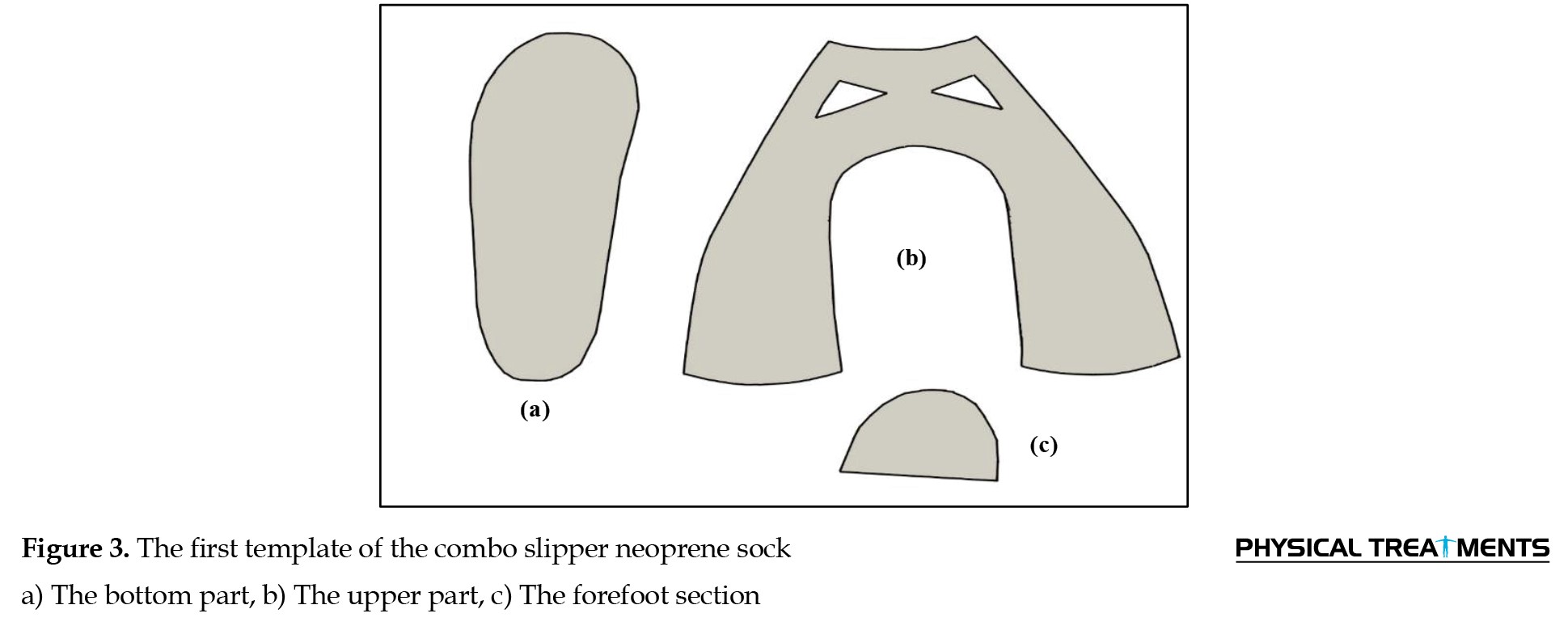
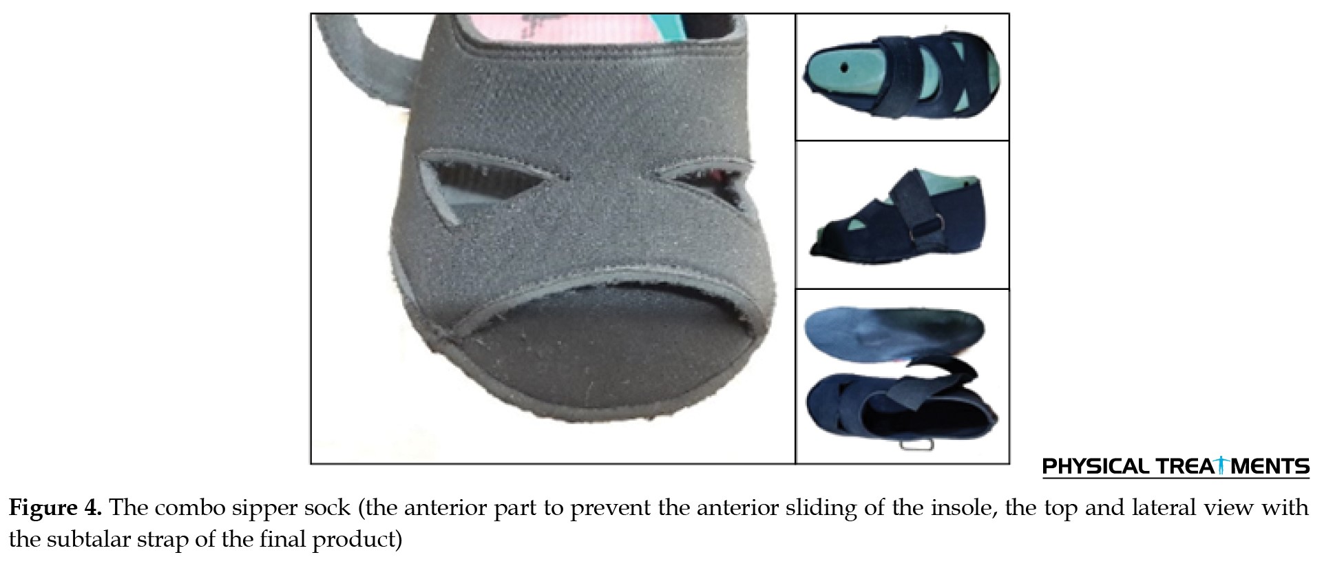
A neoprene slipper sock was designed to fit the insole, following a template created by placing the insole on a standard shoe mold. The sock’s upper was ventilated with three incisions to improve air circulation and reduce sweating. A 4 cm rubber subtalar strap was added, with a Velcro-secured attachment and a metal ring for lateral stability. The design was finalized in CorelDRAW software to standardize sizes across different foot dimensions (Figures 3 and 4).
Statistical analysis
The data were first assessed for normality using the Shapiro-Wilk test. For plantar pressure distribution and related gait parameters, comparisons between the barefoot and combo slipper sock conditions were conducted using a paired-samples t-test, as all data met normality assumptions. SPSS software, version 24 was employed for all statistical analyses, with significance set at P<0.05.
Results
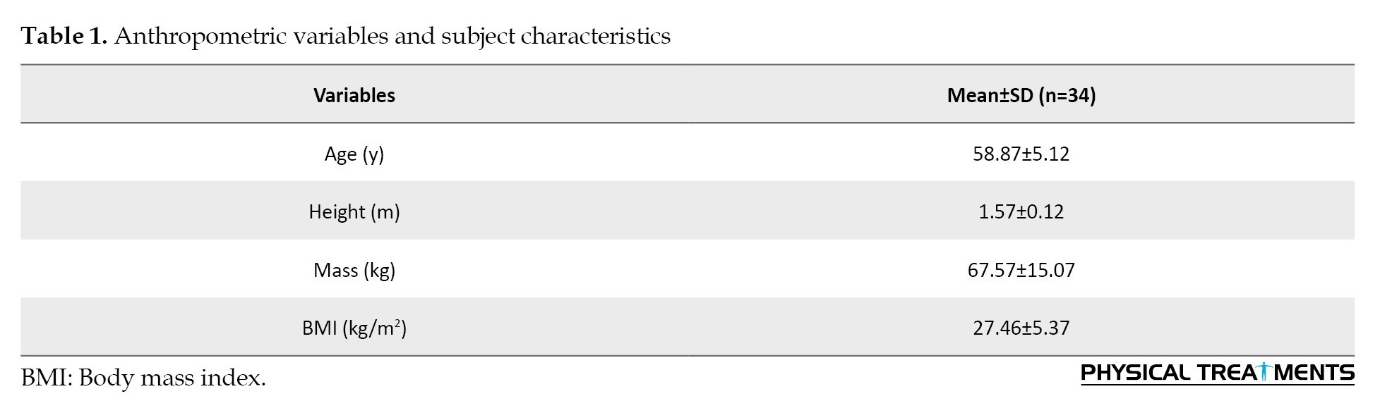
Table 1 presents the Mean±SD of participants’ anthropometric and demographic characteristics.
The results of the paired-samples t-test comparing maximum plantar pressure and relative time for the rollover phases of the quadruple-stance subphases between the two walking conditions (barefoot and wearing combo slippers socks) indicated significant differences. Notably, the two conditions showed significant differences in both plantar pressure variables (P=0.03) and the relative time for the rollover phases (P=0.04). Additionally, VAS results indicated that participants reported less pain when walking with combo slipper socks than when walking barefoot (t=3.04, P=0.01). However, no significant difference was observed in this index at rest.
Table 2 compares the maximum plantar pressure parameters in the ten-foot areas during walking gait, relative time for roll-over phases, and VAS between walking barefoot and with a combo slipper sock.
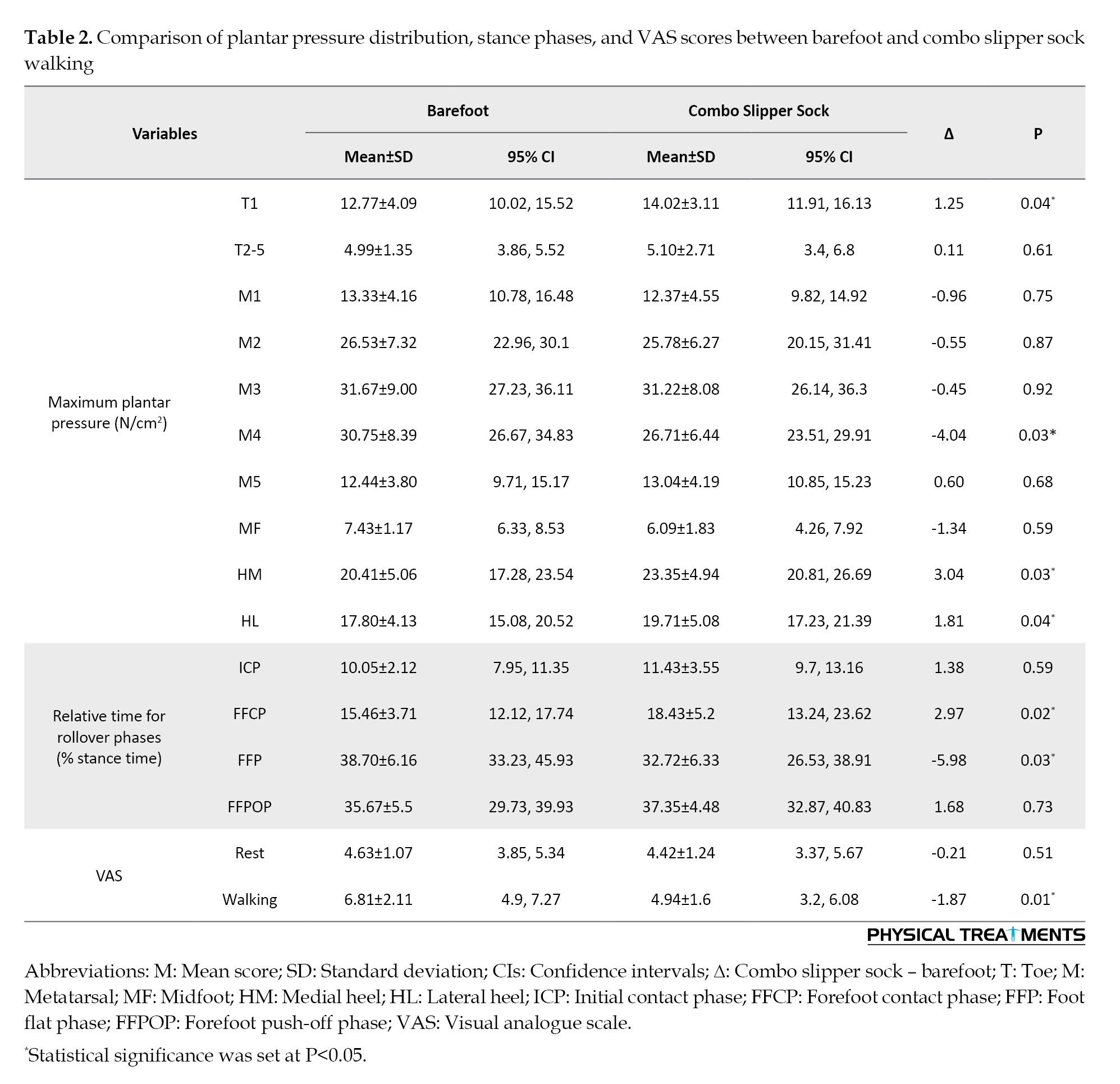
The results show that participants had lower maximum plantar pressure in T1 (t=2.18, P=0.04), lateral heel (HL) (t=2.15, P=0.04), and medial heel (HM) (t=2.15, P=0.03) areas when wearing the combo slipper sock. In contrast, participants experienced higher maximum plantar pressure in the M4 area (t=2.22, P=0.03) when walking barefoot. No significant differences were observed in other values related to plantar pressure distribution across ten zones.
Regarding the relative time spent in the roll-over phases, walking with the combo slipper sock resulted in more time in the initial contact phase (ICP), forefoot contact phase (FFCP), and forefoot push-off phase (FFPOP). Only the FFCP values (t=2.32, P=0.02) showed a significant difference. In contrast, walking barefoot resulted in spending a significantly longer time in the FFP (t=2.21, P=0.03) than walking with the combo slipper sock.
Discussion
This study aimed to design and evaluate a novel combination of a slipper sock and a custom LWI tailored for individuals with KOA. The primary objective was to assess its effectiveness in alleviating pain and optimizing plantar pressure distribution. The integration of a detachable insole, developed using CAD/CAM, enables versatile use both in footwear and independently at home. The slipper sock, constructed from neoprene, ensures comfort, washability, and affordability, offering a cost-effective and accessible solution for patients with KOA.
The innovation behind this design lies in its simplicity and adaptability, distinguishing it from previous orthotic interventions. Earlier approaches, such as the dual-sole sock with an insole pocket designed by Egozi, primarily targeted in-shoe applications and utilized memory foam insoles [30]. Similarly, Saeedi et al. introduced a laminated sock-insole system for flat feet, but its rigid design lacked adaptability [25]. Johnson’s method incorporated a customized wedge slab but failed to address wearability within different footwear settings [31]. Our study represents the first attempt to develop a hybrid slipper sock with integrated orthotic support explicitly designed for patients with KOA, bridging the gap between in-shoe and at-home orthotic solutions.
Our findings revealed significant reductions in pain among individuals with medial KOA while walking with the combo slipper sock and LWIs. These improvements are likely attributed to biomechanical modifications, particularly the lateral shift in the COP, which effectively reduces the KAM arm and, consequently, the external KAM—a key contributor to pain and joint stress [11, 32].
These results align with previous studies demonstrating the efficacy of LWIs in reducing pain and influencing the COP trajectory and plantar pressure distribution in patients with KOA [6, 16, 33-38]. By shifting the COP laterally, LWIs can reduce KAM, a primary contributor to medial compartment loading in KOA. This reduction in KAM helps redistribute plantar loads, mitigating excessive stress on the knee’s medial compartment, potentially decelerating KOA progression and enhancing functional mobility [6, 11].
Beyond pain reduction, our study highlights distinct alterations in plantar pressure distribution and functional mobility between patients with KOA and healthy individuals. Patients with KOA exhibited lower maximum plantar pressure in the medial and lateral heel regions, with compensatory increases in midfoot and fourth metatarsal pressures. These shifts may reflect biomechanical adaptations aimed at minimizing medial knee joint loading, a characteristic feature of KOA progression [39].
Moreover, the observed shortening of initial contact and forefoot push-off phases suggests that patients with KOA adopt gait modifications to reduce weight-bearing duration on the affected knee, likely as a pain-avoidance strategy [27]. This aligns with previous studies reporting reduced stance phase duration and altered CoP trajectories in patients with KOA due to joint instability and discomfort [9]. LWIs may counteract these effects by promoting a more stable stance phase through improved foot pronation control, thereby enhancing weight distribution and reducing excessive medial knee loading [40]. Additionally, LWIs facilitate a smoother COP transition from heel-strike to toe-off, which may improve propulsion mechanics and reduce compensatory forefoot loading [41]. In contrast, the prolonged forefoot plantarflexion phase observed in patients with KOA may represent an adaptive response to enhance balance and stability during weight transfer [27]. By encouraging a more natural gait pattern and redistributing plantar pressures, LWIs may help mitigate these compensatory gait alterations and enhance overall functional mobility [42].
Functional mobility assessments further reinforce these biomechanical alterations. The timed up and go (TUG) test results demonstrated significantly prolonged completion times in KOA patients compared to healthy counterparts, indicative of reduced lower limb strength, balance, and coordination. These impairments heighten the risk of falls and hinder independence in daily activities. Prior research supports the reliability of the TUG test as a performance-based measure for individuals with KOA, with prolonged test times associated with knee pain and quadriceps muscle weakness [26, 27].
The outcomes of this study underscore the potential of the combo slipper sock with LWIs as a practical and conservative intervention for KOA management. Healthcare practitioners should consider an LWI as a non-invasive treatment option, particularly for patients with medial compartment KOA. However, their use should be individualized, as not all patients may respond favorably. Clinical guidance is crucial in ensuring appropriate patient selection and optimizing the therapeutic benefits of lateral wedge orthotics.
The versatility of this design allows patients to seamlessly integrate insoles into their daily routines. Those with unilateral KOA may wear insoles only on the affected side, opting for a non-wedged version on the opposite foot, ensuring greater adaptability. This individualized approach enhances patient compliance and treatment effectiveness.
Conclusion
This study provides compelling evidence supporting the efficacy of a novel combo slipper sock with an LWI in reducing pain and modifying plantar pressure distribution in patients with KOA. These findings contribute to a deeper understanding of the compensatory gait strategies adopted by individuals with KOA and their implications for functional mobility. By identifying specific regions of increased plantar pressure, clinicians can better tailor interventions to address both biomechanical and functional deficits.
Early intervention strategies aimed at redistributing plantar loads, enhancing knee joint stability, and improving overall functional mobility may help slow KOA progression and improve patients’ quality of life. Future research should explore the long-term effects of such interventions and further investigate the interplay between foot alignment, gait biomechanics, and functional mobility in KOA management.
The combo slipper sock and custom-made LWI presented in this study provide a practical, user-friendly solution, allowing patients to easily integrate the insole into their daily routines, whether at home or in a shoe. In cases of unilateral medial compartment KOA, patients can opt to wear the insole on the affected side only, choosing a non-wedged version on the opposite side, enhancing adaptability based on individual needs.
Ethical Considerations
Compliance with ethical guidelines
This study was approved by the Research Ethics Committee of Hamadan University of Medical Sciences, Hamadan, Iran (Code: IR.UMSHA.REC.1401.926).
Funding
This project was funded by the Research Deputy of Hamadan University of Medical Sciences, Hamadan, Iran (Project No.: 14020115183).
Authors' contributions
Conceptualization and supervision: Mehrdad Anbarian and Nahid Tafti; Methodology and investigation: Nahid Tafti, Fereshteh Sabet, and Ehsan Ebrahimpour; Software, data curation, and formal analysis: Fereshteh Sabet and Ehsan Ebrahimpour; Writing the original draft preparation: Fereshteh Sabet and Ehsan Ebrahimpour; Resources, project administration, and funding acquisition: Nahid Tafti; Review, editing and visualization: All authors.
Conflict of interest
The authors declared no conflict of interest.
Acknowledgments
The authors thank all participants in this study.
Full-Text: (280 Views)
Introduction
Knee osteoarthritis (KOA) is a prevalent degenerative joint disorder characterized by progressive cartilage deterioration, resulting in pain, stiffness, and restricted mobility [1]. This condition affects approximately 19.4% of the Iranian population and is often associated with genu varum, a lower-limb malalignment commonly observed among Iranians, Chinese, Koreans, and Pakistanis. This misalignment increases stress on the medial compartment of the knee joint, accelerating cartilage degradation and exacerbating KOA symptoms [1, 2].
Patients with KOA often exhibit altered gait mechanics and distinctive plantar pressure distribution patterns compared to healthy individuals [3]. Research indicates that individuals with KOA experience reduced weight distribution on the hallux and heel, with increased pressure localized to the central foot regions [4, 5]. These deviations in plantar pressure contribute to a forward shift in the center of pressure (CoP) at the start of walking and a backward shift at the end, alongside reductions in anteroposterior CoP path length (%Long) and transverse CoP path width (%Trans) [6]. Understanding these biomechanical alterations is essential for developing effective interventions to improve gait and reduce symptom severity.
Plantar pressure analysis is a widely utilized tool for assessing lower-limb pathologies and gait mechanics, employing either pressure plate systems or in-shoe sensor technology. Lidtke et al. used pressure plate analysis to compare plantar pressure distribution in patients with KOA and healthy individuals, revealing a medial shift in CoP trajectory in the KOA group, suggesting altered weight-bearing patterns [7]. The F-Scan sensor system, introduced in 1998, is another commonly used tool for in-shoe pressure measurement and gait analysis [8]. Zhang et al. further examined plantar pressure data among women with KOA and found increased midfoot and metatarsophalangeal joint loading, which may contribute to foot pronation and gait alterations [9]. Despite these advancements, gait laboratory evaluations remain costly and inaccessible for many patients, necessitating alternative methods for biomechanical assessment [10, 11].
Lateral wedge insoles (LWIs) have been widely recommended as a non-surgical intervention for medial KOA, aiming to redistribute load away from the medial tibiofemoral compartment to alleviate pain and enhance joint function [12]. Initially developed by Sasaki and Yasuda in 1987, these insoles were designed to mitigate the knee adduction moment (KAM) and reduce excessive medial joint loading [13]. While numerous studies have demonstrated their biomechanical effectiveness, their impact on pain relief and functional improvement remains inconclusive, with mixed findings across clinical trials [14-18]. The efficacy of LWIs largely depends on critical design factors, including wedge angle, material properties, and support features, which must be carefully tailored to the needs of KOA patients [19, 20].
The selection of appropriate insoles is often guided by individual comfort preferences; however, research suggests that a full-length lateral wedge with a 6° incline may provide optimal pain relief [21]. The material composition of the insole is also a determining factor: a firmer lateral foam minimizes compression under weight-bearing conditions, while softer medial elements enhance comfort and promote a more even plantar pressure distribution [22]. Furthermore, arch-supporting insoles that stabilize both longitudinal and transverse arches have been shown to optimize weight redistribution [23]. Additional design modifications, such as incorporating a subtalar strap, may improve joint stability and potentially slow KOA progression more effectively than LWIs alone [24]. However, such modifications may necessitate purchasing larger footwear, adding a financial burden for patients.
Previous research has explored sock-integrated insole designs for convenient use in home environments, yet few products successfully combine all essential features necessary for KOA management [25]. An ideal sock-based intervention should include a secure fit, a detachable LWI, a stabilizing subtalar strap, and durable materials for sustained use.
Footwear selection plays a crucial role in the effectiveness of LWIs for patients with KOA. Although closed-toe shoes or sandals are commonly recommended, the angled design of LWIs often requires wearing larger footwear, which imposes financial and practical limitations. Sandals, while potentially more compatible with LWIs in home environments, may not be suitable for populations with soft flooring or sedentary lifestyles, such as elderly individuals in Iranian households. Additionally, sandals generally offer inadequate ankle support and may compromise stability, particularly on uneven surfaces. These factors highlight the need for a more practical and cost-effective alternative to conventional footwear.
To address these limitations, we developed an innovative sock-like footwear solution, featuring integrated LWIs and a supportive subtalar strap. This design provides a lightweight, comfortable, and stable alternative to conventional footwear, accommodating both sedentary and active individuals across diverse household environments while remaining cost-effective.
This technical note introduces a novel slipper sock incorporating a custom-made LWI designed specifically for individuals with KOA. This study aims to evaluate the effectiveness of this innovative intervention in reducing pain and redistributing plantar pressure. We hypothesize that the novel slipper sock with an integrated LWI will significantly reduce pain and improve plantar pressure distribution in patients with KOA.
Materials and Methods
This study recruited 34 female participants diagnosed with bilateral KOA from medical and rehabilitation centers in Hamadan City, Iran. The sample was selected from a larger pool of 133 surveyed individuals with KOA, and the sample size was determined using statistical methods relevant to biomechanical interventions in KOA populations. Each participant provided informed consent before enrollment. The sample size for this study was calculated using G*Power software, version 3.1.9.2 for a paired t-test. An effect size of 0.446 was used, with an alpha level of 0.05 and a desired power of 0.8.
Inclusion and exclusion criteria
Participants eligible for this study were those aged >45 years with clinically diagnosed KOA. The diagnosis was established according to the clinical criteria set forth by the American College of Rheumatology (ACR), with severity determined using the Kellgren-Lawrence (K/L) grading scale; specifically, a K/L grade of >1 was required to confirm KOA. Only participants with K/L grades of 2 or 3 were included, as assessed by a qualified physician through clinical examination and knee radiography [26].
Eligible individuals were required to have no history of cerebrovascular accidents (stroke), uncontrolled hypertension, neuromuscular disorders (such as multiple sclerosis or Parkinson’s disease), or other neurological conditions (including cerebral palsy, dementia, or vertigo). Participants with a body mass index (BMI) greater than 40 kg/m², lower-extremity fractures, concurrent hip osteoarthritis, or awaiting knee arthroplasty were excluded from the study. Additionally, candidates must not require assistance with standing or walking, nor have received any form of treatment—such as medications, injections, or surgical procedures—within six months prior to the commencement of the study [12, 27, 28].
Pain assessment scale (visual analog scale [VAS])
Pain intensity was measured using the VAS, ranging from 0 (“no pain”) to 10 (“unbearable pain”). This scale, with an intraclass correlation coefficient (ICC) of 0.91, is well established for its validity and reliability in chronic pain assessment. Participants completed two VAS assessments under different conditions: walking barefoot and walking with the combo slipper sock. For each condition, pain was measured three times, and the average was used as the final score. Measurements were taken in a standardized left-to-right order, and participants were blinded to their previous scores to avoid bias [29].
Plantar pressure distribution
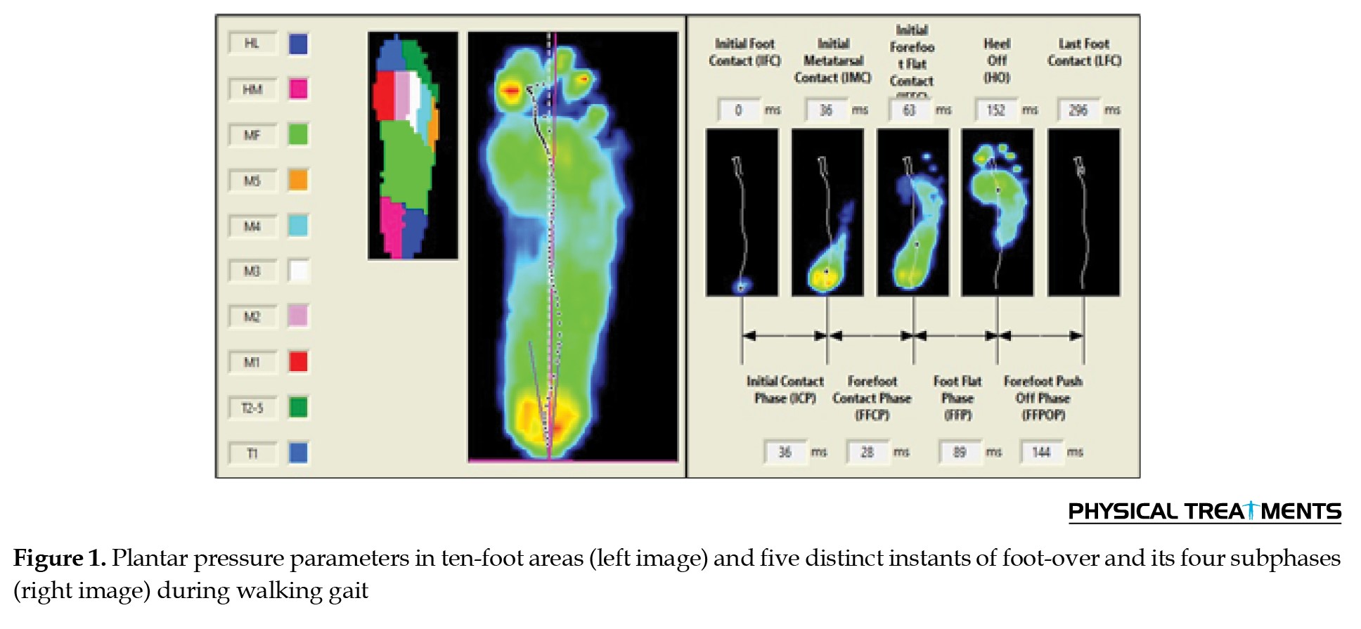
A foot scan pressure plate device (RSscan International, Belgium) with a 40×100 cm surface, 8192 sensors, and a 253 Hz sampling rate was used to assess plantar pressure. The accompanying Footscan software (gait second generation) divided the foot into 10 anatomical zones (medial heel, lateral heel, midfoot, metatarsals M1–M5, hallux, and lesser toes) and measured peak plantar pressure for each. The software further divided the stance phase into four subphases (initial contact, forefoot contact, foot flat, and forefoot push-off) based on distinct stance points (e.g. first foot contact, heel-off) (Figure 1) [27].
Knee osteoarthritis (KOA) is a prevalent degenerative joint disorder characterized by progressive cartilage deterioration, resulting in pain, stiffness, and restricted mobility [1]. This condition affects approximately 19.4% of the Iranian population and is often associated with genu varum, a lower-limb malalignment commonly observed among Iranians, Chinese, Koreans, and Pakistanis. This misalignment increases stress on the medial compartment of the knee joint, accelerating cartilage degradation and exacerbating KOA symptoms [1, 2].
Patients with KOA often exhibit altered gait mechanics and distinctive plantar pressure distribution patterns compared to healthy individuals [3]. Research indicates that individuals with KOA experience reduced weight distribution on the hallux and heel, with increased pressure localized to the central foot regions [4, 5]. These deviations in plantar pressure contribute to a forward shift in the center of pressure (CoP) at the start of walking and a backward shift at the end, alongside reductions in anteroposterior CoP path length (%Long) and transverse CoP path width (%Trans) [6]. Understanding these biomechanical alterations is essential for developing effective interventions to improve gait and reduce symptom severity.
Plantar pressure analysis is a widely utilized tool for assessing lower-limb pathologies and gait mechanics, employing either pressure plate systems or in-shoe sensor technology. Lidtke et al. used pressure plate analysis to compare plantar pressure distribution in patients with KOA and healthy individuals, revealing a medial shift in CoP trajectory in the KOA group, suggesting altered weight-bearing patterns [7]. The F-Scan sensor system, introduced in 1998, is another commonly used tool for in-shoe pressure measurement and gait analysis [8]. Zhang et al. further examined plantar pressure data among women with KOA and found increased midfoot and metatarsophalangeal joint loading, which may contribute to foot pronation and gait alterations [9]. Despite these advancements, gait laboratory evaluations remain costly and inaccessible for many patients, necessitating alternative methods for biomechanical assessment [10, 11].
Lateral wedge insoles (LWIs) have been widely recommended as a non-surgical intervention for medial KOA, aiming to redistribute load away from the medial tibiofemoral compartment to alleviate pain and enhance joint function [12]. Initially developed by Sasaki and Yasuda in 1987, these insoles were designed to mitigate the knee adduction moment (KAM) and reduce excessive medial joint loading [13]. While numerous studies have demonstrated their biomechanical effectiveness, their impact on pain relief and functional improvement remains inconclusive, with mixed findings across clinical trials [14-18]. The efficacy of LWIs largely depends on critical design factors, including wedge angle, material properties, and support features, which must be carefully tailored to the needs of KOA patients [19, 20].
The selection of appropriate insoles is often guided by individual comfort preferences; however, research suggests that a full-length lateral wedge with a 6° incline may provide optimal pain relief [21]. The material composition of the insole is also a determining factor: a firmer lateral foam minimizes compression under weight-bearing conditions, while softer medial elements enhance comfort and promote a more even plantar pressure distribution [22]. Furthermore, arch-supporting insoles that stabilize both longitudinal and transverse arches have been shown to optimize weight redistribution [23]. Additional design modifications, such as incorporating a subtalar strap, may improve joint stability and potentially slow KOA progression more effectively than LWIs alone [24]. However, such modifications may necessitate purchasing larger footwear, adding a financial burden for patients.
Previous research has explored sock-integrated insole designs for convenient use in home environments, yet few products successfully combine all essential features necessary for KOA management [25]. An ideal sock-based intervention should include a secure fit, a detachable LWI, a stabilizing subtalar strap, and durable materials for sustained use.
Footwear selection plays a crucial role in the effectiveness of LWIs for patients with KOA. Although closed-toe shoes or sandals are commonly recommended, the angled design of LWIs often requires wearing larger footwear, which imposes financial and practical limitations. Sandals, while potentially more compatible with LWIs in home environments, may not be suitable for populations with soft flooring or sedentary lifestyles, such as elderly individuals in Iranian households. Additionally, sandals generally offer inadequate ankle support and may compromise stability, particularly on uneven surfaces. These factors highlight the need for a more practical and cost-effective alternative to conventional footwear.
To address these limitations, we developed an innovative sock-like footwear solution, featuring integrated LWIs and a supportive subtalar strap. This design provides a lightweight, comfortable, and stable alternative to conventional footwear, accommodating both sedentary and active individuals across diverse household environments while remaining cost-effective.
This technical note introduces a novel slipper sock incorporating a custom-made LWI designed specifically for individuals with KOA. This study aims to evaluate the effectiveness of this innovative intervention in reducing pain and redistributing plantar pressure. We hypothesize that the novel slipper sock with an integrated LWI will significantly reduce pain and improve plantar pressure distribution in patients with KOA.
Materials and Methods
This study recruited 34 female participants diagnosed with bilateral KOA from medical and rehabilitation centers in Hamadan City, Iran. The sample was selected from a larger pool of 133 surveyed individuals with KOA, and the sample size was determined using statistical methods relevant to biomechanical interventions in KOA populations. Each participant provided informed consent before enrollment. The sample size for this study was calculated using G*Power software, version 3.1.9.2 for a paired t-test. An effect size of 0.446 was used, with an alpha level of 0.05 and a desired power of 0.8.
Inclusion and exclusion criteria
Participants eligible for this study were those aged >45 years with clinically diagnosed KOA. The diagnosis was established according to the clinical criteria set forth by the American College of Rheumatology (ACR), with severity determined using the Kellgren-Lawrence (K/L) grading scale; specifically, a K/L grade of >1 was required to confirm KOA. Only participants with K/L grades of 2 or 3 were included, as assessed by a qualified physician through clinical examination and knee radiography [26].
Eligible individuals were required to have no history of cerebrovascular accidents (stroke), uncontrolled hypertension, neuromuscular disorders (such as multiple sclerosis or Parkinson’s disease), or other neurological conditions (including cerebral palsy, dementia, or vertigo). Participants with a body mass index (BMI) greater than 40 kg/m², lower-extremity fractures, concurrent hip osteoarthritis, or awaiting knee arthroplasty were excluded from the study. Additionally, candidates must not require assistance with standing or walking, nor have received any form of treatment—such as medications, injections, or surgical procedures—within six months prior to the commencement of the study [12, 27, 28].
Pain assessment scale (visual analog scale [VAS])
Pain intensity was measured using the VAS, ranging from 0 (“no pain”) to 10 (“unbearable pain”). This scale, with an intraclass correlation coefficient (ICC) of 0.91, is well established for its validity and reliability in chronic pain assessment. Participants completed two VAS assessments under different conditions: walking barefoot and walking with the combo slipper sock. For each condition, pain was measured three times, and the average was used as the final score. Measurements were taken in a standardized left-to-right order, and participants were blinded to their previous scores to avoid bias [29].
Plantar pressure distribution
A foot scan pressure plate device (RSscan International, Belgium) with a 40×100 cm surface, 8192 sensors, and a 253 Hz sampling rate was used to assess plantar pressure. The accompanying Footscan software (gait second generation) divided the foot into 10 anatomical zones (medial heel, lateral heel, midfoot, metatarsals M1–M5, hallux, and lesser toes) and measured peak plantar pressure for each. The software further divided the stance phase into four subphases (initial contact, forefoot contact, foot flat, and forefoot push-off) based on distinct stance points (e.g. first foot contact, heel-off) (Figure 1) [27].
Participants walked naturally along a 12-meter walkway with the device positioned mid-path. Each participant performed three trials for each condition (barefoot and slipper sock), with a three-minute seated rest between trials to prevent fatigue. Data were collected without participants viewing the recording screen to prevent alterations in gait. Before data collection, a familiarization session was conducted. The order of data collection was randomized using Random.org, with a 48-hour gap between sessions to avoid fatigue [27].
Design and fabrication of the LWI
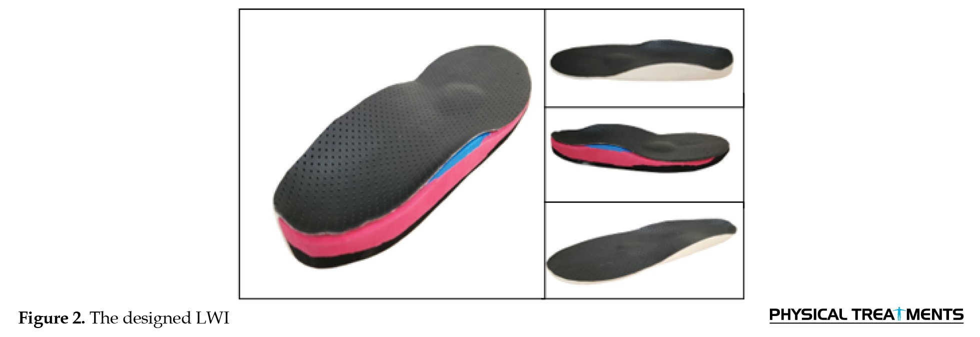
The insole was designed in Rhinoceros 3D® software, incorporating medial and lateral arch support, a metatarsal pad, and a 6° medial heel-and-sole wedge. Separate G-codes were calculated for each side of the insole. To construct the foam block, two layers of Plastazote foam (shore A 70) and ethyl vinyl acetate (shore A 20) were cut, sanded, and adhered to form a 3 cm thickness. The block was then milled with a CNC machine based on the G-code, and the insole’s upper surface was covered in artificial leather (Figure 2).
Design and fabrication of the LWI
The insole was designed in Rhinoceros 3D® software, incorporating medial and lateral arch support, a metatarsal pad, and a 6° medial heel-and-sole wedge. Separate G-codes were calculated for each side of the insole. To construct the foam block, two layers of Plastazote foam (shore A 70) and ethyl vinyl acetate (shore A 20) were cut, sanded, and adhered to form a 3 cm thickness. The block was then milled with a CNC machine based on the G-code, and the insole’s upper surface was covered in artificial leather (Figure 2).
Combo slipper sock prototype
Prototype and Finalization of the Combo Slipper Sock
A neoprene slipper sock was designed to fit the insole, following a template created by placing the insole on a standard shoe mold. The sock’s upper was ventilated with three incisions to improve air circulation and reduce sweating. A 4 cm rubber subtalar strap was added, with a Velcro-secured attachment and a metal ring for lateral stability. The design was finalized in CorelDRAW software to standardize sizes across different foot dimensions (Figures 3 and 4).
Statistical analysis
The data were first assessed for normality using the Shapiro-Wilk test. For plantar pressure distribution and related gait parameters, comparisons between the barefoot and combo slipper sock conditions were conducted using a paired-samples t-test, as all data met normality assumptions. SPSS software, version 24 was employed for all statistical analyses, with significance set at P<0.05.
Results
Table 1 presents the Mean±SD of participants’ anthropometric and demographic characteristics.
The results of the paired-samples t-test comparing maximum plantar pressure and relative time for the rollover phases of the quadruple-stance subphases between the two walking conditions (barefoot and wearing combo slippers socks) indicated significant differences. Notably, the two conditions showed significant differences in both plantar pressure variables (P=0.03) and the relative time for the rollover phases (P=0.04). Additionally, VAS results indicated that participants reported less pain when walking with combo slipper socks than when walking barefoot (t=3.04, P=0.01). However, no significant difference was observed in this index at rest.
Table 2 compares the maximum plantar pressure parameters in the ten-foot areas during walking gait, relative time for roll-over phases, and VAS between walking barefoot and with a combo slipper sock.
The results show that participants had lower maximum plantar pressure in T1 (t=2.18, P=0.04), lateral heel (HL) (t=2.15, P=0.04), and medial heel (HM) (t=2.15, P=0.03) areas when wearing the combo slipper sock. In contrast, participants experienced higher maximum plantar pressure in the M4 area (t=2.22, P=0.03) when walking barefoot. No significant differences were observed in other values related to plantar pressure distribution across ten zones.
Regarding the relative time spent in the roll-over phases, walking with the combo slipper sock resulted in more time in the initial contact phase (ICP), forefoot contact phase (FFCP), and forefoot push-off phase (FFPOP). Only the FFCP values (t=2.32, P=0.02) showed a significant difference. In contrast, walking barefoot resulted in spending a significantly longer time in the FFP (t=2.21, P=0.03) than walking with the combo slipper sock.
Discussion
This study aimed to design and evaluate a novel combination of a slipper sock and a custom LWI tailored for individuals with KOA. The primary objective was to assess its effectiveness in alleviating pain and optimizing plantar pressure distribution. The integration of a detachable insole, developed using CAD/CAM, enables versatile use both in footwear and independently at home. The slipper sock, constructed from neoprene, ensures comfort, washability, and affordability, offering a cost-effective and accessible solution for patients with KOA.
The innovation behind this design lies in its simplicity and adaptability, distinguishing it from previous orthotic interventions. Earlier approaches, such as the dual-sole sock with an insole pocket designed by Egozi, primarily targeted in-shoe applications and utilized memory foam insoles [30]. Similarly, Saeedi et al. introduced a laminated sock-insole system for flat feet, but its rigid design lacked adaptability [25]. Johnson’s method incorporated a customized wedge slab but failed to address wearability within different footwear settings [31]. Our study represents the first attempt to develop a hybrid slipper sock with integrated orthotic support explicitly designed for patients with KOA, bridging the gap between in-shoe and at-home orthotic solutions.
Our findings revealed significant reductions in pain among individuals with medial KOA while walking with the combo slipper sock and LWIs. These improvements are likely attributed to biomechanical modifications, particularly the lateral shift in the COP, which effectively reduces the KAM arm and, consequently, the external KAM—a key contributor to pain and joint stress [11, 32].
These results align with previous studies demonstrating the efficacy of LWIs in reducing pain and influencing the COP trajectory and plantar pressure distribution in patients with KOA [6, 16, 33-38]. By shifting the COP laterally, LWIs can reduce KAM, a primary contributor to medial compartment loading in KOA. This reduction in KAM helps redistribute plantar loads, mitigating excessive stress on the knee’s medial compartment, potentially decelerating KOA progression and enhancing functional mobility [6, 11].
Beyond pain reduction, our study highlights distinct alterations in plantar pressure distribution and functional mobility between patients with KOA and healthy individuals. Patients with KOA exhibited lower maximum plantar pressure in the medial and lateral heel regions, with compensatory increases in midfoot and fourth metatarsal pressures. These shifts may reflect biomechanical adaptations aimed at minimizing medial knee joint loading, a characteristic feature of KOA progression [39].
Moreover, the observed shortening of initial contact and forefoot push-off phases suggests that patients with KOA adopt gait modifications to reduce weight-bearing duration on the affected knee, likely as a pain-avoidance strategy [27]. This aligns with previous studies reporting reduced stance phase duration and altered CoP trajectories in patients with KOA due to joint instability and discomfort [9]. LWIs may counteract these effects by promoting a more stable stance phase through improved foot pronation control, thereby enhancing weight distribution and reducing excessive medial knee loading [40]. Additionally, LWIs facilitate a smoother COP transition from heel-strike to toe-off, which may improve propulsion mechanics and reduce compensatory forefoot loading [41]. In contrast, the prolonged forefoot plantarflexion phase observed in patients with KOA may represent an adaptive response to enhance balance and stability during weight transfer [27]. By encouraging a more natural gait pattern and redistributing plantar pressures, LWIs may help mitigate these compensatory gait alterations and enhance overall functional mobility [42].
Functional mobility assessments further reinforce these biomechanical alterations. The timed up and go (TUG) test results demonstrated significantly prolonged completion times in KOA patients compared to healthy counterparts, indicative of reduced lower limb strength, balance, and coordination. These impairments heighten the risk of falls and hinder independence in daily activities. Prior research supports the reliability of the TUG test as a performance-based measure for individuals with KOA, with prolonged test times associated with knee pain and quadriceps muscle weakness [26, 27].
The outcomes of this study underscore the potential of the combo slipper sock with LWIs as a practical and conservative intervention for KOA management. Healthcare practitioners should consider an LWI as a non-invasive treatment option, particularly for patients with medial compartment KOA. However, their use should be individualized, as not all patients may respond favorably. Clinical guidance is crucial in ensuring appropriate patient selection and optimizing the therapeutic benefits of lateral wedge orthotics.
The versatility of this design allows patients to seamlessly integrate insoles into their daily routines. Those with unilateral KOA may wear insoles only on the affected side, opting for a non-wedged version on the opposite foot, ensuring greater adaptability. This individualized approach enhances patient compliance and treatment effectiveness.
Conclusion
This study provides compelling evidence supporting the efficacy of a novel combo slipper sock with an LWI in reducing pain and modifying plantar pressure distribution in patients with KOA. These findings contribute to a deeper understanding of the compensatory gait strategies adopted by individuals with KOA and their implications for functional mobility. By identifying specific regions of increased plantar pressure, clinicians can better tailor interventions to address both biomechanical and functional deficits.
Early intervention strategies aimed at redistributing plantar loads, enhancing knee joint stability, and improving overall functional mobility may help slow KOA progression and improve patients’ quality of life. Future research should explore the long-term effects of such interventions and further investigate the interplay between foot alignment, gait biomechanics, and functional mobility in KOA management.
The combo slipper sock and custom-made LWI presented in this study provide a practical, user-friendly solution, allowing patients to easily integrate the insole into their daily routines, whether at home or in a shoe. In cases of unilateral medial compartment KOA, patients can opt to wear the insole on the affected side only, choosing a non-wedged version on the opposite side, enhancing adaptability based on individual needs.
Ethical Considerations
Compliance with ethical guidelines
This study was approved by the Research Ethics Committee of Hamadan University of Medical Sciences, Hamadan, Iran (Code: IR.UMSHA.REC.1401.926).
Funding
This project was funded by the Research Deputy of Hamadan University of Medical Sciences, Hamadan, Iran (Project No.: 14020115183).
Authors' contributions
Conceptualization and supervision: Mehrdad Anbarian and Nahid Tafti; Methodology and investigation: Nahid Tafti, Fereshteh Sabet, and Ehsan Ebrahimpour; Software, data curation, and formal analysis: Fereshteh Sabet and Ehsan Ebrahimpour; Writing the original draft preparation: Fereshteh Sabet and Ehsan Ebrahimpour; Resources, project administration, and funding acquisition: Nahid Tafti; Review, editing and visualization: All authors.
Conflict of interest
The authors declared no conflict of interest.
Acknowledgments
The authors thank all participants in this study.
References
- Hochberg MC, Altman RD, April KT, Benkhalti M, Guyatt G, McGowan J, et al. American College of Rheumatology 2012 recommendations for the use of nonpharmacologic and pharmacologic therapies in osteoarthritis of the hand, hip, and knee. Arthritis Care & Research. 2012; 64(4):465-74. [DOI:10.1002/acr.21596] [PMID]
- Bennell KL, Hunter DJ, Hinman RS. Management of osteoarthritis of the knee. BMJ. 2012; 345:e4934. [DOI:10.1136/bmj.e4934] [PMID]
- Katz JN, Arant KR, Loeser RF. Diagnosis and treatment of hip and knee osteoarthritis: A review. JAMA. 2021; 325(6):568-78. [DOI:10.1001/jama.2020.22171] [PMID]
- Saito I, Okada K, Wakasa M, Abe H, Saito A. Foot pressure pattern, hindfoot deformities, and their associations with foot pain in individuals with advanced medial knee osteoarthritis. Gait & Posture. 2018; 59:83-8. [DOI:10.1016/j.gaitpost.2017.09.041] [PMID]
- Saito I, Okada K, Nishi T, Wakasa M, Saito A, Sugawara K, et al. Foot pressure pattern and its correlation with knee range of motion limitations for individuals with medial knee osteoarthritis. Archives of Physical Medicine and Rehabilitation. 2013; 94(12):2502-8. [DOI:10.1016/j.apmr.2013.07.017] [PMID]
- Hsu WC, Chou LW, Chiu HY, Hsieh CW, Hu WP. A study on the effects of Lateral-Wedge Insoles on plantar-pressure pattern for medial knee osteoarthritis using the Wearable Sensing Insole. Sensors. 2022; 23(1):84. [DOI:10.3390/s23010084] [PMID]
- Lidtke RH, Muehleman C, Kwasny M, Block JA. Foot center of pressure and medial knee osteoarthritis. Journal of The American Podiatric Medical Association. 2010; 100(3):178-84. [DOI:10.7547/1000178] [PMID]
- Luo ZP, Berglund LJ, An KN. Validation of F-Scan pressure sensor system: A technical note. Journal of Rehabilitation Research and Development. 1998; 35(2):186-91. [PMID]
- Zhang Z, Wang L, Hu K, Liu Y. Characteristics of plantar loads during walking in patients with knee osteoarthritis. Medical Science Monitor: International Medical Journal of Experimental and Clinical Research. 2017; 23:5714. [DOI:10.12659/MSM.905136] [PMID]
- Felson DT, Parkes M, Carter S, Liu A, Callaghan MJ, Hodgson R, et al. The efficacy of a lateral wedge insole for painful medial knee osteoarthritis after prescreening: A randomized clinical trial. Arthritis & Rheumatology. 2019; 71(6):908-15. [DOI:10.1002/art.40808] [PMID]
- Hinman RS, Bowles KA, Metcalf BB, Wrigley TV, Bennell KL. Lateral wedge insoles for medial knee osteoarthritis: effects on lower limb frontal plane biomechanics. Clinical Biomechanics. 2012; 27(1):27-33. [DOI:10.1016/j.clinbiomech.2011.07.010] [PMID]
- Kolasinski SL, Neogi T, Hochberg MC, Oatis C, Guyatt G, Block J, et al. 2019 American College of Rheumatology/Arthritis Foundation guideline for the management of osteoarthritis of the hand, hip, and knee. Arthritis & Rheumatology. 2020; 72(2):220-33. [DOI:10.1002/art.41142] [PMID]
- Sasaki T, Yasuda K. Clinical evaluation of the treatment of osteoarthritic knees using a newly designed wedged insole. Clinical Orthopaedics and Related Research. 1987(221):181-7. [DOI:10.1097/00003086-198708000-00021]
- Pham T, Maillefert JF, Hudry C, Kieffert P, Bourgeois P, Lechevalier D, et al. Laterally elevated wedged insoles in the treatment of medial knee osteoarthritis: A two-year prospective randomized controlled study. Osteoarthritis and Cartilage. 2004. 12(1):46-55. [DOI:10.1016/j.joca.2003.08.011] [PMID]
- Bennell KL, Bowles KA, Payne C, Cicuttini F, Williamson E, Forbes A, et al. Lateral wedge insoles for medial knee osteoarthritis: 12 month randomised controlled trial. BMJ. 2011; 342:d2912. [DOI:10.1136/bmj.d2912] [PMID]
- Parkes MJ, Maricar N, Lunt M, LaValley MP, Jones RK, Segal NA, et al. Lateral wedge insoles as a conservative treatment for pain in patients with medial knee osteoarthritis: A meta-analysis. JAMA. 2013; 310(7):722-30. [DOI:10.1001/jama.2013.243229] [PMID]
- Zhang J, Wang Q, Zhang C. Ineffectiveness of lateral-wedge insoles on the improvement of pain and function for medial knee osteoarthritis: A meta-analysis of controlled randomized trials. Archives of Orthopaedic and Trauma Surgery. 2018; 138(10):1453-62. [DOI:10.1007/s00402-018-3004-z] [PMID]
- Shaw KE, Charlton JM, Perry CKL, de Vries CM, Redekopp MJ, White JA, et al. The effects of shoe-worn insoles on gait biomechanics in people with knee osteoarthritis: A systematic review and meta-analysis. British Journal of Sports Medicine. 2018; 52(4):238-53. [DOI:10.1136/bjsports-2016-097108] [PMID]
- Ferreira, VMF. The effects of application of lateral wedge insoles on medial osteoarthritis of the knee [PhD dissertation]. Porto: Universidade do Porto; 2020. [Link]
- Barati K, Ebrahimi Takamjani I, Shamsoddini A, Ejraei Dolatabad H. A comparison of the biomechanical and clinical effects of a biaxial ankle-foot orthosis and lateral wedge insole in individuals with medial knee osteoarthritis. Disability and Rehabilitation. 2022; 44(26):8501-8. [DOI:10.1080/09638288.2021.2019841] [PMID]
- Hinman RS, Bowles KA, Payne C, Bennell KL. Effect of length on laterally‐wedged insoles in knee osteoarthritis. Arthritis Care & Research: Official Journal of the American College of Rheumatology. 2008; 59(1):144-7. [DOI:10.1002/art.23249] [PMID]
- Paterson KL, Kasza J, Bennell KL, Wrigley TV, Metcalf BR, Campbell PK, et al. Moderators and mediators of effects of unloading shoes on knee pain in people with knee osteoarthritis: an exploratory analysis of the SHARK randomised controlled trial. Osteoarthritis and Cartilage. 2018; 26(2):227-35. [DOI:10.1016/j.joca.2017.11.002] [PMID]
- Guldemond NA, Leffers P, Schaper NC, Sanders AP, Nieman F, Willems P, et al. The effects of insole configurations on forefoot plantar pressure and walking convenience in diabetic patients with neuropathic feet. Clinical Biomechanics. 2007; 22(1):81-7. [DOI:10.1016/j.clinbiomech.2006.08.004] [PMID]
- Toda Y, Tsukimura N. A 2-year follow-up of a study to compare the efficacy of lateral wedged insoles with subtalar strapping and in-shoe lateral wedged insoles in patients with varus deformity osteoarthritis of the knee. Osteoarthritis and Cartilage. 2006; 14(3):231-7. [DOI:10.1016/j.joca.2005.09.006] [PMID]
- Saeedi H, Javanshir MA, Aboutorabi A. Cushioned stabilizing sock and method. 2016, United States patent application; 2016. [Link]
- Neogi T, Zhang Y. Epidemiology of osteoarthritis. Rheumatic Diseases Clinics of North America. 2013; 39(1):1-19. [DOI:10.1016/j.rdc.2012.10.004] [PMID]
- Etesami AS, Zolaktaf V, Esmaeili H. Effect of knee osteoarthritis on plantar pressure distribution pattern and timing of stance sub-phases in elderly females. Studies in Sport Medicine. 2021; 13(29):97-114. [Link]
- Ebrahimipour E, Anbarian M, Sabet F, Tafti N. Quantifying Spatial-temporal Parameters During Stair Ascent and Descent Among Knee Osteoarthritis Populations. Physical Treatments - Specific Physical Therapy Journal. 2024; 14 (2):147-58. [DOI:10.32598/ptj.14.2.596.1]
- Crichton N. Visual analogue scale (VAS). Journal of Clinical Nursing. 2001; 10(5):706-6. [Link]
- Egozi RD. Sock constructed with an insole. United States patent application. 2011. [Link]
- Johnson LL. Method of treating osteoarthritis using insoles. United States patent. 2012. [Link]
- Yasuda K. Sasaki T. The mechanics of treatment of the osteoarthritic knee with a wedged insole. Clinical Orthopaedics and Related Research. 1987; (215):162-72. [DOI:10.1097/00003086-198702000-00024]
- Tse CTF, Ryan MB, Dien J, Scott A, Hunt MA. An exploration of changes in plantar pressure distributions during walking with standalone and supported lateral wedge insole designs. Journal of Foot and Ankle Research. 2021; 14(1):55. [DOI:10.1186/s13047-021-00493-5] [PMID]
- Ferreira V, Machado L, Roriz P. Center of pressure alterations with the application of lateral wedge insoles. Motricidade. 2019; 15:61-61. [Link]
- Hovanlou F, Sabaghian Rad S, Memar R, Sadeghi H. [The effect of 12 weeks using of customized insoles and exercise in water (front crawl swimming) on plantar pressure distribution and muscle function of girls with flexible flat foot aged 10-14 years (Persin)]. Journal for Research in Sport Rehabilitation. 2019; 6(12):31-43. [Link]
- Haim A, Rozen N, Dekel S, Halperin N, Wolf A. Control of knee coronal plane moment via modulation of center of pressure: A prospective gait analysis study. Journal of Biomechanics. 2008; 41(14):3010-16. [DOI:10.1016/j.jbiomech.2008.07.029] [PMID]
- Moazaz A, Aminian G, Baghaie R, Nabavi H. [The effect of 8 degree lateral heel wedge on energy consumption and displacement of the center of pressure in medial compartment knee osteoarthritis in frontal plane (Persian)]. Iranian Journal of Orthopedic Surgery. 2020; 14(4):7-14. [DOI:10.22034/ijos.2020.121326]
- Moazaz A, Aminian G, Baghaie R, Nabavi H. The effect of 8 degree lateral wedge on energy consumption and displacement of the center of pressure in medial compartment knee osteoarthritis in frontal plane. Iranian Journal of Orthopedic Surgery. 2020; 14(4):7-14.[DOI:10.22034/ijos.2020.121326]
- Kour N, Gupta S, Arora S. A survey of knee osteoarthritis assessment based on gait. Archives of Computational Methods in Engineering. 2021; 28(2):345-85. [DOI:10.1007/s11831-019-09379-z]
- Nakagawa TH, Moriya ET, Maciel CD, Serrão FV. Trunk, pelvis, hip, and knee kinematics, hip strength, and gluteal muscle activation during a single-leg squat in males and females with and without patellofemoral pain syndrome. Journal of Orthopaedic & Sports Physical Therapy. 2012; 42(6):491-501. [DOI:10.2519/jospt.2012.3987] [PMID]
- Kakihana W, Akai M, Nakazawa K, Takashima T, Naito K, Torii S. Effects of laterally wedged insoles on knee and subtalar joint moments. Archives of Physical Medicine and Rehabilitation. 2005; 86(7):1465-71. [DOI:10.1016/j.apmr.2004.09.033] [PMID]
- Erhart JC, Mündermann A, Elspas B, Giori NJ, Andriacchi TP. Changes in knee adduction moment, pain, and functionality with a variable‐stiffness walking shoe after 6 months. Journal of Orthopaedic Research. 2010; 28(7):873-9. [DOI:10.1002/jor.21077] [PMID]
Type of Study: Research |
Subject:
Sport injury and corrective exercises
Received: 2024/11/9 | Accepted: 2025/05/25 | Published: 2026/04/10
Received: 2024/11/9 | Accepted: 2025/05/25 | Published: 2026/04/10
Send email to the article author
| Rights and permissions | |
 |
This work is licensed under a Creative Commons Attribution-NonCommercial 4.0 International License. |

This is an open access article distributed under the terms of the Creative Commons Attribution-NonCommercial 4.0 (CC-By-NC 4.0), which permits use, distribution, and reproduction in any medium, provided the original work is properly cited and is not used for commercial purposes.
Contact Information