Sat, Nov 14, 2026
Volume 16, Issue 4 ( Autumn 2026)
PTJ 2026, 16(4): 419-432 |
Back to browse issues page


Ethics code: IR.SBU.REC.1402.067


Download citation:
BibTeX | RIS | EndNote | Medlars | ProCite | Reference Manager | RefWorks
Send citation to:


BibTeX | RIS | EndNote | Medlars | ProCite | Reference Manager | RefWorks
Send citation to:
Hovanloo F, Matinfard S. Investigating the Impact of Massage and Self-myofascial Release on Shoulder Pain and Mobility in Patients With Trapezius Trigger Points: A Clinical Trial. , PTJ. 2026; 16 (4) :419-432
URL: http://ptj.uswr.ac.ir/article-1-865-en.html
URL: http://ptj.uswr.ac.ir/article-1-865-en.html

1- Department of Health and Sport Rehabilitation, Faculty of Sport Sciences and Health, University of Shahid Beheshti, Tehran, Iran.
Full-Text [PDF 1011 kb]
(419 Downloads)
| Abstract (HTML) (1572 Views)
Full-Text: (90 Views)
Introduction
Myofascial pain syndrome (MPS) is among the most common musculoskeletal disorders [1], caused by hypersensitive spots in the muscle fibers and fascia called trigger points (TPs)[2], and has attracted a lot of attention due to its great impact on people’s quality of life and performance. MPS can be associated with other painful disorders [3]. For instance, myofascial pelvic pain, which is caused by the presence of TPs in pelvic areas [3], can cause various problems such as bladder pain, anxiety, and endometriosis. Some studies reported an increase in MPS and TPs among patients with migraines and tension-type headaches [4]. Others have argued that TPs commonly occur after whiplash injuries, lumbar disc protrusion, and idiopathic neck pain [5]. In addition, MPS is a painful condition that can imitate other painful conditions and musculoskeletal disorders [6]. For example, the TPs in the upper angle of the scapula in the joint area with the posterior wall of the chest can create pain similar to angina pectoris while occurring on only one side without symmetry [6]. Even though MPS is one of the most well-known pain syndromes, its exact initial cause, which can cause TPs to first appear, is still under research [2]. It must be clarified that TPs can occur in different areas for various reasons. Studies have shown that 85% of people experience the presence of TPs at least once in their life. The trapezius muscle is among the most common areas of TPs; therefore, inactive TPs are reported by 78.8% of healthy individuals [7]. Apart from the high prevalence of TPs, the complications arising from their presence include hyperalgesia, limited range of motion (ROM), stiffness, weakness, proprioceptive interference, and coordination disorders [7]. The trapezius muscle needs special attention due to its area and function. The trapezius muscle is regarded as a key muscle in the shoulder girdle that provides the required stability of the scapula through its joints. The location of the scapula plays a critical role in the optimal function of the glenohumeral joint [8]. Considering impaired normal muscle function in the presence of TPs [9] and the muscle’s role in the shoulder girdle, treating TPs in this muscle is crucial. The high prevalence of TPs, in addition to the complications arising from their presence, such as hyperalgesia, limited ROM, stiffness, weakness, proprioceptive interference, and coordination disorder [7]. This puts the decision of choosing the right treatment method in a highly important position.
Several researchers have sought to find effective strategies for releasing TPs, as well as evaluate invasive and non-invasive methods. Invasive methods include injection of non-steroidal anti-inflammatory drugs or botulinum toxin, dry needling, and acupuncture [2]. In contrast, non-invasive techniques include massage, self-myofascial release (SMR), tai chi, pressure and stretching techniques, biofeedback and meditation [10]. Non-invasive methods are preferred to manage conditions, such as MPS. Some of the above-mentioned invasive methods reduce pain quickly, despite their side effects, such as addiction and abuse [2]. However, alternative methods with fewer side effects should be used to improve the pain resulting from this disorder [11]. SMR and massage have attracted considerable attention in recent decades.
SMR, which is widely applied by active people and specialists in dealing with patients, is considered useful for athletes in all sports fields at all levels of competition [12]. SMR includes a set of self-massage methods in which certain areas are subjected to pressure and tension employing the weight of the person’s own body using instruments, such as foam rolling, lacrosse, tennis balls, and special release sticks [13]. A person can adjust the amount of pressure based on his/her tolerance and comfort, which makes this approach bearable. Several benefits of SMR have been proposed, the most significant of which include ROM improvement [14], improved proprioception, increased recovery speed [15], and performance enhancement, relieving the pain created by TPs [16].
Among these methods, massage therapy and SMR are among the most critical interventions. Massage is described as a method to manipulate soft tissue, which helps create relaxation [17]. This method can relieve pain and improve ROM through mechanisms such as increasing blood flow in the area following pressure application, which occurs after temporary ischemia, resulting in enhanced oxygen supply to the area, breaking the vicious cycle of energy [18], and discharging accumulated lactic acid after repeated motions [19]. In addition, the release of parasympathetic hormones during the massage can help reduce muscle tone.
No study has been conducted to compare the above-mentioned methods in reducing complications created by TPs in the trapezius muscle, including decreased pain and ROM following their presence, despite the increasing attention of researchers and athletes in this field. A more effective method should be found to improve the aforementioned areas and their complications to return the person to a normal life faster and improve the quality of his/her performance, considering the significance of the trapezius muscle in shoulder stability and the high prevalence of TPs in these areas. This study aimed to investigate the above-mentioned methods in terms of their efficiency in reducing the aforementioned complications.
Materials and Methods
To conduct the study, a notice was distributed to academic women (students) at Shahid Beheshti University to recruit participants. To this end, eligible subjects were invited to cooperate after being informed of MPS and its symptoms. Then, the OSIPOW psychological strain questionnaire and visual analog scale (VAS) were distributed among those interested in participating in the study. The sample size was determined based on previous interventional studies with similar methodologies, including the study by Jafari et al. [20]. Furthermore, an a priori power analysis was conducted using G*Power software, version 3.1 for a one-way analysis of covariance (ANCOVA). Assuming a medium effect size (f=0.25), a significance level of 0.05, and a statistical power of 0.80, the minimum required sample size was calculated to be 42 participants. To account for possible dropouts, 45 participants (15 per group) were included in this study.
All 45 participants were recruited from a single center (Shahid Beheshti University, Tehran); therefore, between-city comparisons were not applicable.
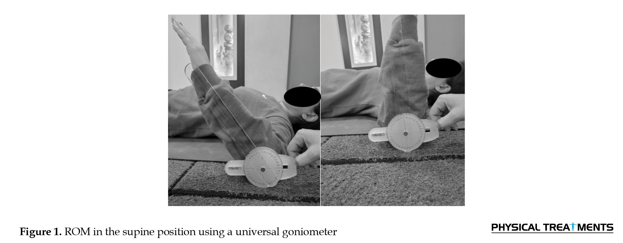
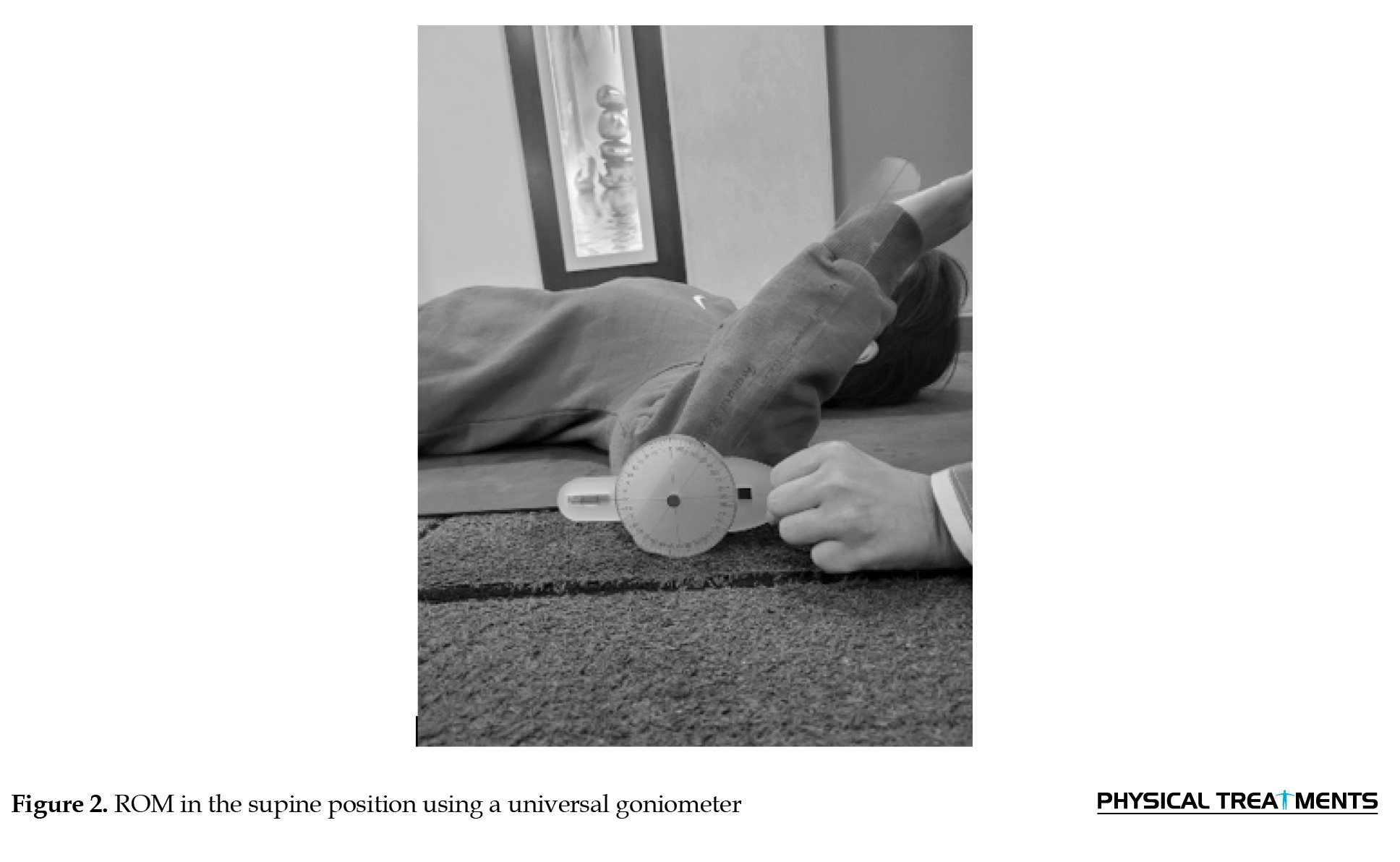
In the next step, 45 subjects were randomly divided into two experimental groups, including massage (n=15) and SMR (n=15), as well as one control group, based on the information obtained from the distributed questionnaires and an expert’s confirmation regarding the presence of MPS in the patients’ trapezius muscles. Next, the primary diagnosis indices proposed by Simon were used to confirm the presence of TPs [2] .The indices included a palpable stiff band in the muscle, identification of sensitive nodules in the stiff band, and reappearance of the patient’s symptoms by applying constant pressure in the sensitive areas in the stiff muscle band. A lottery method was used to randomly divide people into different groups, as follows: The people’s names were written on sheets of paper of the same size and color, placed in a container, and randomly drawn. In this method, the first 15 participants were placed in the massage group, the second 15 in the self-release group, and the last 15 in the control group. It must be mentioned that the control group did not receive any specific intervention during the intervention process and only participated in the pre-test and post-test. Then, briefing sessions were held, and the consent form for participating in the study was distributed among the subjects after explaining MPS and the type of treatment protocols. During the pre-test, the subjects were assessed in terms of height and body mass index (BMI), and ROM was measured using a universal goniometer (neck: Flexion, extension, bilateral rotation, bilateral lateral flexion; shoulder: Flexion, extension, abduction, internal/external rotation [Figures 1 and 2]) [21].
Myofascial pain syndrome (MPS) is among the most common musculoskeletal disorders [1], caused by hypersensitive spots in the muscle fibers and fascia called trigger points (TPs)[2], and has attracted a lot of attention due to its great impact on people’s quality of life and performance. MPS can be associated with other painful disorders [3]. For instance, myofascial pelvic pain, which is caused by the presence of TPs in pelvic areas [3], can cause various problems such as bladder pain, anxiety, and endometriosis. Some studies reported an increase in MPS and TPs among patients with migraines and tension-type headaches [4]. Others have argued that TPs commonly occur after whiplash injuries, lumbar disc protrusion, and idiopathic neck pain [5]. In addition, MPS is a painful condition that can imitate other painful conditions and musculoskeletal disorders [6]. For example, the TPs in the upper angle of the scapula in the joint area with the posterior wall of the chest can create pain similar to angina pectoris while occurring on only one side without symmetry [6]. Even though MPS is one of the most well-known pain syndromes, its exact initial cause, which can cause TPs to first appear, is still under research [2]. It must be clarified that TPs can occur in different areas for various reasons. Studies have shown that 85% of people experience the presence of TPs at least once in their life. The trapezius muscle is among the most common areas of TPs; therefore, inactive TPs are reported by 78.8% of healthy individuals [7]. Apart from the high prevalence of TPs, the complications arising from their presence include hyperalgesia, limited range of motion (ROM), stiffness, weakness, proprioceptive interference, and coordination disorders [7]. The trapezius muscle needs special attention due to its area and function. The trapezius muscle is regarded as a key muscle in the shoulder girdle that provides the required stability of the scapula through its joints. The location of the scapula plays a critical role in the optimal function of the glenohumeral joint [8]. Considering impaired normal muscle function in the presence of TPs [9] and the muscle’s role in the shoulder girdle, treating TPs in this muscle is crucial. The high prevalence of TPs, in addition to the complications arising from their presence, such as hyperalgesia, limited ROM, stiffness, weakness, proprioceptive interference, and coordination disorder [7]. This puts the decision of choosing the right treatment method in a highly important position.
Several researchers have sought to find effective strategies for releasing TPs, as well as evaluate invasive and non-invasive methods. Invasive methods include injection of non-steroidal anti-inflammatory drugs or botulinum toxin, dry needling, and acupuncture [2]. In contrast, non-invasive techniques include massage, self-myofascial release (SMR), tai chi, pressure and stretching techniques, biofeedback and meditation [10]. Non-invasive methods are preferred to manage conditions, such as MPS. Some of the above-mentioned invasive methods reduce pain quickly, despite their side effects, such as addiction and abuse [2]. However, alternative methods with fewer side effects should be used to improve the pain resulting from this disorder [11]. SMR and massage have attracted considerable attention in recent decades.
SMR, which is widely applied by active people and specialists in dealing with patients, is considered useful for athletes in all sports fields at all levels of competition [12]. SMR includes a set of self-massage methods in which certain areas are subjected to pressure and tension employing the weight of the person’s own body using instruments, such as foam rolling, lacrosse, tennis balls, and special release sticks [13]. A person can adjust the amount of pressure based on his/her tolerance and comfort, which makes this approach bearable. Several benefits of SMR have been proposed, the most significant of which include ROM improvement [14], improved proprioception, increased recovery speed [15], and performance enhancement, relieving the pain created by TPs [16].
Among these methods, massage therapy and SMR are among the most critical interventions. Massage is described as a method to manipulate soft tissue, which helps create relaxation [17]. This method can relieve pain and improve ROM through mechanisms such as increasing blood flow in the area following pressure application, which occurs after temporary ischemia, resulting in enhanced oxygen supply to the area, breaking the vicious cycle of energy [18], and discharging accumulated lactic acid after repeated motions [19]. In addition, the release of parasympathetic hormones during the massage can help reduce muscle tone.
No study has been conducted to compare the above-mentioned methods in reducing complications created by TPs in the trapezius muscle, including decreased pain and ROM following their presence, despite the increasing attention of researchers and athletes in this field. A more effective method should be found to improve the aforementioned areas and their complications to return the person to a normal life faster and improve the quality of his/her performance, considering the significance of the trapezius muscle in shoulder stability and the high prevalence of TPs in these areas. This study aimed to investigate the above-mentioned methods in terms of their efficiency in reducing the aforementioned complications.
Materials and Methods
To conduct the study, a notice was distributed to academic women (students) at Shahid Beheshti University to recruit participants. To this end, eligible subjects were invited to cooperate after being informed of MPS and its symptoms. Then, the OSIPOW psychological strain questionnaire and visual analog scale (VAS) were distributed among those interested in participating in the study. The sample size was determined based on previous interventional studies with similar methodologies, including the study by Jafari et al. [20]. Furthermore, an a priori power analysis was conducted using G*Power software, version 3.1 for a one-way analysis of covariance (ANCOVA). Assuming a medium effect size (f=0.25), a significance level of 0.05, and a statistical power of 0.80, the minimum required sample size was calculated to be 42 participants. To account for possible dropouts, 45 participants (15 per group) were included in this study.
All 45 participants were recruited from a single center (Shahid Beheshti University, Tehran); therefore, between-city comparisons were not applicable.
In the next step, 45 subjects were randomly divided into two experimental groups, including massage (n=15) and SMR (n=15), as well as one control group, based on the information obtained from the distributed questionnaires and an expert’s confirmation regarding the presence of MPS in the patients’ trapezius muscles. Next, the primary diagnosis indices proposed by Simon were used to confirm the presence of TPs [2] .The indices included a palpable stiff band in the muscle, identification of sensitive nodules in the stiff band, and reappearance of the patient’s symptoms by applying constant pressure in the sensitive areas in the stiff muscle band. A lottery method was used to randomly divide people into different groups, as follows: The people’s names were written on sheets of paper of the same size and color, placed in a container, and randomly drawn. In this method, the first 15 participants were placed in the massage group, the second 15 in the self-release group, and the last 15 in the control group. It must be mentioned that the control group did not receive any specific intervention during the intervention process and only participated in the pre-test and post-test. Then, briefing sessions were held, and the consent form for participating in the study was distributed among the subjects after explaining MPS and the type of treatment protocols. During the pre-test, the subjects were assessed in terms of height and body mass index (BMI), and ROM was measured using a universal goniometer (neck: Flexion, extension, bilateral rotation, bilateral lateral flexion; shoulder: Flexion, extension, abduction, internal/external rotation [Figures 1 and 2]) [21].
Pain intensity was assessed using the VAS [22]. Functional disability was assessed using the Persian-validated disabilities of the arm, shoulder and hand (DASH) questionnaire [23]. In addition, the subjects in the massage group were analyzed for skin sensitivity or lack of sensitivity to massage oil. Further, two experimental groups were intervened in for a specified period of time (five days). Finally, all subjects were again assessed in terms of pain, shoulder ROM, and upper limb function, and the results were compared with the control group and initial results.
The inclusion criteria included having active TPs in the trapezius muscle based on Simon’s indices [2], being female, not being an athlete, not having undergone surgery or having disorders in the cervical and thoracic spine [24], not suffering from skin diseases and open wounds, abnormal neck lordosis (normal range: 31-40 degrees) [25], back kyphosis (normal range: 20-50 degrees) [26], allergy to massage oils, and high mental tension.
This study was designed as a randomized controlled trial with blinded outcome assessment and blinded data analysis. To reduce measurement bias, the outcome assessor had no access to baseline measurements during post-intervention assessments. Additionally, prior to statistical analysis, group assignments were concealed, and datasets were coded (A, B, and C), ensuring that the statistician remained blinded until the completion of the analysis.
It must also be mentioned that the intervention protocols were researcher-developed and were approved by two content experts: A corrective exercise specialist and [2] an associate professor from Shahid Beheshti University, Tehran, Iran. Pilot testing was also conducted (n=5 participants with similar characteristics), with qualitative feedback confirming protocol feasibility and acceptability. No modifications were required.
To measure ROM using a goniometer, the following methods were used:
For shoulder abduction measurement, the participant lay supine, and the examiner abducted the arm until the endrange sensation or until upward rotation/elevation of the scapula was perceived. The goniometer axis was positioned over the acromion process, the stationary arm was aligned parallel to the sternum, and the moving arm was aligned with the midline of the humerus. To measure internal shoulder rotation, the participant lay supine with the shoulder abducted to 90°. The examiner internally rotated the arm until an endrange sensation occurred or until compensatory scapular protraction or anterior tilt was observed. The goniometer axis was placed over the olecranon process, the stationary arm was held perpendicular to the floor, and the moving arm was aligned with the ulna. For shoulder external rotation, the participant and goniometer were positioned as for internal rotation; the movement was stopped when the participant perceived the end of range or when compensatory scapular posterior tilt or retraction occurred [27].
The shoulder was positioned at 90° of abduction and the elbow at 90° of flexion. Internal rotation was measured actively.
The shoulder was positioned at 90° of abduction and the elbow at 90° of flexion. External rotation was measured actively.
Statistical method
Descriptive and inferential statistics were used to sort and analyze the data, respectively. In addition, the Shapiro-Wilk test was applied to check whether the data are considered parametric or non-parametric. The results indicated that the data were regarded as parametric. Further, a one-way ANCOVA was conducted using SPSS to compare post-intervention outcomes among the three groups (massage, SMR, and control), using baseline values as covariates. Assumptions of normality, homogeneity of variances, and homogeneity of regression slopes were examined prior to analysis. The homogeneity of regression slopes assumption was tested and met.
Self-myofascial release
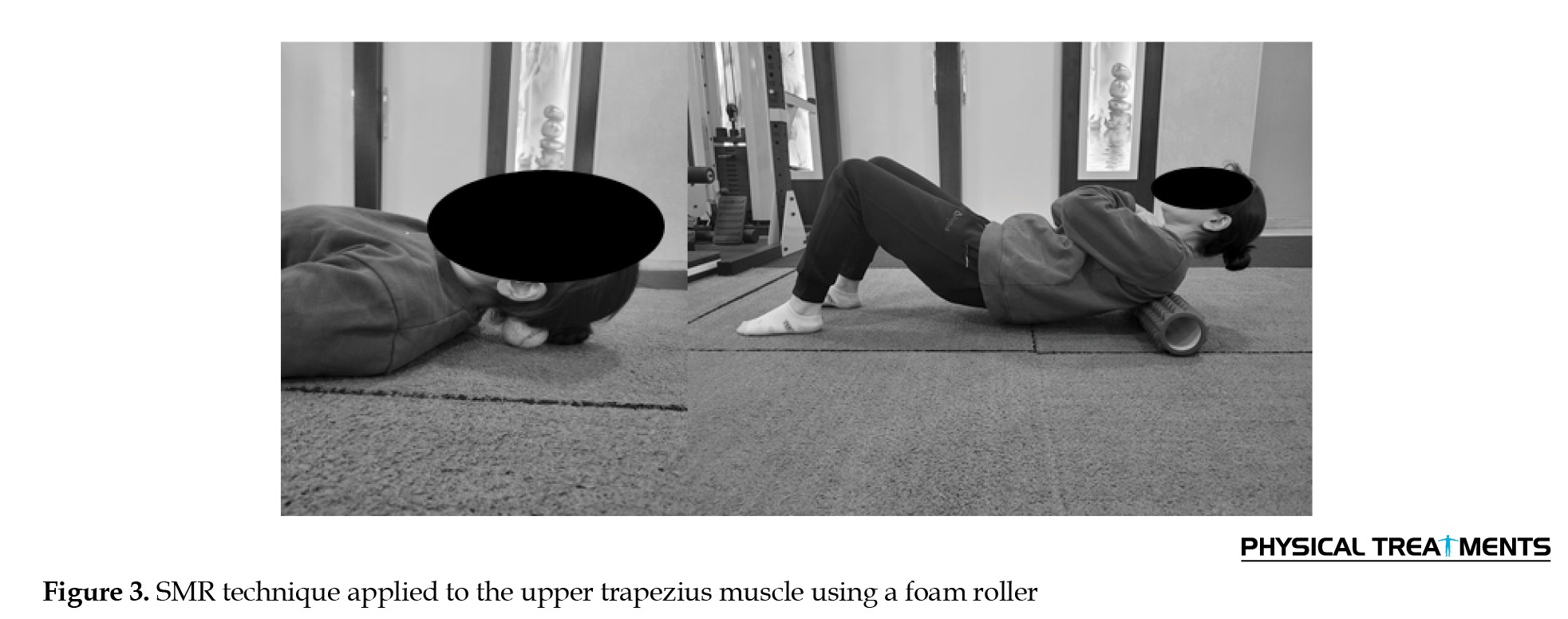
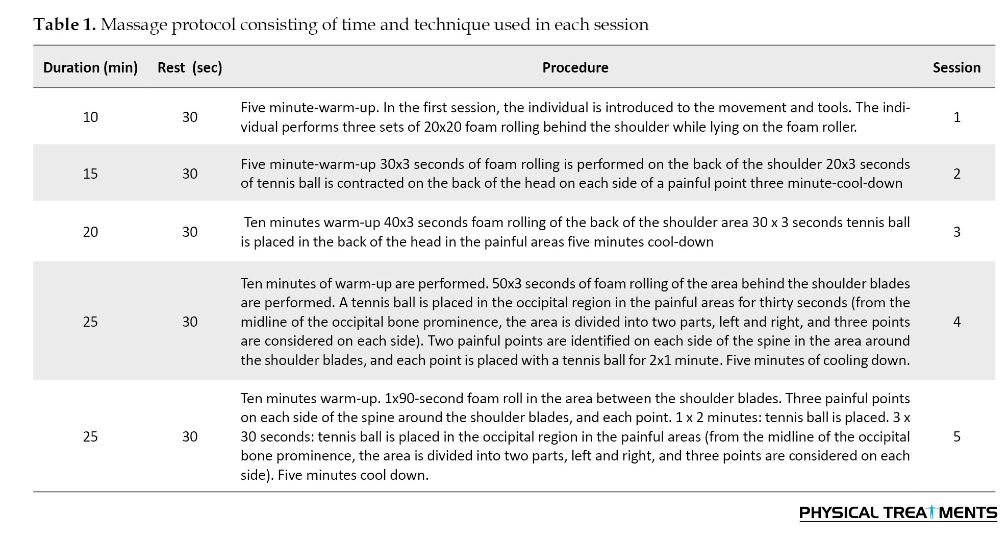
The SMR intervention (Figure 3) lasted five days, during which the motion performance increased by five minutes every day.
The inclusion criteria included having active TPs in the trapezius muscle based on Simon’s indices [2], being female, not being an athlete, not having undergone surgery or having disorders in the cervical and thoracic spine [24], not suffering from skin diseases and open wounds, abnormal neck lordosis (normal range: 31-40 degrees) [25], back kyphosis (normal range: 20-50 degrees) [26], allergy to massage oils, and high mental tension.
This study was designed as a randomized controlled trial with blinded outcome assessment and blinded data analysis. To reduce measurement bias, the outcome assessor had no access to baseline measurements during post-intervention assessments. Additionally, prior to statistical analysis, group assignments were concealed, and datasets were coded (A, B, and C), ensuring that the statistician remained blinded until the completion of the analysis.
It must also be mentioned that the intervention protocols were researcher-developed and were approved by two content experts: A corrective exercise specialist and [2] an associate professor from Shahid Beheshti University, Tehran, Iran. Pilot testing was also conducted (n=5 participants with similar characteristics), with qualitative feedback confirming protocol feasibility and acceptability. No modifications were required.
To measure ROM using a goniometer, the following methods were used:
For shoulder abduction measurement, the participant lay supine, and the examiner abducted the arm until the endrange sensation or until upward rotation/elevation of the scapula was perceived. The goniometer axis was positioned over the acromion process, the stationary arm was aligned parallel to the sternum, and the moving arm was aligned with the midline of the humerus. To measure internal shoulder rotation, the participant lay supine with the shoulder abducted to 90°. The examiner internally rotated the arm until an endrange sensation occurred or until compensatory scapular protraction or anterior tilt was observed. The goniometer axis was placed over the olecranon process, the stationary arm was held perpendicular to the floor, and the moving arm was aligned with the ulna. For shoulder external rotation, the participant and goniometer were positioned as for internal rotation; the movement was stopped when the participant perceived the end of range or when compensatory scapular posterior tilt or retraction occurred [27].
The shoulder was positioned at 90° of abduction and the elbow at 90° of flexion. Internal rotation was measured actively.
The shoulder was positioned at 90° of abduction and the elbow at 90° of flexion. External rotation was measured actively.
Statistical method
Descriptive and inferential statistics were used to sort and analyze the data, respectively. In addition, the Shapiro-Wilk test was applied to check whether the data are considered parametric or non-parametric. The results indicated that the data were regarded as parametric. Further, a one-way ANCOVA was conducted using SPSS to compare post-intervention outcomes among the three groups (massage, SMR, and control), using baseline values as covariates. Assumptions of normality, homogeneity of variances, and homogeneity of regression slopes were examined prior to analysis. The homogeneity of regression slopes assumption was tested and met.
Self-myofascial release
The SMR intervention (Figure 3) lasted five days, during which the motion performance increased by five minutes every day.
The performance lasted ten minutes during the first session, which increased to 25 minutes during the last session. Time intervals most frequently examined in foam rolling research are 30, 60, and 120 seconds, and these durations have commonly been adopted by researchers. This is likely because manual fascial techniques applied in clinical settings are typically performed within similar timeframes [28]. Patel et al. [29] reported that most participants experienced therapeutic effects when the intervention lasted between 60 and 120 seconds. In this context, the study by Sullivan et al. [28] is particularly noteworthy, as the number of repetitions and their duration were manipulated while pressure and rolling frequency were kept constant. In the sit-and-reach test, a 4.3% increase in ROM was observed in participants who rolled for 10 seconds compared with those who rolled for 5 seconds. However, the authors acknowledged that these findings were insufficient for drawing definitive conclusions and emphasized the need for further investigation into the relationship between treatment duration, intensity, and the resulting outcomes. Based on these findings, the duration of foam rolling in the present study ranged from 30 to 120 seconds. The intervention time was progressively increased across sessions to enhance the potential effectiveness of the protocol. Applying pressure and performing the SMR process increased relatively during each session. Accordingly, most of the intervention was performed through the foam rolling process during the first session, while more penetrating and concentrated pressure was used by applying point pressure with a tennis ball in different areas with TPs during the last session.
It must be mentioned that a researcher-made protocol was utilized after implementing the pilot process.
The participant was positioned in supine with knees flexed, and slow, controlled rolling was performed over the trigger point region using a tennis ball with sustained pressure over the identified trigger point in supine position.
Massage
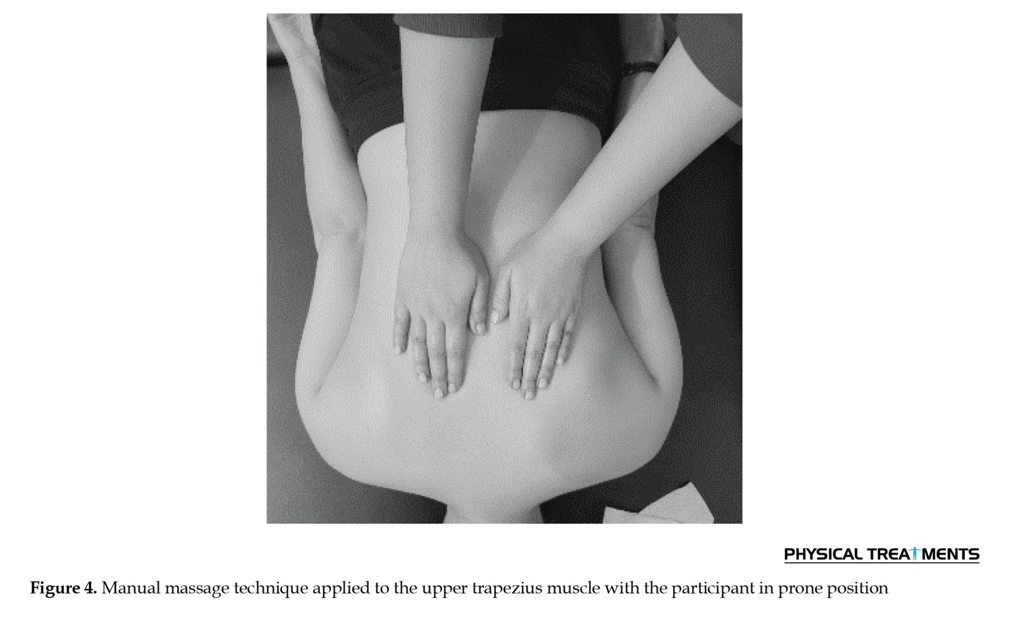
Similar to SMR, a researcher-made massage protocol (Figure 4) was applied after the pilot process.
It must be mentioned that a researcher-made protocol was utilized after implementing the pilot process.
The participant was positioned in supine with knees flexed, and slow, controlled rolling was performed over the trigger point region using a tennis ball with sustained pressure over the identified trigger point in supine position.
Massage
Similar to SMR, a researcher-made massage protocol (Figure 4) was applied after the pilot process.
The duration of the intervention increased during each session. Accordingly, the intervention lasted 10 minutes during the first session, increasing to 25 minutes by the last session. The techniques employed during the initial sessions included Swedish massage with adjusted pressures. A friction technique was applied for one minute using the palms at a high speed to increase the temperature of the targeted areas. The kneading technique was then performed, particularly to identify tender points, in combination with massage movements for variable intervals of 2-4 minutes. This technique was also used to apply stretching to the identified TPs. Pressure technique constituted the primary method for trigger point release in this protocol. The TPs identified during kneading were subjected to ischemic compression. In the initial sessions, mild pressure was applied using the palms. Gradually, deeper pressure was introduced using the thumbs and subsequently the elbows, with intensity progressively increasing from moderate to strong. The duration of pressure application increased from 90 seconds in the first session to 10 minutes in the final session, with pressure delivered in repeated sets of 90 seconds. To increase efficiency, pressure techniques were used in the TP areas during each session. More penetrating pressure was applied from the palm to the tip of the thumb and elbow during the final sessions, with a duration of 60-90 seconds in three sets. Bilateral thumb and palm pressure was applied over the identified myofascial trigger point region.
Results
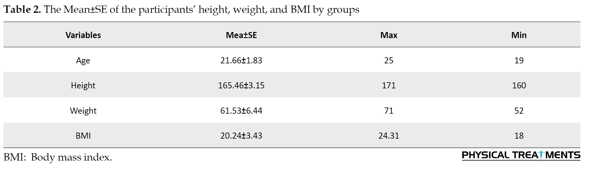
Table 1 presents the Mean±SE of the participants’ height, weight, and BMI by groups.
Tables 2 and 3 present the results related to the description of the pre-test, post-test, and adjusted post-test.
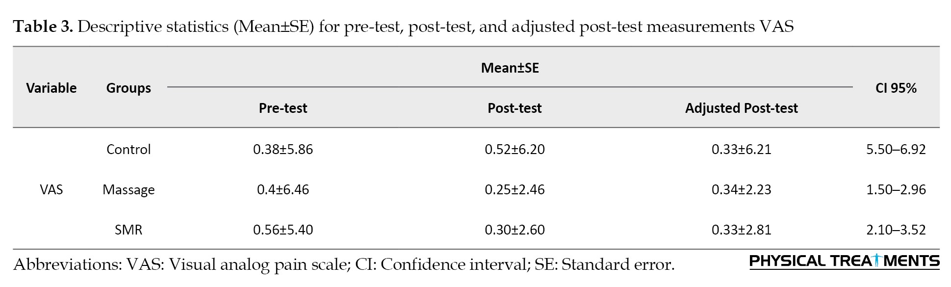
Tables 3 and 4 presents the results related to the one-way ANCOVA test utilized to compare the adjusted mean between the groups.
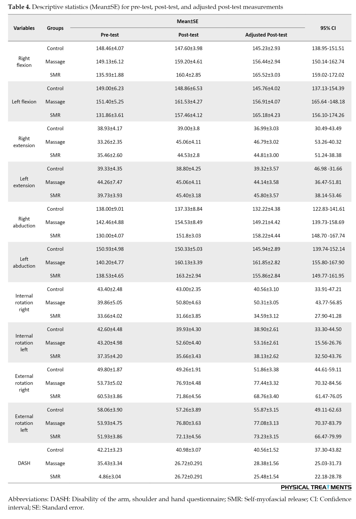
Table 5 presents present the ANCOVA test results.
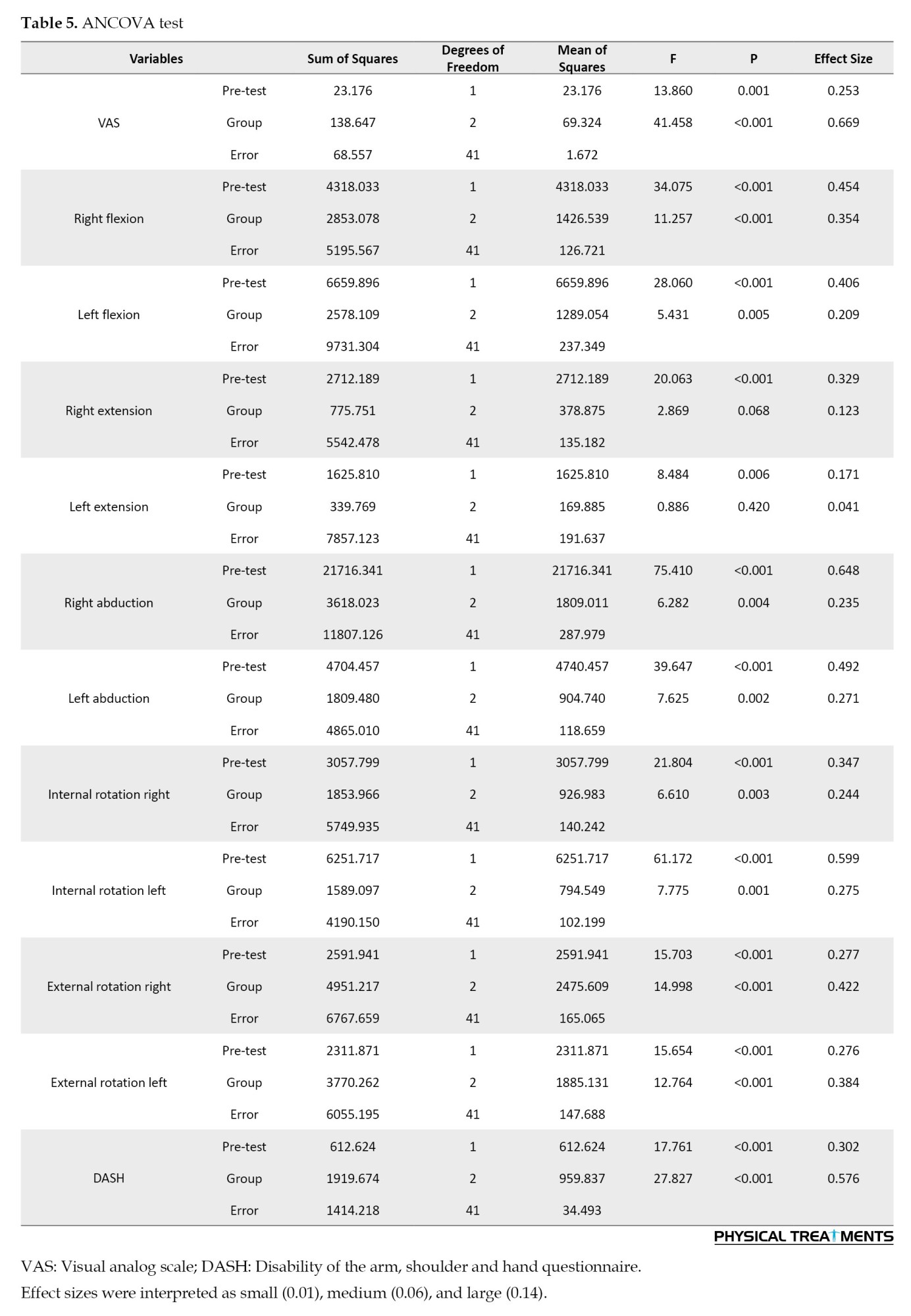
Table 6 presents the results associated with the Bonferroni Post Hoc test.
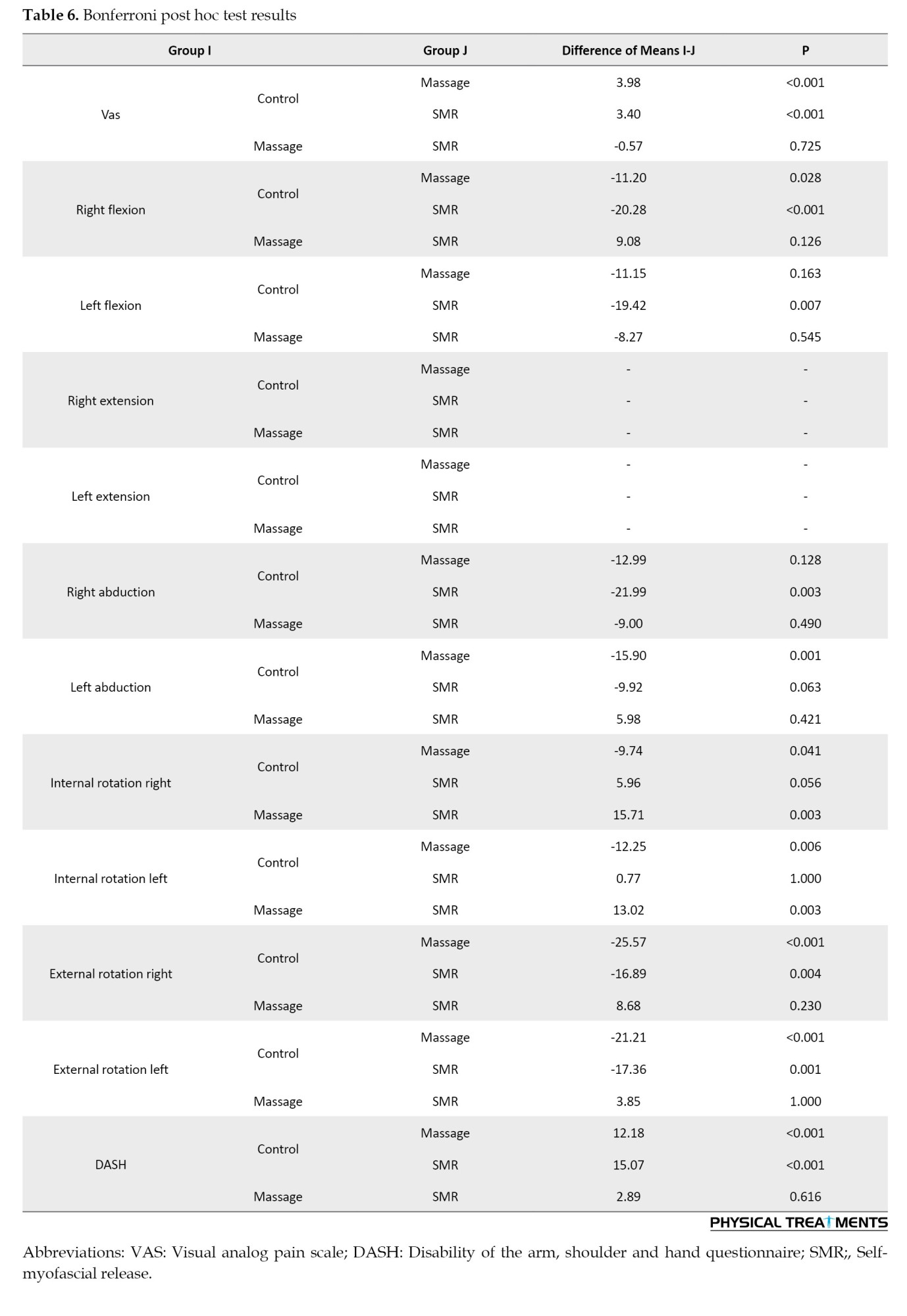
Based on the VAS, massage and SMR significantly reduced the pain created by TPs (P<0.001). Regarding ROM, none of the interventions significantly affected the right (P>0.001) and left (P>0.001) shoulder stretch. However, massage significantly affected the right internal (P>0.001), right external (P>0.001), left internal (P>0.001), and left external rotation (P<0.001) between and within the groups. In addition, SMR significantly affected the right (P>0.001) and left (P<0.001) abduction, as well as right (P<0.001) and left (P>0.001) flexion. Finally, both interventions significantly improved the score of the DASH test (P<0.001).
After adjustment for baseline values, ANCOVA demonstrated significant group effects for VAS (F(2, 41)=41.458, P<0.001, partial η²=0.669), indicating a large effect size.
Significant group differences with «large effect sizes» were also observed for right flexion (F=11.257, P<0.001, η²=0.354), left flexion (F=5.431, P=0.005, η²=0.209), right abduction (F=6.282, P=0.004, η²=0.235), left abduction (F=7.625, P=0.002, η²=0.271), internal rotation right (F=6.610, p=0.003, η²=0.244), internal rotation left (F=7.775, P=0.001, η²=0.275), external rotation right (F=14.998, P<0.001, η²=0.422), external rotation left (F=12.764, P<0.001, η²=0.384), and DASH (F=27.827, P<0.001, η²=0.576).
No significant group effects were found for right extension (P=0.068) or left extension (P=0.420). Overall, the magnitude of the partial eta squared values indicates predominantly «large practical effects», supporting the clinical relevance of the interventions.
Discussion
The present study aimed to evaluate the effect of SMR and massage on the symptoms created by TPs in the trapezius muscle among women suffering from MPS. The results showed that the five sessions of massage and SMR protocols significantly reduced pain among patients with TPs, as measured by VAS.
Several theories have been proposed to explain the effectiveness of SMR processes and massage in reducing the pain of TPs in MPS, including changes in elasticity due to thixotropic properties of soft tissue [28], piezoelectric effects, release of fascia adhesions, cellular responses to pressure, stimulation of tissue fluid flow, nerve inhibition, and release of TPs [30].
Existing theories that describe the effect of SMR include gate control theory of pain, intrapersonal attention, parasympathetic response of the autonomic nervous system, and serotonin release [31]. Based on the gate control theory of pain, sensory stimuli, such as pressure, are transmitted in the pathways of the nervous system faster than pain. The pressure stimulus, which acts faster, intervenes in the process of transferring painful stimuli to the brain, resulting in the elimination of the gate of pain perception [31]. Intrapersonal attention refers to the level of attention of a person while receiving a massage. Some claim that human attention to touch reduces the perception of pain by the brain. Stimulating the parasympathetic response reduces the release of stress hormones, resulting in decreased anxiety, depression, and pain. The release of serotonin prevents the transmission of harmful stimuli to the brain. Other inhibitory neurotransmitters such as endorphins may be released following the application of pressure through the therapeutic method.
The results revealed that SMR increased the ROM in right and left shoulder flexion, as well as right and left abduction, more than the massage. Generally, the trapezius muscle provides stability, as well as facilitating the motion of the scapula. The elevators, upper scapular rotators, and neck extensors form the upper fibers in the trapezius muscle. The medial fibers pull the scapula back, while the inferior fibers lower the scapula and assist in its upward rotation. This superficial and extensive muscle, which is mostly posterior, actively participates in motions, such as lateral neck flexion, head rotation, shoulder elevation and lowering, and arm internal rotation. Apparently, releasing the trapezius muscle without focusing on partner and opposing muscles improves the shoulder ROM only to a limited extent.
Based on the results, the massage protocol can significantly increase the range of internal and external rotation in the right and left arms after five sessions on the trapezius muscle among women with MPS.
It should be noted that these findings align with studies by Bethers et al. [32] and Li and Guan [33], which examined the immediate effects of massage. In fact, TPs can occur in muscle fibers and fascia tissue. Damage to fascia tissue can lead to its disruption in different ways. Limitation of motion in the fascia tissue can reduce its shock absorption process and lead to the peripheral involvement of blood vessels and nerves, resulting in decreased ROM throughout the body. The functional role of fascia may be impaired after repeated injuries, physical trauma, and inflammation, which can alter the natural biomechanics in the body, increase tension in the system, develop myofascial pain, and decrease the ROM. In addition, mechanical pressure application can reduce adhesions between tissue layers, enhance muscle compliance, and decrease muscle fiber stiffness. Prolonged pressure on muscle tissue can induce muscle relaxation. Pressure on myofascial TrPs increases sarcomere length, thereby reducing abnormal tension, overall pain, and improving ROM [34]. Additionally, localized pressure enhances hyperemia and blood perfusion, promoting oxygenation, reducing production of inflammatory and nociceptive mediators, and consequently minimizing muscle fiber damage while optimizing muscle strength and length [34].
A decrease in the DASH questionnaire score was observed in both massage and SMR groups, indicating an effective result. The musculoskeletal system is considered a network of interconnected tissues that should work together to produce effective motion. The restrictions created by the fascia can disrupt the normal function of the muscle when the muscle and fascia are subjected to microtrauma. Myofascial TPs can interfere with the normal function of the fascia and lead to muscle dysfunction, which manifests as reduced ROM, neuromuscular components, and decreased strength. Improved ROM allows for better motion patterns, which can improve performance and reduce the risk of musculoskeletal injuries.
Ethical Considerations
Compliance with ethical guidelines
This study was conducted under the supervision of the Ethics Committee of Shahid Beheshti University, Tehran, Iran (Code: IR.SBU.REC.1402.067 and Iranian Registry of Clinical Trials (IRCT) (ID: IRCT20230914059432N1).
Funding
This article was extracted from a master thesis of Sara MatinFard, approved by Shahid Beheshti University, Tehran, Iran.
Authors' contributions
Methodology, validation, supervision, review and editing: Fariborz Hovanloo; Conceptualization, investigation, data curation, and writing the original draft preparation: Sara MatinFard.
Conflict of interest
The authors declared no conflicts of interest.
Acknowledgments
The authors express their sincere gratitude to all the people who helped and supported this research.
Results
Table 1 presents the Mean±SE of the participants’ height, weight, and BMI by groups.
Tables 2 and 3 present the results related to the description of the pre-test, post-test, and adjusted post-test.
Tables 3 and 4 presents the results related to the one-way ANCOVA test utilized to compare the adjusted mean between the groups.
Table 5 presents present the ANCOVA test results.
Table 6 presents the results associated with the Bonferroni Post Hoc test.
Based on the VAS, massage and SMR significantly reduced the pain created by TPs (P<0.001). Regarding ROM, none of the interventions significantly affected the right (P>0.001) and left (P>0.001) shoulder stretch. However, massage significantly affected the right internal (P>0.001), right external (P>0.001), left internal (P>0.001), and left external rotation (P<0.001) between and within the groups. In addition, SMR significantly affected the right (P>0.001) and left (P<0.001) abduction, as well as right (P<0.001) and left (P>0.001) flexion. Finally, both interventions significantly improved the score of the DASH test (P<0.001).
After adjustment for baseline values, ANCOVA demonstrated significant group effects for VAS (F(2, 41)=41.458, P<0.001, partial η²=0.669), indicating a large effect size.
Significant group differences with «large effect sizes» were also observed for right flexion (F=11.257, P<0.001, η²=0.354), left flexion (F=5.431, P=0.005, η²=0.209), right abduction (F=6.282, P=0.004, η²=0.235), left abduction (F=7.625, P=0.002, η²=0.271), internal rotation right (F=6.610, p=0.003, η²=0.244), internal rotation left (F=7.775, P=0.001, η²=0.275), external rotation right (F=14.998, P<0.001, η²=0.422), external rotation left (F=12.764, P<0.001, η²=0.384), and DASH (F=27.827, P<0.001, η²=0.576).
No significant group effects were found for right extension (P=0.068) or left extension (P=0.420). Overall, the magnitude of the partial eta squared values indicates predominantly «large practical effects», supporting the clinical relevance of the interventions.
Discussion
The present study aimed to evaluate the effect of SMR and massage on the symptoms created by TPs in the trapezius muscle among women suffering from MPS. The results showed that the five sessions of massage and SMR protocols significantly reduced pain among patients with TPs, as measured by VAS.
Several theories have been proposed to explain the effectiveness of SMR processes and massage in reducing the pain of TPs in MPS, including changes in elasticity due to thixotropic properties of soft tissue [28], piezoelectric effects, release of fascia adhesions, cellular responses to pressure, stimulation of tissue fluid flow, nerve inhibition, and release of TPs [30].
Existing theories that describe the effect of SMR include gate control theory of pain, intrapersonal attention, parasympathetic response of the autonomic nervous system, and serotonin release [31]. Based on the gate control theory of pain, sensory stimuli, such as pressure, are transmitted in the pathways of the nervous system faster than pain. The pressure stimulus, which acts faster, intervenes in the process of transferring painful stimuli to the brain, resulting in the elimination of the gate of pain perception [31]. Intrapersonal attention refers to the level of attention of a person while receiving a massage. Some claim that human attention to touch reduces the perception of pain by the brain. Stimulating the parasympathetic response reduces the release of stress hormones, resulting in decreased anxiety, depression, and pain. The release of serotonin prevents the transmission of harmful stimuli to the brain. Other inhibitory neurotransmitters such as endorphins may be released following the application of pressure through the therapeutic method.
The results revealed that SMR increased the ROM in right and left shoulder flexion, as well as right and left abduction, more than the massage. Generally, the trapezius muscle provides stability, as well as facilitating the motion of the scapula. The elevators, upper scapular rotators, and neck extensors form the upper fibers in the trapezius muscle. The medial fibers pull the scapula back, while the inferior fibers lower the scapula and assist in its upward rotation. This superficial and extensive muscle, which is mostly posterior, actively participates in motions, such as lateral neck flexion, head rotation, shoulder elevation and lowering, and arm internal rotation. Apparently, releasing the trapezius muscle without focusing on partner and opposing muscles improves the shoulder ROM only to a limited extent.
Based on the results, the massage protocol can significantly increase the range of internal and external rotation in the right and left arms after five sessions on the trapezius muscle among women with MPS.
It should be noted that these findings align with studies by Bethers et al. [32] and Li and Guan [33], which examined the immediate effects of massage. In fact, TPs can occur in muscle fibers and fascia tissue. Damage to fascia tissue can lead to its disruption in different ways. Limitation of motion in the fascia tissue can reduce its shock absorption process and lead to the peripheral involvement of blood vessels and nerves, resulting in decreased ROM throughout the body. The functional role of fascia may be impaired after repeated injuries, physical trauma, and inflammation, which can alter the natural biomechanics in the body, increase tension in the system, develop myofascial pain, and decrease the ROM. In addition, mechanical pressure application can reduce adhesions between tissue layers, enhance muscle compliance, and decrease muscle fiber stiffness. Prolonged pressure on muscle tissue can induce muscle relaxation. Pressure on myofascial TrPs increases sarcomere length, thereby reducing abnormal tension, overall pain, and improving ROM [34]. Additionally, localized pressure enhances hyperemia and blood perfusion, promoting oxygenation, reducing production of inflammatory and nociceptive mediators, and consequently minimizing muscle fiber damage while optimizing muscle strength and length [34].
A decrease in the DASH questionnaire score was observed in both massage and SMR groups, indicating an effective result. The musculoskeletal system is considered a network of interconnected tissues that should work together to produce effective motion. The restrictions created by the fascia can disrupt the normal function of the muscle when the muscle and fascia are subjected to microtrauma. Myofascial TPs can interfere with the normal function of the fascia and lead to muscle dysfunction, which manifests as reduced ROM, neuromuscular components, and decreased strength. Improved ROM allows for better motion patterns, which can improve performance and reduce the risk of musculoskeletal injuries.
Ethical Considerations
Compliance with ethical guidelines
This study was conducted under the supervision of the Ethics Committee of Shahid Beheshti University, Tehran, Iran (Code: IR.SBU.REC.1402.067 and Iranian Registry of Clinical Trials (IRCT) (ID: IRCT20230914059432N1).
Funding
This article was extracted from a master thesis of Sara MatinFard, approved by Shahid Beheshti University, Tehran, Iran.
Authors' contributions
Methodology, validation, supervision, review and editing: Fariborz Hovanloo; Conceptualization, investigation, data curation, and writing the original draft preparation: Sara MatinFard.
Conflict of interest
The authors declared no conflicts of interest.
Acknowledgments
The authors express their sincere gratitude to all the people who helped and supported this research.
References
- Tedeschi R, Giorgi F, Platano D, Berti L. Classifying low back pain through pain mechanisms: A scoping review for physiotherapy practice. Journal of Clinical Medicine. 2025; 14(2):412. [DOI:10.3390/jcm14020412] [PMID] [PMCID]
- Galasso A, Urits I, An D, Nguyen D, Borchart M, Yazdi C, et al. A comprehensive review of the treatment and management of myofascial pain syndrome. Current Pain and Headache Reports. 2020; 24(8):43. [DOI:10.1007/s11916-020-00877-5] [PMID]
- Bonder JH, Chi M, Rispoli L. Myofascial pelvic pain and related disorders. Physical Medicine and Rehabilitation Clinics. 2017; 28(3):501-15. [DOI:10.1016/j.pmr.2017.03.005] [PMID]
- Do TP, Heldarskard GF, Kolding LT, Hvedstrup J, Schytz HW. Myofascial trigger points in migraine and tension-type headache. The Journal of Headache and Pain. 2018; 19(1):84. [DOI:10.1186/s10194-018-0913-8] [PMID] [PMCID]
- Chiarotto A, Clijsen R, Fernandez-de-Las-Penas C, Barbero M. Prevalence of myofascial trigger points in spinal disorders: A systematic review and meta-analysis. Archives of Physical Medicine and Rehabilitation. 2016; 97(2):316-37. [DOI:10.1016/j.apmr.2015.09.021] [PMID]
- Travell JG, Simons DG. Myofascial pain and dysfunction: The trigger point manual. Philadelphia: Lippincott Williams & Wilkins; 1992. [Link]
- Li L, Stoop R, Clijsen R, Hohenauer E, Fernández-de-Las-Peñas C, Huang Q, et al. Criteria used for the diagnosis of myofascial trigger points in clinical trials on physical therapy: Updated systematic review. The Clinical Journal of Pain. 2020; 36(12):955-67. [DOI:10.1097/AJP.0000000000000875] [PMID]
- Briel S, Olivier B, Mudzi W. Scapular force: Couple ratios in healthy shoulders - An observational study reflecting typical values. South African Journal of Physiotherapy. 2022; 78(1):1619. [DOI:10.4102/sajp.v78i1.1619] [PMID] [PMCID]
- Lucas KR. The impact of latent trigger points on regional muscle function. Current Pain and Headache Reports. 2008; 12(5):344-9. [DOI:10.1007/s11916-008-0058-6] [PMID]
- Malanga GA, Gwynn MW, Smith R, Miller D. Tizanidine is effective in the treatment of myofascial pain syndrome. Pain Physician Journal. 2002; 5(4):422. [DOI:10.36076/ppj.2002/5/422] [PMID]
- Manchikanti L, Sanapati J, Benyamin RM, Atluri S, Kaye AD, Hirsch JA. Reframing the prevention strategies of the opioid crisis: Focusing on prescription opioids, fentanyl, and heroin epidemic. Pain Physician Journal. 2018;21(4):309. [DOI:10.36076/ppj.2018.4.309] [PMID]
- Couture G, Karlik D, Glass SC, Hatzel BM. The effect of foam rolling duration on hamstring range of motion. The Open Orthopaedics Journal. 2015; 9:450. [DOI:10.2174/1874325001509010450] [PMID] [PMCID]
- Cheatham SW, Kolber MJ, Cain M, Lee M. The effects of self-myofascial release using a foam roll or roller massager on joint range of motion, muscle recovery, and performance: a systematic review. International Journal of Sports Physical Therapy. 2015; 10(6):827. [PMID]
- Bradbury-Squires DJ, Noftall JC, Sullivan KM, Behm DG, Power KE, Button DC. Roller-massager application to the quadriceps and knee-joint range of motion and neuromuscular efficiency during a lunge. Journal of Athletic Training. 2015; 50(2):133-40. [DOI:10.4085/1062-6050-49.5.03] [PMID] [PMCID]
- Pearcey GE, Bradbury-Squires DJ, Kawamoto J-E, Drinkwater EJ, Behm DG, Button DC. Foam rolling for delayed-onset muscle soreness and recovery of dynamic performance measures. Journal of Athletic Training. 2015; 50(1):5-13. [DOI:10.4085/1062-6050-50.1.01] [PMID] [PMCID]
- Wilke J, Vogt L, Banzer W. Immediate effects of self-myofascial release on latent trigger point sensitivity: A randomized, placebo-controlled trial. Biology of Sport. 2018; 35(4):349-54. [DOI:10.5114/biolsport.2018.78055] [PMID]
- Moyer CA, Rounds J, Hannum JW. A meta-analysis of massage therapy research. Psychological Bulletin. 2004; 130(1):3. [DOI:10.1037/0033-2909.130.1.3] [PMID]
- Dębski P, Białas E, Gnat R. The parameters of foam rolling, self-myofascial release treatment: A review of the literature. Biomedical Human Kinetics. 2019; 11(1):36-46. [DOI:10.2478/bhk-2019-0005]
- Kamali F, Abolahrari Shirazi S, Besharati M, Shams Salehi S. [The comparison of the therapeutic effect of ischemic compression on upper trapezius trigger points in neutral and passive stretched positions in computer’s operators (Persian)]. Journal of Research in Rehabilitation Sciences. 2014; 10(1):1-11. [Link]
- Jafari B, Hovanloo F, Mohammadi F. [Comparison of the effect of six weeks of virtual reality rehabilitation program and reflexology massage on the strength and range of motion of athletes with paraplegia (Persian)]. Sport Medicine Studies. 2022; 13(30):135-58. [Link]
- Riddle DL, Rothstein JM, Lamb RL. Goniometric reliability in a clinical setting: Shoulder measurements. Physical Therapy. 1987; 67(5):668-73. [DOI:10.1093/ptj/67.5.668] [PMID]
- Begum MR, Hossain MA. Validity and reliability of visual analogue scale (VAS) for pain measurement. Journal of Medical Case Reports and Reviews. 2019; 2(11):394-402. [Link]
- Mousavi SJ, Parnianpour M, Abedi M, Askary-Ashtiani A, Karimi A, Khorsandi A, et al. Cultural adaptation and validation of the Persian version of the Disabilities of the Arm, Shoulder and Hand (DASH) outcome measure. Clinical Rehabilitation. 2008; 22(8):749-57. [DOI:10.1177/0269215508085821] [PMID]
- Safavi SN, Taghizadeh S, Ahmadi Ashan E. Mildly kyphotic students had more shoulder trigger points than students with normal spinal posture. Iranian Rehabilitation Journal. 2024; 22(2):285-94. [DOI:10.32598/irj.22.2.2036.2]
- McAviney J, Schulz D, Bock R, Harrison DE, Holland B. Determining the relationship between cervical lordosis and neck complaints. Journal of Manipulative and Physiological Therapeutics. 2005; 28(3):187-93. [DOI:10.1016/j.jmpt.2005.02.015] [PMID]
- Abrisham SMJ, Ardekani MRS, Mzarch MAB. Evaluation of the normal range of thoracic kyphosis and lumbar lordosis angles using EOS imaging. Maedica. 2020; 15(1):87. [DOI:10.26574/maedica.2020.15.1.87] [PMID] [PMCID]
- Norkin CC WD. Measurement of joint motion: A guide to goniometry. FA Davis: FA Davis; 2016. [Link]
- Sullivan KM, Silvey DB, Button DC, Behm DG. Roller-massager application to the hamstrings increases sit and reach range of motion within five to ten seconds without performance impairments. International Journal of Sports Physical Therapy. 2013; 8(3):228. [PMID]
- Grieve R, Goodwin F, Alfaki M, Bourton AJ, Jeffries C, Scott H. The immediate effect of bilateral self myofascial release on the plantar surface of the feet on hamstring and lumbar spine flexibility: A pilot randomised controlled trial. ournal of Bodywork and Movement Therapies. 2015; 19(3):544-52. [DOI:10.1016/j.jbmt.2014.12.004] [PMID]
- Patel DG, Vyas NJ, Sheth MS. Immediate effect of application of bilateral self myo-fascial release on the plantar surface of the foot on hamstring and lumbar spine flexibility: A quasi experimental study. Foot. 2016; 3(7):10.1016. [DOI:10.20530/IJTA_32_94-99]
- Paolini J. Review of myofascial release as an effective massage therapy technique. International Journal of Athletic Therapy and Training. 2009; 14(5):30-4. [DOI:10.1123/att.14.5.30]
- Bethers AH, Swanson DC, Sponbeck JK, Mitchell UH, Draper DO, Feland JB, et al. Positional release therapy and therapeutic massage reduce muscle trigger and tender points. Journal of Bodywork and Movement Therapies. 2021; 28:264-70. [DOI:10.1016/j.jbmt.2021.07.005]
- Li X, Guan R. How does agricultural mechanization service affect agricultural green transformation in China?. International Journal of Environmental Research and Public Health. 2023; 20(2):1655. [DOI:10.3390/ijerph20021655]
- Cagnie B DV, Coppieters I, Van Oosterwijck J, Cools A, Danneels L. Effect of ischemic compression on trigger points in the neck and shoulder muscles in office workers: A cohort study. Journal of Manipulative and Physiological Therapeutics. 2013; 36(8):482-9. [DOI:10.1016/j.jmpt.2013.07.001] [PMID]
Type of Study: Research |
Subject:
Sport injury and corrective exercises
Received: 2025/11/24 | Accepted: 2026/05/4 | Published: 2026/10/18
Received: 2025/11/24 | Accepted: 2026/05/4 | Published: 2026/10/18
Send email to the article author
| Rights and permissions | |
 |
This work is licensed under a Creative Commons Attribution-NonCommercial 4.0 International License. |

This is an open access article distributed under the terms of the Creative Commons Attribution-NonCommercial 4.0 (CC-By-NC 4.0), which permits use, distribution, and reproduction in any medium, provided the original work is properly cited and is not used for commercial purposes.
Contact Information