Sun, Nov 8, 2026
Volume 16, Issue 4 ( Autumn 2026)
PTJ 2026, 16(4): 465-472 |
Back to browse issues page


Ethics code: IR.MUI.MED.REC.1402.264


Download citation:
BibTeX | RIS | EndNote | Medlars | ProCite | Reference Manager | RefWorks
Send citation to:


BibTeX | RIS | EndNote | Medlars | ProCite | Reference Manager | RefWorks
Send citation to:
Maghroori R, Haghighat S, Izadi F. Efficacy of TECAR Therapy in the Management of Plantar Fasciitis: A Randomized Clinical Trial. PTJ 2026; 16 (4) :465-472
URL: http://ptj.uswr.ac.ir/article-1-862-en.html
URL: http://ptj.uswr.ac.ir/article-1-862-en.html

1- Department of Physical Medicine and Rehabilitation, School of Medicine, Isfahan University of Medical Sciences, Isfahan, Iran.
Keywords: Plantar fasciitis (PF), Physical therapy modalities, Transfer of energy capacitive and resistive (TECAR) therapy, Heel pain, Noninvasive therapy
Full-Text [PDF 499 kb]
(1107 Downloads)
| Abstract (HTML) (4587 Views)
Full-Text: (67 Views)
Introduction
Plantar fasciitis (PF) commonly occurs in individuals regardless of age or physical activity status and represents a major musculoskeletal complaint [1]. This condition is characterized by discomfort in the inner heel, which worsens during weight-bearing activities [2]. The lifetime incidence is approximately 10%. In one-third of the cases, PF may manifest bilaterally [3]. This disease frequently becomes chronic, with symptoms persisting for up to one year. PF is estimated to account for nearly one million annual visits to healthcare providers across the United States [1].
PF is mostly diagnosed by physical examination and clinical history [4]. Patients experience severe pain during the initial steps taken upon rising from bed in the morning. Although pain tends to subside as activities commence, it may worsen by the end of the day. Tenderness may be observed at the medial calcaneal tubercle. The windlass test is helpful in establishing the pathology with a high specificity (100%) but low sensitivity (32%) [3]. Ultrasound is also considered a sensitive instrument for PF assessment, with significant findings including increased plantar fascia thickness (PFT) and hypoechoic appearance of plantar fascia. X-ray imaging is also useful for ruling out other pathologies [5].
Although PF is self-limiting and resolves within a year, its impact on routine activities drives patients to pursue treatment prior to the resolution of the pain. Approximately 70% of patients experience reduced pain with conservative treatment alone [6]. However, some may require a combination of conservative management and other therapies [6]. Different therapeutic options have been suggested for PF, including night splint use [7], shock wave therapy [8], laser therapy [9], corticosteroid injections [10], and surgical interventions [11]. Nevertheless, based on the current studies, none of the aforementioned therapeutic options have demonstrated clear superiority, and with ongoing technological advancements, the optimal approach remains a matter of debate [3].
Recently, studies have utilized a novel electrotherapy modality in the management of musculoskeletal pain known as transfer of energy capacitive and resistive (TECAR) [12–14]. TECAR therapy delivers high-frequency energy (300 kHz–1 MHz) in a non-invasive manner, stimulating the body's innate capacity for regeneration [15]. TECAR therapy operates via two modes of electric charge transfer: capacitive and resistive. The capacitive mode primarily affects soft tissues and muscles rich in electrolytes. In contrast, the resistive mode targets denser structures, such as tendons, bones, and joints [14]. The findings of a recent meta-analysis suggested TECAR therapy as an effective method for management of sports-related pains [13].
Given the therapeutic mechanisms of TECAR therapy, including enhanced blood flow and tissue oxygenation, enhanced tissue metabolism, and reduced tissue edema [16], it is expected that this treatment would improve tissue perfusion and minimize edema in patients with PF. Considering the lack of prior surveys on the effectiveness of TECAR therapy in managing PF, coupled with the fact that PF has a high prevalence and clinical significance and that effective non-surgical management is required, this research was conducted to assess how TECAR treatment influences pain relief and functional improvement in individuals with PF.
Materials and Methods
Study design and protocol
This was a double-blind randomized clinical trial (in this investigation, both participants and therapists were blinded using a sham TECAR device that looked and felt identical to the active device but did not deliver therapeutic current). In accordance with the Declaration of Helsinki, all participants received a detailed explanation of the study protocol and voluntarily signed a written informed consent form. Group assignment was determined using a computer-generated randomization list prepared by an independent researcher with no role in participant recruitment or evaluation. Concealment of allocation was achieved by employing consecutively numbered, sealed, and opaque envelopes. Simple randomization was applied, and no block randomization or stratification was used.
Study participants
All patients with PF referred to the Physical Medicine and Rehabilitation Clinic, Isfahan University of Medical Sciences, Isfahan, Iran, were eligible for inclusion in the trial. The included patients were adults (aged 18-68 years), with at least a history of heel pain for 4 weeks. PF was diagnosed by a physiatrist through ultrasonographic evaluation. The severity of heel pain was assessed using the visual analog scale (VAS). Patients with a VAS score of >3 were included in this trial. The VAS cutoff (>3) was selected based on prior literature [17, 18] and expert consensus among the study investigators, all of whom were specialists in Physical Medicine and Rehabilitation. Pain scores ≤3 are generally considered mild, less likely to cause functional impairment, and typically do not require specific interventional treatment. The exclusion criteria were history of diabetes, arthritis, Achilles tendon injury, history of trauma, injections or surgeries on the affected heel, and any contraindications for TECAR therapy (including pregnancy, use of a pacemaker, neoplasms, open wounds and skin lesions, skin sensitivity, and lack of heat sensation). Individuals with an intense level of physical activity who were unable to reduce it were also excluded. During the study, patients were excluded if their symptoms worsened and required more vigorous interventions.
Study design and intervention
During the study period, participants were enrolled consecutively from patients referred to the physical medicine and rehabilitation clinic. Individuals meeting the specified eligibility criteria and agreeing to participate were included in this study. Participants were then allocated to either the intervention or control group through computer-based randomization with concealed allocation. The required sample size was calculated using an independent two-sample mean comparison formula with a two-tailed significance level of 0.05 and 80% power. Drawing on prior evidence [19], a minimum of 26 participants in each group were required. To account for potential dropouts, 30 participants were enrolled in each group. Finally, 30 patients were eligible to be included in each category. In the intervention group, individuals underwent TECAR therapy (twice per week for 4 weeks), a set of exercises (stretching the calf muscle and plantar fascia, passive dorsiflexion of the toes, strengthening intrinsic foot muscles, and rolling the sole three times daily (morning, afternoon, and evening) each set lasting 30 seconds), and a silicone heel pad was also prescribed for them. They additionally consumed celecoxib 200mg per day for 15 days. The control group performed the same set of exercises using a silicone heel pad and consumed the same medication.
TECAR therapy was delivered using the WINBACK 3 device (France) with a frequency of 500 Hz and an intensity range of 20–40%, adjusted according to patient tolerance at the physiotherapy section of Amin Hospital, Isfahan, Iran. Treatment was applied using a medium-sized CET electrode (60 mm). The capacitive and resistive modes were applied as described above. The patient was placed in the prone position, and conductive gel was applied near the Achilles tendon insertion. TECAR therapy was administered for 15 minutes per session (5 minutes of resistive, 10 minutes of capacitive) for 8 sessions, two sessions per week.
Data collection and measurements
At the beginning, demographic characteristics of participants, including age, sex, occupation (homemaker, unemployed, employed), severity of pain, severity of symptoms and patient’s function, were recorded. Ultrasonography (Alpinion E-CUBE 9, Republic of Korea) was employed to evaluate PFT. Ultrasonography was performed by a single experienced physiatrist who was blinded to group allocation and clinical data. To reduce measurement variability, PFT was measured twice at each assessment, and the average value was used for analysis. PF was diagnosed by an experienced physiatrist based on clinical presentation and ultrasonographic findings. No strict cut-off value for PFT was used for diagnosis; instead, ultrasonography was applied as a supportive imaging tool to confirm the clinical diagnosis and to quantify PFT for outcome assessment. The patients were placed in a prone position, ensuring knees were fully extended, and ankles dorsiflexed to 90°, with legs allowed to hang naturally. Using a 5–13 MHz probe on the Alpinion E-CUBE 9 (Republic of Korea), PFT was measured twice at the standard location crossing the anterior border of the calcaneus, with the average taken to minimize potential errors from the transducer. At the end of intervention and 2 months post-intervention, the severity of the pain and symptoms and the patient’s function were assessed. VAS score was utilized to assess the severity of pain [20]. This 10-point scale rated pain from 0 (no pain) to 10 (worst possible pain), with 1–3 indicating mild, 4–7 moderate, and 8–10 severe pain levels [21]. The reliability of the Iranian version of this scale has been approved [22]. The modified roles and Maudsley (RM) score was used to categorize participants into four levels: excellent, good, fair, and poor, based on their pain levels and ability to perform daily activities. This score was a functional evaluation method used to assess pain and activity limitations, often in the context of foot and ankle conditions, such as PF [23]. The main outcome of interest was the level of pain, determined using the VAS. Secondary outcomes included PFT measured using ultrasonography and functional status assessed using the modified RM score. Outcomes were evaluated at baseline, at the end of the intervention, and two months after the intervention.
Statistical analysis
Continuous data are expressed as Mean±SD, and categorical variables as No. (%). Data normality was assessed using the Shapiro–Wilk test and inspection of box plots. For within-group comparisons, repeated measures analysis of variance (ANOVA) or Friedman tests were applied, whereas independent t-tests, ANOVA, chi-square, or Fisher’s exact tests were used for between-group analyses. All analyses were performed using SPSS software, version 21.0 (SPSS Inc., Chicago, IL, USA), with statistical significance set at P<0.05.
Results
In each arm of the study, 30 individuals were enrolled. The mean age of all participants was 41.68±13.17, ranging from 18-63 years. Most patients were male (51.7%), and the majority were employed (31.7%). Table 1 presents the baseline characteristics of each group.
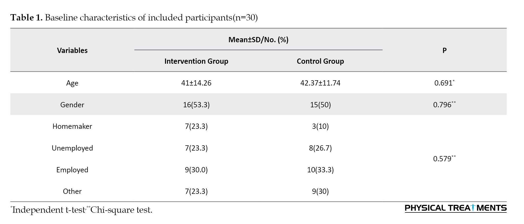
The groups did not differ significantly with respect to age, PFT, gender, or employment status (P>0.05 for all comparisons).
The evaluation of the VAS scores (an indicator of pain severity) demonstrated no significant difference between groups at baseline (P=0.693). Immediately after the intervention, the mean VAS scores in both groups decreased significantly compared to baseline levels (P<0.001). Two months after the intervention, the scores in both groups continued to diminish compared to the values immediately after intervention (P<0.001). In both arms of the study (immediately after intervention and 2 months later), the VAS scores were significantly lower among patients who underwent TECAR therapy (P<0.001). Table 2 presents the mean VAS scores in each group, before and after the intervention.
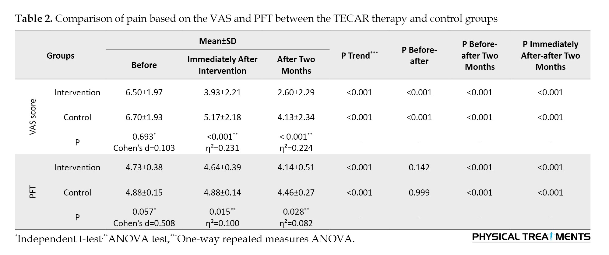
No significant difference in PFT was observed between the two groups at baseline (P=0.057). Immediately after the intervention, and after 2 months, the PFT was significantly lower in the TECAR therapy group than in the control group (P=0.015 and P=0.028, respectively). No significant change was observed in the fascia thickness immediately after the intervention in both groups (P=0.142 for the intervention group, and P=0.999 for the control group). However, measurements in both arms of the study, 2 months after the intervention, revealed a significant decrease in fascia thickness compared to the baseline levels and levels immediately after the intervention (all P<0.001). Table 2 presents the mean PFT in each group, before and after the intervention.
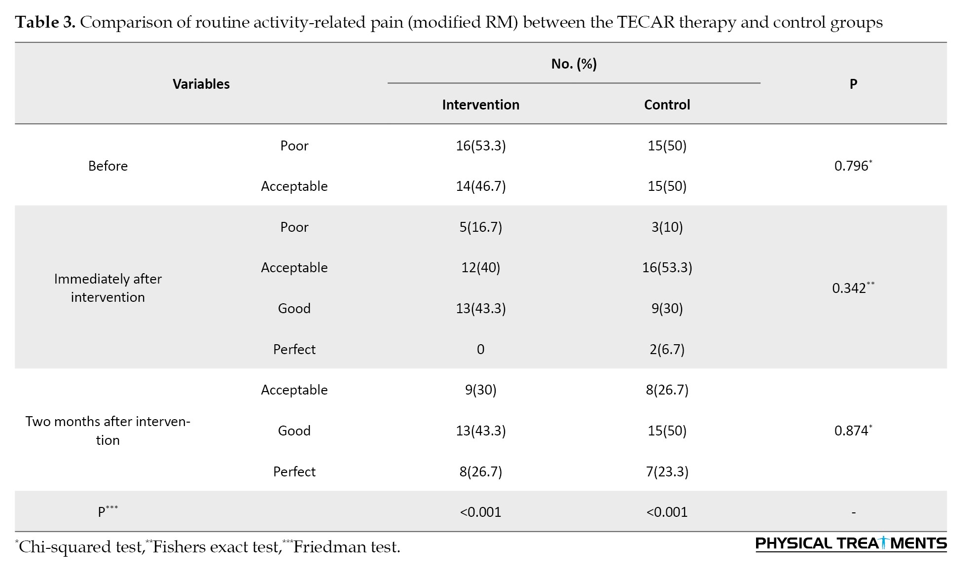
Regarding the RM scores, there was no significant difference between the two groups before the intervention (P=0.796), immediately after the intervention (P=0.342), and 2 months later (P=0.874). The Friedman test revealed a significant improvement in RM score distribution over time in both the TECAR therapy (P<0.001) and control groups (P<0.001). The pairwise Wilcoxon test showed a significant improvement in pain intensity both immediately after treatment compared to before (P<0.001) and two months after treatment compared to baseline (P<0.001). Additionally, a significant improvement in the distribution of RM categories was observed 2 months after the intervention compared to immediately after (P<0.001). Table 3 presents the distribution of RM scores in each group, before and after the intervention.
Discussion
In this double-blind randomized clinical trial, we aimed to assess the efficacy of TECAR therapy in the management of PF. Our findings showed that both TECAR therapy and standard conservative management considerably diminished pain intensity in patients with PF over time. Although the groups did not differ significantly at baseline, the TECAR therapy group demonstrated better pain relief than the control group, as evidenced by significantly lower VAS scores both immediately following the intervention and two months later. Accordingly, TECAR therapy may provide better short- and mid-term analgesic effects than traditional management alone. Additionally, within-group analyses showed that both groups’ functional status significantly improved over time, despite the fact that there were no discernible differences in RM scores between groups at any point in time. PFT did not differ significantly between groups at baseline; however, it was significantly lower in the TECAR therapy group both in immediate post-treatment measurements and at the two-month follow-up. Both groups showed a significant reduction in thickness only at the two-month mark compared to baseline and immediate post-treatment measurements. This highlights that the effect of treatments on PFT may be delayed.
PF, also known as jogger’s heel, policeman’s heel, or tennis heel, is frequently classified as an overuse injury, mainly resulting from repetitive strain that leads to micro-tears in the plantar fascia [24]. Nearly half of individuals suffering from this condition will additionally experience heel spurs; however, these spurs are not the underlying cause. PF is frequently linked to runners and older individuals; however, additional risk factors include obesity, heel pad atrophy, aging, jobs that necessitate extended periods of standing, and weight-bearing activities [25]. Either chronic inflammation, which involves sustained immune cell activity, or chronic degeneration, which is characterized by fibroblastic proliferation without immune involvement, can be the course of PF. Histological investigations that demonstrate tissue alterations, such as collagen degradation and angiofibroblastic hyperplasia in the absence of inflammatory cells, frequently lend credence to the degenerative pathway [26].
There is currently no universally accepted treatment for PF, as its management typically involves a combination of conservative and adjunctive therapies tailored to individual patient needs [27]. Conservative measures, such as stretching exercises, orthotics, non-steroidal anti-inflammatory drugs, and physical therapy remain the first-line approach, particularly in the early stages of the condition. Adjunctive treatments, including corticosteroid injections, extracorporeal shockwave therapy, platelet-rich plasma, and newer modalities, such as TECAR therapy, have shown varying degrees of success, particularly in patients with persistent symptoms [3, 28]. Surgical intervention is generally reserved for chronic, refractory cases that do not respond to non-surgical treatment [11].
TECAR therapy is a newly emerged physical therapy technique that utilizes radiofrequency energy (300 kHz to 1 MHz) to generate heat within biological tissues, enhancing the body’s natural healing processes. It works through two main mechanisms: Capacitive energy transfer (CET), which targets superficial tissues with high water content using an insulated electrode, and resistive energy transfer (RET), which targets deeper, denser tissues using a non-insulated electrode. CET promotes local heating to muscles and soft tissues, improving blood flow, metabolic activity, and tissue flexibility, and reduces muscle tension. RET delivers energy to tissues, such as bone and tendons, promoting collagen synthesis, tissue healing, and reducing inflammation [12]. Results of a meta-analysis on patients with diverse musculoskeletal pain, such as low back pain, shoulder pain, knee pain, and leg pain, demonstrated beneficial effects of TECAR therapy in the management of musculoskeletal pain, particularly in long-term follow-ups [29]. This aligns with the findings of this study. Results of our trial indicated that TECAR therapy significantly affects the healing process of PF. However, its beneficial effects were not considerably more than those of traditional treatments.
Although the diagnosis of PF is primarily based on physical examination and patient history, PFT assessment using ultrasonography has been identified as a potential diagnostic tool [30]. It has been noted that the PFT among patients with PF is higher than in normal individuals. Values above 4 mm are considered diagnostic for PF [31]. Our findings indicated that therapeutic interventions decreased the thickness of plantar fascia in PF patients, but did not return it to the normal range. This thickness may return to normal levels during long-term follow-up.
This study has several strengths. Notably, to our knowledge, this is the first study to examine the effectiveness of TECAR therapy in patients with PF. Second, due to the double-blinded manner of this study, selection and performance bias have been minimized. Third, utilizing standardized outcome measures, including the VAS and RM scores, and objective assessment of PFT adds robustness to the data collection and interpretation. However, the limitations should be mentioned. Due to the modest sample size, the study’s findings may have limited generalizability and reduced statistical power, especially for functional outcome measures. Participants were recruited consecutively from a single center, which may limit the generalizability of the findings. However, random sequence generation, allocation concealment, and blinding were applied to minimize selection and performance bias. Although ultrasonography was performed by a single blinded evaluator with repeated measurements to reduce variability, formal inter- or intra-rater reliability analysis was not conducted. Additionally, the short follow-up duration may not capture the long-term therapeutic effects, particularly in terms of returning PFT to normal levels. Moreover, the absence of a third group receiving sham TECAR therapy in addition to medication and exercise prevented a more precise isolation of the specific effects attributable to TECAR therapy.
Conclusion
This trial indicated that TECAR therapy, in combination with traditional conservative treatment, offers better short- and mid-term pain relief and a more significant reduction in PFT than conservative treatment alone in patients with PF. Both groups exhibited improved functional outcomes, with no significant differences between them. TECAR therapy is a promising adjuvant treatment, but larger studies with longer follow-up are needed to establish its long-term effectiveness.
Ethical Considerations
Compliance with ethical guidelines
This study was approved by the Ethics Committee of Isfahan University of Medical Sciences, Isfahan, Iran (Code: IR.MUI.MED.REC.1402.264) and subsequently recorded in the Iranian Registry of Clinical Trials (Code: IRCT20231105059968N1, Registration date: January 22, 2024).
Funding
This research did not receive any grant from funding agencies in the public, commercial, or non-profit sectors.
Authors' contributions
Conceptualization and methodology: Razieh Maghroori and Shila Haghighat; Investigation: Fatemeh Izadi; Writing the original draft: Razieh Maghroori and Fatemeh Izadi; Supervision: Shila Haghighat; Review and editing: All authors.
Conflict of interest
The authors declared no conflicts of interest.
Plantar fasciitis (PF) commonly occurs in individuals regardless of age or physical activity status and represents a major musculoskeletal complaint [1]. This condition is characterized by discomfort in the inner heel, which worsens during weight-bearing activities [2]. The lifetime incidence is approximately 10%. In one-third of the cases, PF may manifest bilaterally [3]. This disease frequently becomes chronic, with symptoms persisting for up to one year. PF is estimated to account for nearly one million annual visits to healthcare providers across the United States [1].
PF is mostly diagnosed by physical examination and clinical history [4]. Patients experience severe pain during the initial steps taken upon rising from bed in the morning. Although pain tends to subside as activities commence, it may worsen by the end of the day. Tenderness may be observed at the medial calcaneal tubercle. The windlass test is helpful in establishing the pathology with a high specificity (100%) but low sensitivity (32%) [3]. Ultrasound is also considered a sensitive instrument for PF assessment, with significant findings including increased plantar fascia thickness (PFT) and hypoechoic appearance of plantar fascia. X-ray imaging is also useful for ruling out other pathologies [5].
Although PF is self-limiting and resolves within a year, its impact on routine activities drives patients to pursue treatment prior to the resolution of the pain. Approximately 70% of patients experience reduced pain with conservative treatment alone [6]. However, some may require a combination of conservative management and other therapies [6]. Different therapeutic options have been suggested for PF, including night splint use [7], shock wave therapy [8], laser therapy [9], corticosteroid injections [10], and surgical interventions [11]. Nevertheless, based on the current studies, none of the aforementioned therapeutic options have demonstrated clear superiority, and with ongoing technological advancements, the optimal approach remains a matter of debate [3].
Recently, studies have utilized a novel electrotherapy modality in the management of musculoskeletal pain known as transfer of energy capacitive and resistive (TECAR) [12–14]. TECAR therapy delivers high-frequency energy (300 kHz–1 MHz) in a non-invasive manner, stimulating the body's innate capacity for regeneration [15]. TECAR therapy operates via two modes of electric charge transfer: capacitive and resistive. The capacitive mode primarily affects soft tissues and muscles rich in electrolytes. In contrast, the resistive mode targets denser structures, such as tendons, bones, and joints [14]. The findings of a recent meta-analysis suggested TECAR therapy as an effective method for management of sports-related pains [13].
Given the therapeutic mechanisms of TECAR therapy, including enhanced blood flow and tissue oxygenation, enhanced tissue metabolism, and reduced tissue edema [16], it is expected that this treatment would improve tissue perfusion and minimize edema in patients with PF. Considering the lack of prior surveys on the effectiveness of TECAR therapy in managing PF, coupled with the fact that PF has a high prevalence and clinical significance and that effective non-surgical management is required, this research was conducted to assess how TECAR treatment influences pain relief and functional improvement in individuals with PF.
Materials and Methods
Study design and protocol
This was a double-blind randomized clinical trial (in this investigation, both participants and therapists were blinded using a sham TECAR device that looked and felt identical to the active device but did not deliver therapeutic current). In accordance with the Declaration of Helsinki, all participants received a detailed explanation of the study protocol and voluntarily signed a written informed consent form. Group assignment was determined using a computer-generated randomization list prepared by an independent researcher with no role in participant recruitment or evaluation. Concealment of allocation was achieved by employing consecutively numbered, sealed, and opaque envelopes. Simple randomization was applied, and no block randomization or stratification was used.
Study participants
All patients with PF referred to the Physical Medicine and Rehabilitation Clinic, Isfahan University of Medical Sciences, Isfahan, Iran, were eligible for inclusion in the trial. The included patients were adults (aged 18-68 years), with at least a history of heel pain for 4 weeks. PF was diagnosed by a physiatrist through ultrasonographic evaluation. The severity of heel pain was assessed using the visual analog scale (VAS). Patients with a VAS score of >3 were included in this trial. The VAS cutoff (>3) was selected based on prior literature [17, 18] and expert consensus among the study investigators, all of whom were specialists in Physical Medicine and Rehabilitation. Pain scores ≤3 are generally considered mild, less likely to cause functional impairment, and typically do not require specific interventional treatment. The exclusion criteria were history of diabetes, arthritis, Achilles tendon injury, history of trauma, injections or surgeries on the affected heel, and any contraindications for TECAR therapy (including pregnancy, use of a pacemaker, neoplasms, open wounds and skin lesions, skin sensitivity, and lack of heat sensation). Individuals with an intense level of physical activity who were unable to reduce it were also excluded. During the study, patients were excluded if their symptoms worsened and required more vigorous interventions.
Study design and intervention
During the study period, participants were enrolled consecutively from patients referred to the physical medicine and rehabilitation clinic. Individuals meeting the specified eligibility criteria and agreeing to participate were included in this study. Participants were then allocated to either the intervention or control group through computer-based randomization with concealed allocation. The required sample size was calculated using an independent two-sample mean comparison formula with a two-tailed significance level of 0.05 and 80% power. Drawing on prior evidence [19], a minimum of 26 participants in each group were required. To account for potential dropouts, 30 participants were enrolled in each group. Finally, 30 patients were eligible to be included in each category. In the intervention group, individuals underwent TECAR therapy (twice per week for 4 weeks), a set of exercises (stretching the calf muscle and plantar fascia, passive dorsiflexion of the toes, strengthening intrinsic foot muscles, and rolling the sole three times daily (morning, afternoon, and evening) each set lasting 30 seconds), and a silicone heel pad was also prescribed for them. They additionally consumed celecoxib 200mg per day for 15 days. The control group performed the same set of exercises using a silicone heel pad and consumed the same medication.
TECAR therapy was delivered using the WINBACK 3 device (France) with a frequency of 500 Hz and an intensity range of 20–40%, adjusted according to patient tolerance at the physiotherapy section of Amin Hospital, Isfahan, Iran. Treatment was applied using a medium-sized CET electrode (60 mm). The capacitive and resistive modes were applied as described above. The patient was placed in the prone position, and conductive gel was applied near the Achilles tendon insertion. TECAR therapy was administered for 15 minutes per session (5 minutes of resistive, 10 minutes of capacitive) for 8 sessions, two sessions per week.
Data collection and measurements
At the beginning, demographic characteristics of participants, including age, sex, occupation (homemaker, unemployed, employed), severity of pain, severity of symptoms and patient’s function, were recorded. Ultrasonography (Alpinion E-CUBE 9, Republic of Korea) was employed to evaluate PFT. Ultrasonography was performed by a single experienced physiatrist who was blinded to group allocation and clinical data. To reduce measurement variability, PFT was measured twice at each assessment, and the average value was used for analysis. PF was diagnosed by an experienced physiatrist based on clinical presentation and ultrasonographic findings. No strict cut-off value for PFT was used for diagnosis; instead, ultrasonography was applied as a supportive imaging tool to confirm the clinical diagnosis and to quantify PFT for outcome assessment. The patients were placed in a prone position, ensuring knees were fully extended, and ankles dorsiflexed to 90°, with legs allowed to hang naturally. Using a 5–13 MHz probe on the Alpinion E-CUBE 9 (Republic of Korea), PFT was measured twice at the standard location crossing the anterior border of the calcaneus, with the average taken to minimize potential errors from the transducer. At the end of intervention and 2 months post-intervention, the severity of the pain and symptoms and the patient’s function were assessed. VAS score was utilized to assess the severity of pain [20]. This 10-point scale rated pain from 0 (no pain) to 10 (worst possible pain), with 1–3 indicating mild, 4–7 moderate, and 8–10 severe pain levels [21]. The reliability of the Iranian version of this scale has been approved [22]. The modified roles and Maudsley (RM) score was used to categorize participants into four levels: excellent, good, fair, and poor, based on their pain levels and ability to perform daily activities. This score was a functional evaluation method used to assess pain and activity limitations, often in the context of foot and ankle conditions, such as PF [23]. The main outcome of interest was the level of pain, determined using the VAS. Secondary outcomes included PFT measured using ultrasonography and functional status assessed using the modified RM score. Outcomes were evaluated at baseline, at the end of the intervention, and two months after the intervention.
Statistical analysis
Continuous data are expressed as Mean±SD, and categorical variables as No. (%). Data normality was assessed using the Shapiro–Wilk test and inspection of box plots. For within-group comparisons, repeated measures analysis of variance (ANOVA) or Friedman tests were applied, whereas independent t-tests, ANOVA, chi-square, or Fisher’s exact tests were used for between-group analyses. All analyses were performed using SPSS software, version 21.0 (SPSS Inc., Chicago, IL, USA), with statistical significance set at P<0.05.
Results
In each arm of the study, 30 individuals were enrolled. The mean age of all participants was 41.68±13.17, ranging from 18-63 years. Most patients were male (51.7%), and the majority were employed (31.7%). Table 1 presents the baseline characteristics of each group.
The groups did not differ significantly with respect to age, PFT, gender, or employment status (P>0.05 for all comparisons).
The evaluation of the VAS scores (an indicator of pain severity) demonstrated no significant difference between groups at baseline (P=0.693). Immediately after the intervention, the mean VAS scores in both groups decreased significantly compared to baseline levels (P<0.001). Two months after the intervention, the scores in both groups continued to diminish compared to the values immediately after intervention (P<0.001). In both arms of the study (immediately after intervention and 2 months later), the VAS scores were significantly lower among patients who underwent TECAR therapy (P<0.001). Table 2 presents the mean VAS scores in each group, before and after the intervention.
No significant difference in PFT was observed between the two groups at baseline (P=0.057). Immediately after the intervention, and after 2 months, the PFT was significantly lower in the TECAR therapy group than in the control group (P=0.015 and P=0.028, respectively). No significant change was observed in the fascia thickness immediately after the intervention in both groups (P=0.142 for the intervention group, and P=0.999 for the control group). However, measurements in both arms of the study, 2 months after the intervention, revealed a significant decrease in fascia thickness compared to the baseline levels and levels immediately after the intervention (all P<0.001). Table 2 presents the mean PFT in each group, before and after the intervention.
Regarding the RM scores, there was no significant difference between the two groups before the intervention (P=0.796), immediately after the intervention (P=0.342), and 2 months later (P=0.874). The Friedman test revealed a significant improvement in RM score distribution over time in both the TECAR therapy (P<0.001) and control groups (P<0.001). The pairwise Wilcoxon test showed a significant improvement in pain intensity both immediately after treatment compared to before (P<0.001) and two months after treatment compared to baseline (P<0.001). Additionally, a significant improvement in the distribution of RM categories was observed 2 months after the intervention compared to immediately after (P<0.001). Table 3 presents the distribution of RM scores in each group, before and after the intervention.
Discussion
In this double-blind randomized clinical trial, we aimed to assess the efficacy of TECAR therapy in the management of PF. Our findings showed that both TECAR therapy and standard conservative management considerably diminished pain intensity in patients with PF over time. Although the groups did not differ significantly at baseline, the TECAR therapy group demonstrated better pain relief than the control group, as evidenced by significantly lower VAS scores both immediately following the intervention and two months later. Accordingly, TECAR therapy may provide better short- and mid-term analgesic effects than traditional management alone. Additionally, within-group analyses showed that both groups’ functional status significantly improved over time, despite the fact that there were no discernible differences in RM scores between groups at any point in time. PFT did not differ significantly between groups at baseline; however, it was significantly lower in the TECAR therapy group both in immediate post-treatment measurements and at the two-month follow-up. Both groups showed a significant reduction in thickness only at the two-month mark compared to baseline and immediate post-treatment measurements. This highlights that the effect of treatments on PFT may be delayed.
PF, also known as jogger’s heel, policeman’s heel, or tennis heel, is frequently classified as an overuse injury, mainly resulting from repetitive strain that leads to micro-tears in the plantar fascia [24]. Nearly half of individuals suffering from this condition will additionally experience heel spurs; however, these spurs are not the underlying cause. PF is frequently linked to runners and older individuals; however, additional risk factors include obesity, heel pad atrophy, aging, jobs that necessitate extended periods of standing, and weight-bearing activities [25]. Either chronic inflammation, which involves sustained immune cell activity, or chronic degeneration, which is characterized by fibroblastic proliferation without immune involvement, can be the course of PF. Histological investigations that demonstrate tissue alterations, such as collagen degradation and angiofibroblastic hyperplasia in the absence of inflammatory cells, frequently lend credence to the degenerative pathway [26].
There is currently no universally accepted treatment for PF, as its management typically involves a combination of conservative and adjunctive therapies tailored to individual patient needs [27]. Conservative measures, such as stretching exercises, orthotics, non-steroidal anti-inflammatory drugs, and physical therapy remain the first-line approach, particularly in the early stages of the condition. Adjunctive treatments, including corticosteroid injections, extracorporeal shockwave therapy, platelet-rich plasma, and newer modalities, such as TECAR therapy, have shown varying degrees of success, particularly in patients with persistent symptoms [3, 28]. Surgical intervention is generally reserved for chronic, refractory cases that do not respond to non-surgical treatment [11].
TECAR therapy is a newly emerged physical therapy technique that utilizes radiofrequency energy (300 kHz to 1 MHz) to generate heat within biological tissues, enhancing the body’s natural healing processes. It works through two main mechanisms: Capacitive energy transfer (CET), which targets superficial tissues with high water content using an insulated electrode, and resistive energy transfer (RET), which targets deeper, denser tissues using a non-insulated electrode. CET promotes local heating to muscles and soft tissues, improving blood flow, metabolic activity, and tissue flexibility, and reduces muscle tension. RET delivers energy to tissues, such as bone and tendons, promoting collagen synthesis, tissue healing, and reducing inflammation [12]. Results of a meta-analysis on patients with diverse musculoskeletal pain, such as low back pain, shoulder pain, knee pain, and leg pain, demonstrated beneficial effects of TECAR therapy in the management of musculoskeletal pain, particularly in long-term follow-ups [29]. This aligns with the findings of this study. Results of our trial indicated that TECAR therapy significantly affects the healing process of PF. However, its beneficial effects were not considerably more than those of traditional treatments.
Although the diagnosis of PF is primarily based on physical examination and patient history, PFT assessment using ultrasonography has been identified as a potential diagnostic tool [30]. It has been noted that the PFT among patients with PF is higher than in normal individuals. Values above 4 mm are considered diagnostic for PF [31]. Our findings indicated that therapeutic interventions decreased the thickness of plantar fascia in PF patients, but did not return it to the normal range. This thickness may return to normal levels during long-term follow-up.
This study has several strengths. Notably, to our knowledge, this is the first study to examine the effectiveness of TECAR therapy in patients with PF. Second, due to the double-blinded manner of this study, selection and performance bias have been minimized. Third, utilizing standardized outcome measures, including the VAS and RM scores, and objective assessment of PFT adds robustness to the data collection and interpretation. However, the limitations should be mentioned. Due to the modest sample size, the study’s findings may have limited generalizability and reduced statistical power, especially for functional outcome measures. Participants were recruited consecutively from a single center, which may limit the generalizability of the findings. However, random sequence generation, allocation concealment, and blinding were applied to minimize selection and performance bias. Although ultrasonography was performed by a single blinded evaluator with repeated measurements to reduce variability, formal inter- or intra-rater reliability analysis was not conducted. Additionally, the short follow-up duration may not capture the long-term therapeutic effects, particularly in terms of returning PFT to normal levels. Moreover, the absence of a third group receiving sham TECAR therapy in addition to medication and exercise prevented a more precise isolation of the specific effects attributable to TECAR therapy.
Conclusion
This trial indicated that TECAR therapy, in combination with traditional conservative treatment, offers better short- and mid-term pain relief and a more significant reduction in PFT than conservative treatment alone in patients with PF. Both groups exhibited improved functional outcomes, with no significant differences between them. TECAR therapy is a promising adjuvant treatment, but larger studies with longer follow-up are needed to establish its long-term effectiveness.
Ethical Considerations
Compliance with ethical guidelines
This study was approved by the Ethics Committee of Isfahan University of Medical Sciences, Isfahan, Iran (Code: IR.MUI.MED.REC.1402.264) and subsequently recorded in the Iranian Registry of Clinical Trials (Code: IRCT20231105059968N1, Registration date: January 22, 2024).
Funding
This research did not receive any grant from funding agencies in the public, commercial, or non-profit sectors.
Authors' contributions
Conceptualization and methodology: Razieh Maghroori and Shila Haghighat; Investigation: Fatemeh Izadi; Writing the original draft: Razieh Maghroori and Fatemeh Izadi; Supervision: Shila Haghighat; Review and editing: All authors.
Conflict of interest
The authors declared no conflicts of interest.
References
- Rhim HC, Kwon J, Park J, Borg-Stein J, Tenforde AS. A systematic review of systematic reviews on the epidemiology, evaluation, and treatment of plantar fasciitis. Life. 2021; 11(12):1287. [DOI:10.3390/life11121287] [PMID] [PMCID]
- Cutts S, Obi N, Pasapula C, Chan W. Plantar fasciitis. Annals. 2012; 94(8):539-42. [DOI:10.1308/003588412X13171221592456] [PMID] [PMCID]
- Tseng WC, Chen YC, Lee TM, Chen WS. Plantar fasciitis: An updated review. Journal of Medical Ultrasound. 2023 Dec;31(4):268. [DOI:10.4103/jmu.jmu_2_23] [PMID] [PMCID]
- Goff JD, Crawford R. Diagnosis and treatment of plantar fasciitis. American Family Physician. 2011; 84(6):676-82. [PMID]
- Johnson RE, Haas K, Lindow K, Shields R. Plantar fasciitis: What is the diagnosis and treatment?. Orthopaedic Nursing. 2014; 33(4):198-204. [DOI:10.1097/NOR.0000000000000073]
- Luffy L, Grosel J, Thomas R, So E. Plantar fasciitis: A review of treatments. JAAPA. 2018; 31(1):20-4. [PMID]
- Pereira Bendo AJ, Guimarães Barros MI, Flor Bertolini GR, de Carvalho AR, Buzanello Donin C, Rosângela Buzanello Azevedo M. Night splints in plantar fasciitis: A systematic review. Muscles, Ligaments & Tendons Journal. 2023; 13(2):283. [DOI:10.32098/mltj.02.2023.12]
- Charles R, Fang L, Zhu R, Wang J. The effectiveness of shockwave therapy on patellar tendinopathy, Achilles tendinopathy, and plantar fasciitis: A systematic review and meta-analysis. Frontiers in Immunology. 2023; 14:1193835. [DOI:10.3389/fimmu.2023.1193835] [PMID] [PMCID]
- Yadav S, Sharma S, Chatterjee S, Sharma A, Thakur S. Effect of LASER therapy on plantar fasciitis pain: illuminating a promising treatment approach - A systematic review. Lasers in Medical Science. 2025; 40(1):18. [DOI:10.1007/s10103-025-04289-y] [PMID]
- Whittaker GA, Munteanu SE, Menz HB, Bonanno DR, Gerrard JM, Landorf KB. Corticosteroid injection for plantar heel pain: A systematic review and meta-analysis. BMC BMC Musculoskeletal Disorders. 2019; 20(1):378. [DOI:10.1186/s12891-019-2749-z] [PMID] [PMCID]
- Nayar SK, Alcock H, Vemulapalli K. Surgical treatment options for plantar fasciitis and their effectiveness: A systematic review and network meta-analysis. Archives of Orthopaedic and Trauma Surgery. 2023; 143(8):4641-51. [DOI:10.1007/s00402-022-04739-0] [PMID]
- Lupowitz LG, Ramus L, Delacour F, Johnson K. TECAR therapy: A clinical commentary on its evolution, application, and future in rehabilitation. International Journal of Sports Physical Therapy. 2025; 20(4):632.[DOI:10.26603/001c.130909] [PMID] [PMCID]
- Kudich E, Miha V, Kozinc Ž. Effectiveness of TECAR therapy for managing pain in sports-related musculoskeletal pathologies: A systematic review with meta-analysis. SN Comprehensive Clinical Medicine. 2025; 7(1):28. [DOI:10.1007/s42399-025-01794-7]
- Szabo DA, Neagu N, Teodorescu S, Predescu C, Sopa IS, Panait L. TECAR therapy associated with high-intensity laser therapy (Hilt) and manual therapy in the treatment of muscle disorders: A literature review on the theorised effects supporting their use. Journal of Clinical Medicine. 2022; 11(20):6149. [DOI:10.3390/jcm11206149] [PMID] [PMCID]
- Ribeiro S, Henriques B, Cardoso R. The effectiveness of tecar therapy in musculoskeletal disorders. International Journal of Public Health and Health Systems. 2018; 3(5):77-83. [Link]
- Clijsen R, Leoni D, Schneebeli A, Cescon C, Soldini E, Li L, et al. Does the application of tecar therapy affect temperature and perfusion of skin and muscle microcirculation? A pilot feasibility study on healthy subjects. Journal of Alternative and Complementary Medicine. 2020; 26(2):147. [DOI:10.1089/acm.2019.0165] [PMID] [PMCID]
- Taheri P, Maghroori R, Arzaghi S. The assessment of TECAR therapy efficacy to manage lateral epicondylitis: A randomized clinical-trial. Advanced Biomedical Research. 2025; 14(1):129. [DOI:10.4103/abr.abr_202_24]
- Boonstra AM, Schiphorst Preuper HR, Balk GA, Stewart RE. Cut-off points for mild, moderate, and severe pain on the visual analogue scale for pain in patients with chronic musculoskeletal pain. Pain. 2014; 155(12):2545. [DOI:10.1016/j.pain.2014.09.014] [PMID]
- Davari A, Mansour Sohani S, Sarrafzadeh J, Nikjoui A. Evaluation of the effects of tecar therapy on acute symptoms of athletes following lateral ankle ligament sprain. Function and Disability Journal. 2021; 4(1):0-0. [DOI:10.32598/fdj.4.31]
- Vickers ER, Cousins MJ, Woodhouse A. Pain description and severity of chronic orofacial pain conditions.Australian Dental Journal. 1998; 43(6):403-9. [DOI:10.1111/j.1834-7819.1998.tb00200.x] [PMID]
- Aicher B, Peil H, Peil B, Diener HC. Pain measurement: Visual analogue scale (VAS) and verbal rating scale (VRS) in clinical trials with OTC analgesics in headache. Cephalalgia. 2012; 32(3):185-97. [DOI:10.1177/03331024111430856] [PMID]
- Amin MR, Siratinayer M, Abadi A, Moradyan T. Correlation between visual analogue scale and short form of McGill questionnaire in patients with chronic low back pain. Majallah-i Dānishgāh-i ̒Ulūm-i Pizishkī-i Qum. 2012; 6(1):31-4. [DOI:10.5555/20123144997]
- Ibrahim MI, Donatelli RA, Schmitz C, Hellman MA, Buxbaum F. Chronic plantar fasciitis treated with two sessions of radial extracorporeal shock wave therapy. Foot & Ankle International. 2010; 31(5):391-7. [DOI:10.3113/FAI.2010.0391] [PMID]
- Ali Q, Long Y, Ali M. Prevalence, causes, and treatment of plantar fasciitis in young females of a medical college. Bulletin of Faculty of Physical Therapy. 2024; 29(1):31. [DOI:10.1186/s43161-024-00195-6]
- Buchanan BK, Sina RE, Kushner D. Plantar fasciitis. Treasure Island (FL): StatPearls Publishing; 2025. [Link]
- Liebmann K, Kimbrough DW, Best TM, Kouroupis D, Rodriguez Materon S. Plantar fasciitis pathophysiology and the potential role of mesenchymal stem cell-derived extracellular vesicles as therapy. Biomedicines. 2025; 13(7):1528. [DOI:10.3390/biomedicines13071528] [PMID] [PMCID]
- Koc TA, Bise CG, Neville C, Carreira D, Martin RL, McDonough CM. Heel pain - plantar fasciitis: Revision 2023. Journal of Orthopaedic & Sports Physical Therapy. 2023; 53(12):CPG1-39. [DOI:10.2519/jospt.2023.0303] [PMID]
- Guimarães J de S, Arcanjo FL, Leporace G, Metsavaht LF, Conceição CS, Moreno MVMG, et al. Effects of therapeutic interventions on pain due to plantar fasciitis: A systematic review and meta-analysis.Clinical Rehabilitation. 2023; 37(6):727-46. [DOI:10.1177/02692155221143865] [PMID]
- Vahdatpour B, Haghighat S, Sadri L, Taghian M, Sadri S. Effects of transfer energy capacitive and resistive on musculoskeletal pain: A systematic review and meta-analysis. Galen Medical Journal. 2022; 11:e2407. [DOI:10.31661/gmj.v11i.2407] [PMID] [PMCID]
- Thanwisate T, Siriwanarangsun P, Piyaselakul S, Tharmviboonsri T, Chuckpaiwong B. Plantar fascia thickness and stiffness in healthy individuals vs patients with plantar fasciitis. Foot and Ankle International. 2024; 45(11):1270-8. [DOI:10.1177/10711007241274765] [PMID]
- Ermutlu C, Aksakal M, Gümüştaş A, Özkaya G, Kovalak E, Özkan Y. Thickness of plantar fascia is not predictive of functional outcome in plantar fasciitis treatment. Acta Orthopaedica et Traumatologica Turcica. 2018; 52(6):442-6. [DOI:10.1016/j.aott.2018.01.002] [PMID] [PMCID]
Type of Study: Research |
Subject:
Physical Therapy
Received: 2025/11/18 | Accepted: 2026/02/12 | Published: 2026/10/18
Received: 2025/11/18 | Accepted: 2026/02/12 | Published: 2026/10/18
Send email to the article author
| Rights and permissions | |
 |
This work is licensed under a Creative Commons Attribution-NonCommercial 4.0 International License. |

This is an open access article distributed under the terms of the Creative Commons Attribution-NonCommercial 4.0 (CC-By-NC 4.0), which permits use, distribution, and reproduction in any medium, provided the original work is properly cited and is not used for commercial purposes.
Contact Information