Sun, Nov 8, 2026
Volume 16, Issue 4 ( Autumn 2026)
PTJ 2026, 16(4): 453-464 |
Back to browse issues page


Download citation:
BibTeX | RIS | EndNote | Medlars | ProCite | Reference Manager | RefWorks
Send citation to:


BibTeX | RIS | EndNote | Medlars | ProCite | Reference Manager | RefWorks
Send citation to:
Dsouza C J, Rebello S R, Ashraf M, Alankar A S, Lewis C P, Mendonca G A. Toe Grip Strength and Its Relationship With Balance and Mobility in Knee Osteoarthritis: Implications for Physical Therapy Assessment and Intervention. PTJ 2026; 16 (4) :453-464
URL: http://ptj.uswr.ac.ir/article-1-793-en.html
URL: http://ptj.uswr.ac.ir/article-1-793-en.html
Cyanna Joseph Dsouza1  , Sydney Roshan Rebello2 , Miqdad Ashraf *2 , Amith Sridhar Alankar3 , Charissa Patson Lewis2 , Glevisha Ann Mendonca2
1- Father Muller College of Physiotherapy, A Unit of Father Muller Charitable Institutions, Mangalore, India. & Srinivas Institute of Physiotherapy, Mangalore, India.
2- Father Muller College of Physiotherapy, A Unit of Father Muller Charitable Institutions, Mangalore, India.
3- Department of Orthopaedics, Father Muller Medical College, A Unit of Father Muller Charitable Institutions, Mangalore, India.
2- Father Muller College of Physiotherapy, A Unit of Father Muller Charitable Institutions, Mangalore, India.
3- Department of Orthopaedics, Father Muller Medical College, A Unit of Father Muller Charitable Institutions, Mangalore, India.
Full-Text [PDF 1062 kb]
(993 Downloads)
| Abstract (HTML) (2818 Views)
Full-Text: (67 Views)
Introduction
Recent global estimates indicate that knee OA affects nearly 375 million people in 2021, and its prevalence is projected to approach 1 billion worldwide by 2050 with ageing populations and rising obesity [1, 2]. However, knee OA poses a significant public health issue in India, particularly among individuals aged 40 and older, with data also suggesting an earlier onset influenced by genetic, lifestyle, and environmental factors [3, 4]. These studies also indicate that more than one-third of the population in rural South India are diagnosed with this condition, with significant associations with obesity, diabetes, hypertension, sedentary lifestyle, menopause, and the practice of squatting for toileting.
Excessive mechanical loading on joint surfaces significantly contributes to the onset and progression of knee OA, often resulting from abnormal knee joint loading due to disrupted kinematic interactions between the foot and knee [5]. Abnormal foot posture or altered foot movement may contribute to degenerative changes in knee joint cartilage by causing improper loading [6]. As a result, the foot is increasingly considered in lower-limb assessments.
The first metatarsal joint plays a crucial role in maintaining the structural integrity of the foot arch, supporting body weight, and evenly distributing mechanical stress during ambulation [7, 8]. Dysfunction at this joint, such as reduced TGS due to hallux impairment, may disrupt the biomechanical chain influencing proximal joints. When TGS is weak, individuals tend to compensate by increasing ankle dorsiflexion, leading to higher muscular demand around the ankle and elevated torque at the knee and hip during walking [9, 10].
TGS plays a vital role in mobility among older adults and may contribute to dysfunction of the proximal joints [11, 12]. Balance impairments are particularly pronounced in individuals with knee OA, likely due to reduced muscle strength and joint position sense. These deficits lead to greater difficulty in walking and an increased risk of falls compared to those without OA [13]. Furthermore, falls in this population can result in severe injuries, fear of movement, or even fatal outcomes, adding a substantial financial strain to healthcare systems [14, 15]. Therefore, assessing balance impairments is essential for optimizing rehabilitation strategies.
A common gait abnormality in individuals with knee OA is the failure to maintain consistent toe-ground contact throughout the stance phase until toe-off, suggesting a possible relationship between compromised toe flexor strength and altered gait mechanics in this population. Given the association between the forefoot, hallux, and rearfoot through the midfoot, hallux dysfunction may indirectly influence knee kinematics in OA by altering rearfoot motion patterns during gait [16]. Uritani et al. highlighted the importance of assessing toe flexor strength as a predictive and preventive measure for walking dysfunction and falls [17]. They also observed a link between preoperative TGS and postoperative functional mobility in patients who underwent total knee replacement (TKR) [18]. Because TKR is an end-stage intervention, this link underscores the critical role of TGS in mobility and recovery.
Nevertheless, a notable gap remains in the mechanical linkage between TGS and knee biomechanics. While some studies have examined broad association trends, few have explored how toe strength directly contributes to dynamic balance and mobility impairments in knee OA. Moreover, biomechanical explanations tying TGS to compensatory movement patterns in proximal joints are underrepresented, particularly in resource-limited settings where early intervention could be pivotal.
To the best of our current understanding, published evidence examining the correlation among these factors in individuals with knee OA in an Indian context is lacking. As life expectancy rises and obesity becomes more prevalent, India is on the brink of a substantial OA burden, emphasizing the need for region-specific research. In rural areas, limited access to diagnostic imaging and specialized orthopaedic services frequently contributes to delayed identification and management of the condition, often until it has progressed to more advanced stages. The growing prevalence of OA is set to escalate disability and deteriorate quality of life, presenting a significant challenge for India’s resource-limited public health system and imposing a heavy economic strain on the country [3, 19].
Accordingly, the present study aimed to investigate the relationship between TGS and dynamic balance and mobility in patients with knee OA. We hypothesized that TGS deficits would correlate with diminished performance in assessments of dynamic balance and mobility. Understanding these associations, especially their functional and mechanistic underpinnings, may help develop targeted physical therapy strategies to enhance lower limb function and mobility in this population.
Materials and Methods
Study design and setting
This observational study employed a cross-sectional design and was conducted over a three-month period, from December 2023 to February 2024, recruiting participants from a tertiary care hospital and community setting in a South Indian city. Individuals meeting the inclusion criteria were enrolled after written informed consent was obtained following a comprehensive explanation of the study’s objectives, potential benefits, and associated risks. Reporting adhered to the Strengthening the Reporting of Observational Studies in Epidemiology criteria for observational research.
Eligibility criteria
Participants included individuals aged 45 to 75 years with a diagnosis of knee OA confirmed by an orthopaedic specialist, based on the American College of Rheumatology criteria [20], and a history of the condition for more than three months. The orthopaedic surgeon also assessed the severity of knee OA from an anteroposterior weight-bearing film using the Kellgren and Lawrence scale [21], and only those with Grade 2, characterized by definite osteophytes and possible joint space narrowing, were included. This reduced heterogeneity, as advanced grades, is associated with substantial structural damage and functional loss, potentially confounding the relationship between TGS and mobility. Eligible participants were required to ambulate independently without assistive devices or caregiver support and to have a minimum education level equivalent to completion of 4th grade. Exclusion criteria encompassed any neuromuscular or musculoskeletal disorders, prior TKR, lumbar spine surgery, or surgical procedures involving the lower extremities.
After screening, demographic information was recorded, and all outcome measures were evaluated on the same day by a single researcher (MA) to reduce inter-rater variability. The most affected knee was identified through participant-reported symptoms of pain and functional limitation, further confirmed by radiographic findings of grade 2 OA. In cases of bilateral OA, the knee with greater symptom severity as reported by the participant was selected for assessment.
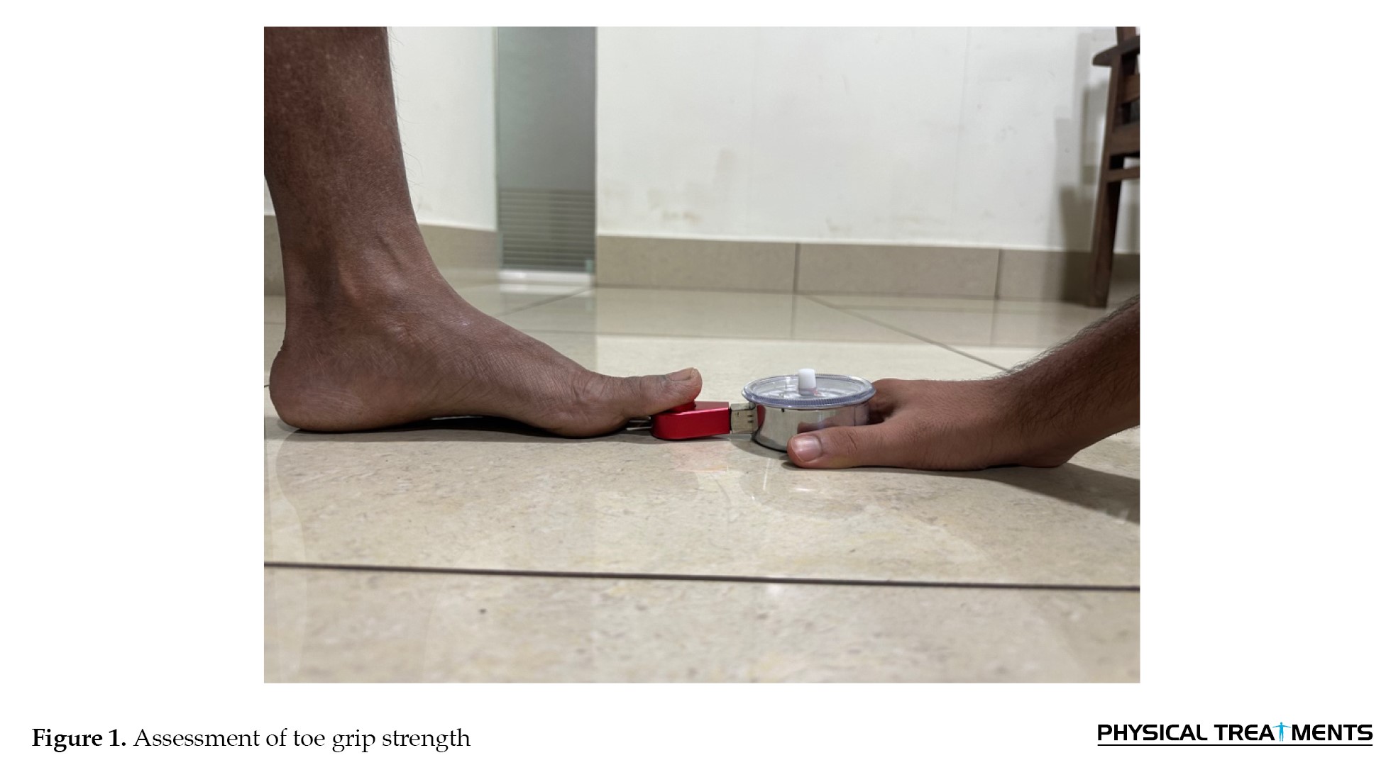
TGS: The strength of the flexor hallucis longus was assessed with a pinch gauge dynamometer [22, 23], which has demonstrated excellent intra-rater reliability (ICC=0.94–0.98) in previous studies [24, 25]. Participants were seated with their feet flat on the floor, and the dynamometer was positioned beneath the hallux, with its proximal end aligned just below the distal crease of the first metatarsophalangeal joint (Figure 1).
Recent global estimates indicate that knee OA affects nearly 375 million people in 2021, and its prevalence is projected to approach 1 billion worldwide by 2050 with ageing populations and rising obesity [1, 2]. However, knee OA poses a significant public health issue in India, particularly among individuals aged 40 and older, with data also suggesting an earlier onset influenced by genetic, lifestyle, and environmental factors [3, 4]. These studies also indicate that more than one-third of the population in rural South India are diagnosed with this condition, with significant associations with obesity, diabetes, hypertension, sedentary lifestyle, menopause, and the practice of squatting for toileting.
Excessive mechanical loading on joint surfaces significantly contributes to the onset and progression of knee OA, often resulting from abnormal knee joint loading due to disrupted kinematic interactions between the foot and knee [5]. Abnormal foot posture or altered foot movement may contribute to degenerative changes in knee joint cartilage by causing improper loading [6]. As a result, the foot is increasingly considered in lower-limb assessments.
The first metatarsal joint plays a crucial role in maintaining the structural integrity of the foot arch, supporting body weight, and evenly distributing mechanical stress during ambulation [7, 8]. Dysfunction at this joint, such as reduced TGS due to hallux impairment, may disrupt the biomechanical chain influencing proximal joints. When TGS is weak, individuals tend to compensate by increasing ankle dorsiflexion, leading to higher muscular demand around the ankle and elevated torque at the knee and hip during walking [9, 10].
TGS plays a vital role in mobility among older adults and may contribute to dysfunction of the proximal joints [11, 12]. Balance impairments are particularly pronounced in individuals with knee OA, likely due to reduced muscle strength and joint position sense. These deficits lead to greater difficulty in walking and an increased risk of falls compared to those without OA [13]. Furthermore, falls in this population can result in severe injuries, fear of movement, or even fatal outcomes, adding a substantial financial strain to healthcare systems [14, 15]. Therefore, assessing balance impairments is essential for optimizing rehabilitation strategies.
A common gait abnormality in individuals with knee OA is the failure to maintain consistent toe-ground contact throughout the stance phase until toe-off, suggesting a possible relationship between compromised toe flexor strength and altered gait mechanics in this population. Given the association between the forefoot, hallux, and rearfoot through the midfoot, hallux dysfunction may indirectly influence knee kinematics in OA by altering rearfoot motion patterns during gait [16]. Uritani et al. highlighted the importance of assessing toe flexor strength as a predictive and preventive measure for walking dysfunction and falls [17]. They also observed a link between preoperative TGS and postoperative functional mobility in patients who underwent total knee replacement (TKR) [18]. Because TKR is an end-stage intervention, this link underscores the critical role of TGS in mobility and recovery.
Nevertheless, a notable gap remains in the mechanical linkage between TGS and knee biomechanics. While some studies have examined broad association trends, few have explored how toe strength directly contributes to dynamic balance and mobility impairments in knee OA. Moreover, biomechanical explanations tying TGS to compensatory movement patterns in proximal joints are underrepresented, particularly in resource-limited settings where early intervention could be pivotal.
To the best of our current understanding, published evidence examining the correlation among these factors in individuals with knee OA in an Indian context is lacking. As life expectancy rises and obesity becomes more prevalent, India is on the brink of a substantial OA burden, emphasizing the need for region-specific research. In rural areas, limited access to diagnostic imaging and specialized orthopaedic services frequently contributes to delayed identification and management of the condition, often until it has progressed to more advanced stages. The growing prevalence of OA is set to escalate disability and deteriorate quality of life, presenting a significant challenge for India’s resource-limited public health system and imposing a heavy economic strain on the country [3, 19].
Accordingly, the present study aimed to investigate the relationship between TGS and dynamic balance and mobility in patients with knee OA. We hypothesized that TGS deficits would correlate with diminished performance in assessments of dynamic balance and mobility. Understanding these associations, especially their functional and mechanistic underpinnings, may help develop targeted physical therapy strategies to enhance lower limb function and mobility in this population.
Materials and Methods
Study design and setting
This observational study employed a cross-sectional design and was conducted over a three-month period, from December 2023 to February 2024, recruiting participants from a tertiary care hospital and community setting in a South Indian city. Individuals meeting the inclusion criteria were enrolled after written informed consent was obtained following a comprehensive explanation of the study’s objectives, potential benefits, and associated risks. Reporting adhered to the Strengthening the Reporting of Observational Studies in Epidemiology criteria for observational research.
Eligibility criteria
Participants included individuals aged 45 to 75 years with a diagnosis of knee OA confirmed by an orthopaedic specialist, based on the American College of Rheumatology criteria [20], and a history of the condition for more than three months. The orthopaedic surgeon also assessed the severity of knee OA from an anteroposterior weight-bearing film using the Kellgren and Lawrence scale [21], and only those with Grade 2, characterized by definite osteophytes and possible joint space narrowing, were included. This reduced heterogeneity, as advanced grades, is associated with substantial structural damage and functional loss, potentially confounding the relationship between TGS and mobility. Eligible participants were required to ambulate independently without assistive devices or caregiver support and to have a minimum education level equivalent to completion of 4th grade. Exclusion criteria encompassed any neuromuscular or musculoskeletal disorders, prior TKR, lumbar spine surgery, or surgical procedures involving the lower extremities.
After screening, demographic information was recorded, and all outcome measures were evaluated on the same day by a single researcher (MA) to reduce inter-rater variability. The most affected knee was identified through participant-reported symptoms of pain and functional limitation, further confirmed by radiographic findings of grade 2 OA. In cases of bilateral OA, the knee with greater symptom severity as reported by the participant was selected for assessment.
TGS: The strength of the flexor hallucis longus was assessed with a pinch gauge dynamometer [22, 23], which has demonstrated excellent intra-rater reliability (ICC=0.94–0.98) in previous studies [24, 25]. Participants were seated with their feet flat on the floor, and the dynamometer was positioned beneath the hallux, with its proximal end aligned just below the distal crease of the first metatarsophalangeal joint (Figure 1).
The examiner manually stabilized the device. Participants were instructed to press downward with maximal effort for three seconds, ensuring the heel remained in contact with the ground. Three trials were conducted, with a rest period of less than five seconds between attempts.
Functional reach test (FRT): Participants stood near a wall without touching it and raised the arm closest to the wall to 90° of shoulder flexion while maintaining a closed fist [26]. The initial position of the third metacarpal head was marked on a yardstick affixed to the wall. The participants then reached as far as possible without taking a step. The final position was recorded, and the reach distance was calculated as the difference between the start and end positions. We averaged three trials.
Timed up and go test (TUG): Participants wore regular footwear and did not use assistive devices during testing. Initially, each participant was seated in a chair with an armrest [27, 28]. At the therapist’s cue, they stood up, started the timer, walked three meters at a comfortable pace, turned, walked back, and sat down completely, at which point timing stopped. The timing ended when the participant was completely seated. Two practice trials were conducted prior to recording one final measurement for statistical analysis.
Visual analog scale (VAS): Pain intensity was assessed using a 100mm horizontal line, marked with the descriptors “no pain” at 0mm and “worst possible pain” at 100mm [29]. The participants indicated their current pain level by placing a mark on the line. The pain score was quantified by measuring the distance in millimeters from the 0mm point to the participant’s mark using a standard ruler.
Western ontario and mcmaster university (WOMAC) OA index: This questionnaire consists of three scales and 24 items, each scored on a 5-point ordinal scale. Higher scores reflect increased symptom severity [30].
Sample size
The a priori sample size for testing the association between TGS and mobility/balance outcomes was calculated for a two-tailed correlation test using the Fisher z transformation, assuming an expected effect size of r=0.23 (based on the smallest relevant correlation reported by Uritani et al. [31]), with a significance level of 5% and a statistical power of 80%. This indicated a minimum requirement of 146 participants; we recruited 147, thereby exceeding the target.
Statistical methods
Descriptive statistics, including means and standard deviations, were used to summarize the demographic variables and outcome measures. We applied the Kolmogorov–Smirnov test to assess the normality of the data distribution. Because the variables were not normally distributed, Spearman’s rank correlation was used to examine the relationship between TGS, TUG, and FRT. The correlation coefficient (r) was interpreted as: 0–0.25 (negligible), 0.25–0.50 (fair), 0.50–0.75 (moderate to good), and ≥0.75 (good to excellent) [32]. A partial correlation analysis was conducted to explore the associations between outcome measures while controlling for age, body mass index (BMI), and duration of OA. Scatter plots were used to depict the relationships between TGS and TUG and FRT. Simple linear regression was used to assess the predictive value of FRT and TUG on TGS, while a multiple regression model including both TUG and FRT was constructed to evaluate their combined effect on TGS. Multicollinearity diagnostics were performed using the variance inflation factor (VIF) and tolerance statistics to ensure the validity of regression models. Statistical analyses were performed using IBM SPSS software, version 25.0 (IBM Corp., Armonk, NY, USA), and a P<0.05 was considered statistically significant.
Results
Of the 186 participants assessed for eligibility, 39 did not meet the inclusion criteria. A total of 147 participants with knee OA were included in the analysis. The mean age was 59.3±7.8 years, and the mean BMI was 25.3±4.2 kg/m². The mean duration of knee OA was 3.8 ± 3.9 years (Table 1).

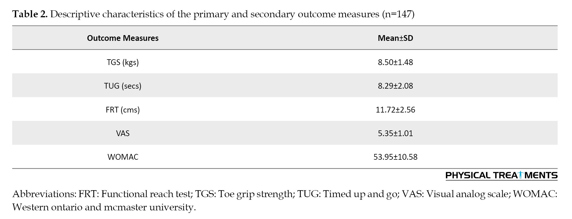
Consequently, statistical analyses were conducted on data from 147 participants, comprising 70 males (47.65%) and 77 females (52.4%). Of the participants, 38% had right knee involvement, 46% had left knee involvement, and 16% had bilateral knee involvement. Table 2 presents descriptive statistics for outcome measures.
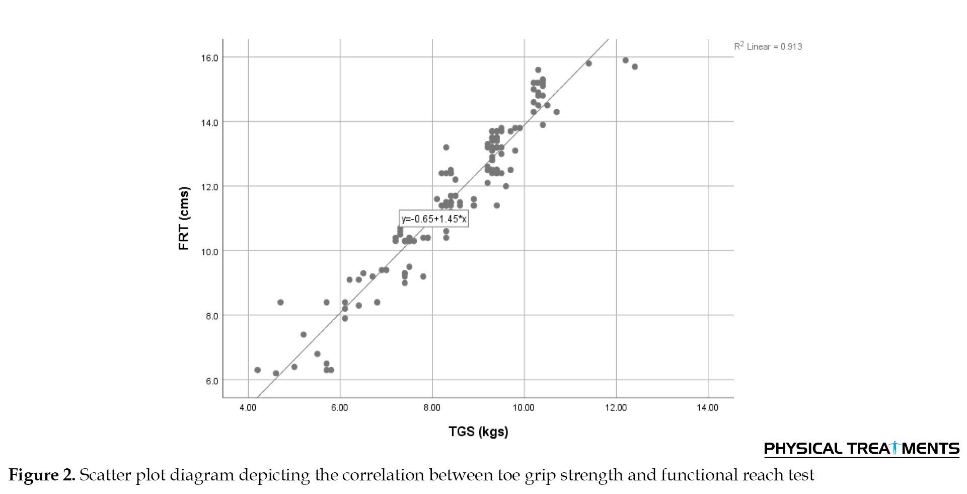
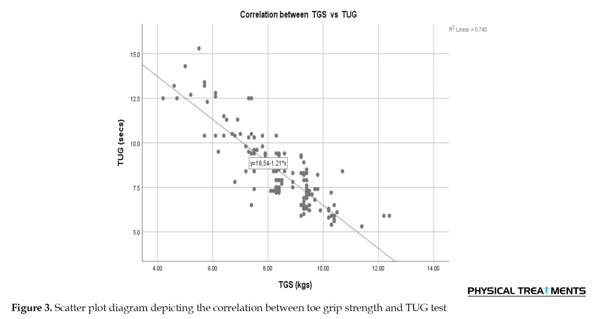
A good-to-excellent positive correlation was observed between TGS and the FRT (r=0.942; P<0.001), indicating that individuals with lower TGS achieved shorter distances in the reach test. Similarly, a strong negative correlation was found between TGS and the TUG (r=-0.832; P<0.001), suggesting that individuals with greater TGS completed the TUG in less time (Table 3).
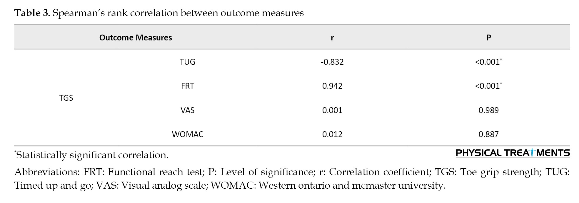
Although TGS showed a negligible correlation with both VAS (r=0.001; P=0.989) and WOMAC (r=0.012; P=0.887), these correlations were not statistically significant (Table 3).
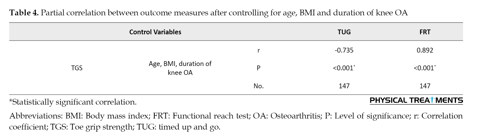
After controlling for age, BMI, and knee OA duration, TGS remained strongly associated with TUG (r=–0.735; P<0.001) and FRT (r=0.892; P<0.001), confirming that these relationships were independent of confounding variables (Table 4).
The scatter plot diagrams illustrate good to excellent linear relationships: A positive association between TGS and FRT (Figure 1), and a negative association between TGS and TUG (Figure 2).
Functional reach test (FRT): Participants stood near a wall without touching it and raised the arm closest to the wall to 90° of shoulder flexion while maintaining a closed fist [26]. The initial position of the third metacarpal head was marked on a yardstick affixed to the wall. The participants then reached as far as possible without taking a step. The final position was recorded, and the reach distance was calculated as the difference between the start and end positions. We averaged three trials.
Timed up and go test (TUG): Participants wore regular footwear and did not use assistive devices during testing. Initially, each participant was seated in a chair with an armrest [27, 28]. At the therapist’s cue, they stood up, started the timer, walked three meters at a comfortable pace, turned, walked back, and sat down completely, at which point timing stopped. The timing ended when the participant was completely seated. Two practice trials were conducted prior to recording one final measurement for statistical analysis.
Visual analog scale (VAS): Pain intensity was assessed using a 100mm horizontal line, marked with the descriptors “no pain” at 0mm and “worst possible pain” at 100mm [29]. The participants indicated their current pain level by placing a mark on the line. The pain score was quantified by measuring the distance in millimeters from the 0mm point to the participant’s mark using a standard ruler.
Western ontario and mcmaster university (WOMAC) OA index: This questionnaire consists of three scales and 24 items, each scored on a 5-point ordinal scale. Higher scores reflect increased symptom severity [30].
Sample size
The a priori sample size for testing the association between TGS and mobility/balance outcomes was calculated for a two-tailed correlation test using the Fisher z transformation, assuming an expected effect size of r=0.23 (based on the smallest relevant correlation reported by Uritani et al. [31]), with a significance level of 5% and a statistical power of 80%. This indicated a minimum requirement of 146 participants; we recruited 147, thereby exceeding the target.
Statistical methods
Descriptive statistics, including means and standard deviations, were used to summarize the demographic variables and outcome measures. We applied the Kolmogorov–Smirnov test to assess the normality of the data distribution. Because the variables were not normally distributed, Spearman’s rank correlation was used to examine the relationship between TGS, TUG, and FRT. The correlation coefficient (r) was interpreted as: 0–0.25 (negligible), 0.25–0.50 (fair), 0.50–0.75 (moderate to good), and ≥0.75 (good to excellent) [32]. A partial correlation analysis was conducted to explore the associations between outcome measures while controlling for age, body mass index (BMI), and duration of OA. Scatter plots were used to depict the relationships between TGS and TUG and FRT. Simple linear regression was used to assess the predictive value of FRT and TUG on TGS, while a multiple regression model including both TUG and FRT was constructed to evaluate their combined effect on TGS. Multicollinearity diagnostics were performed using the variance inflation factor (VIF) and tolerance statistics to ensure the validity of regression models. Statistical analyses were performed using IBM SPSS software, version 25.0 (IBM Corp., Armonk, NY, USA), and a P<0.05 was considered statistically significant.
Results
Of the 186 participants assessed for eligibility, 39 did not meet the inclusion criteria. A total of 147 participants with knee OA were included in the analysis. The mean age was 59.3±7.8 years, and the mean BMI was 25.3±4.2 kg/m². The mean duration of knee OA was 3.8 ± 3.9 years (Table 1).
Consequently, statistical analyses were conducted on data from 147 participants, comprising 70 males (47.65%) and 77 females (52.4%). Of the participants, 38% had right knee involvement, 46% had left knee involvement, and 16% had bilateral knee involvement. Table 2 presents descriptive statistics for outcome measures.
A good-to-excellent positive correlation was observed between TGS and the FRT (r=0.942; P<0.001), indicating that individuals with lower TGS achieved shorter distances in the reach test. Similarly, a strong negative correlation was found between TGS and the TUG (r=-0.832; P<0.001), suggesting that individuals with greater TGS completed the TUG in less time (Table 3).
Although TGS showed a negligible correlation with both VAS (r=0.001; P=0.989) and WOMAC (r=0.012; P=0.887), these correlations were not statistically significant (Table 3).
After controlling for age, BMI, and knee OA duration, TGS remained strongly associated with TUG (r=–0.735; P<0.001) and FRT (r=0.892; P<0.001), confirming that these relationships were independent of confounding variables (Table 4).
The scatter plot diagrams illustrate good to excellent linear relationships: A positive association between TGS and FRT (Figure 1), and a negative association between TGS and TUG (Figure 2).
Regression analysis (Table 5) showed that both FRT and TUG strongly predicted TGS.
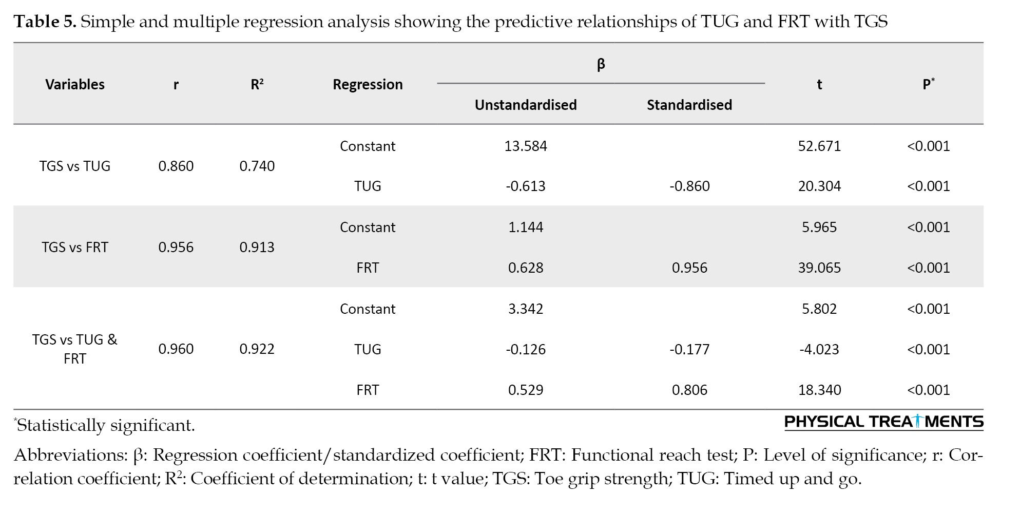
FRT alone explained 91.3% of the variance in TGS (β=0.628; P<0.001), while TUG accounted for 74% (β=–0.613; P<0.001). In the combined model, both remained significant (R²=0.922), with FRT emerging as the stronger predictor, indicating that balance has a greater influence on TGS than mobility. Collinearity diagnostics confirmed that multicollinearity was not present (all VIF values<2.0; tolerance>0.5). Thus, the high R² values reflect robust associations between functional balance, mobility, and TGS.
Discussion
This study investigated the association between TGS and functional measures of mobility and balance in individuals with knee OA. We found that both the TUG and FRT were significant predictors of TGS. Among these, FRT explained more variance in TGS than TUG, suggesting that balance capacity contributes more strongly to toe flexor strength than mobility performance. The results highlight the potential impact of TGS on functional ability and mobility in patients with knee OA.
A systematic review of observational studies found that greater toe flexor strength is associated with improved postural stability in individuals aged 60 and above [33]. Yong-Wook et al. showed that elderly non-fallers had significantly stronger muscles in the hallux and second toe, as well as better balance than fallers; toe flexor strength also correlated moderately with FRT, suggesting a role in fall prevention [34]. Similarly, Menz et al. highlighted that toe flexor strength, along with other ankle and foot features, can potentially predict functional performance and balance in older adults [10]. Unsurprisingly, interventions aimed at strengthening toe grip in younger adults have also improved performance on tasks requiring dynamic balance [35, 36].
The foot, a complex and adaptable structure with multiple degrees of freedom, also controls the center of mass. Extrinsic and intrinsic toe flexors play a crucial role in foot function, whether the foot is unloaded or in contact with the ground. In an upright stance, the toe flexors must produce adequate, timely plantar flexor torque to help prevent excessive forward displacement of the centre of mass beyond the base of support [9]. Consequently, balance stability depends on the effective activation of the extrinsic toe flexors to produce plantar flexor force at the ankle joint. This highlights the significance of TGS in maintaining postural control and preventing falls [33]. Toe flexors not only generate mechanical force but also contribute sensory feedback through proprioceptors, aiding neuromuscular control of standing balance and gait. Therefore, impaired proprioception may partly explain the strong association observed between FRT and TUG [37, 38].
FRT alone explained 91.3% of the variance in TGS (β=0.628; P<0.001), while TUG accounted for 74% (β=–0.613; P<0.001). In the combined model, both remained significant (R²=0.922), with FRT emerging as the stronger predictor, indicating that balance has a greater influence on TGS than mobility. Collinearity diagnostics confirmed that multicollinearity was not present (all VIF values<2.0; tolerance>0.5). Thus, the high R² values reflect robust associations between functional balance, mobility, and TGS.
Discussion
This study investigated the association between TGS and functional measures of mobility and balance in individuals with knee OA. We found that both the TUG and FRT were significant predictors of TGS. Among these, FRT explained more variance in TGS than TUG, suggesting that balance capacity contributes more strongly to toe flexor strength than mobility performance. The results highlight the potential impact of TGS on functional ability and mobility in patients with knee OA.
A systematic review of observational studies found that greater toe flexor strength is associated with improved postural stability in individuals aged 60 and above [33]. Yong-Wook et al. showed that elderly non-fallers had significantly stronger muscles in the hallux and second toe, as well as better balance than fallers; toe flexor strength also correlated moderately with FRT, suggesting a role in fall prevention [34]. Similarly, Menz et al. highlighted that toe flexor strength, along with other ankle and foot features, can potentially predict functional performance and balance in older adults [10]. Unsurprisingly, interventions aimed at strengthening toe grip in younger adults have also improved performance on tasks requiring dynamic balance [35, 36].
The foot, a complex and adaptable structure with multiple degrees of freedom, also controls the center of mass. Extrinsic and intrinsic toe flexors play a crucial role in foot function, whether the foot is unloaded or in contact with the ground. In an upright stance, the toe flexors must produce adequate, timely plantar flexor torque to help prevent excessive forward displacement of the centre of mass beyond the base of support [9]. Consequently, balance stability depends on the effective activation of the extrinsic toe flexors to produce plantar flexor force at the ankle joint. This highlights the significance of TGS in maintaining postural control and preventing falls [33]. Toe flexors not only generate mechanical force but also contribute sensory feedback through proprioceptors, aiding neuromuscular control of standing balance and gait. Therefore, impaired proprioception may partly explain the strong association observed between FRT and TUG [37, 38].
Interestingly, the lack of significant correlation between TGS and WOMAC may highlight a dissociation between neuromuscular performance and subjective symptom perception in knee OA. This aligns with prior literature suggesting that functional impairments may not always parallel pain reports [39]. This observation underscores the need to explore both biomechanical and psychosocial dimensions of OA.
Our findings align with previous research showing that TGS is significantly weaker in individuals with knee OA than in control subjects. While the causal link between TGS and knee OA has not yet been established, diminished TGS may compromise dynamic balance during standing and reduce propulsive force, which could subsequently increase mechanical stress on the knee joint [40]. This observation underscores the importance of TGS in the functional biomechanics of osteoarthritic knees. While diminished TGS may contribute to altered biomechanics and increased knee joint loading, this hypothesis requires confirmation in mechanistic and longitudinal studies. A previous investigation in 665 Japanese older adults found that TGS was independently correlated with TUG performance but not with FRT. These findings highlight the potential of TGS-focused physical therapy to improve functional mobility [17]. Evidence from European and North American cohorts similarly supports the association between foot muscle strength and functional performance, suggesting that the observed relationships are not restricted to Asian populations [37, 41].
Prior studies, such as those by Poojari et al. have also demonstrated a correlation between reduced TGS and fear of pain in patients with knee OA, suggesting a potential psychosomatic link that influences TGS [23]. Furthermore, a multicenter cross-sectional study found that TGS negatively correlated with age and positively with isometric strength of the knee extensors in patients with OA. In the control group, TGS was similarly associated with age and the TUG test, emphasizing the interplay between forefoot function and knee OA progression [31]. These findings suggest that improving TGS could be a practical therapeutic strategy to mitigate knee OA progression.
Our findings align with emerging evidence supporting functional, muscle-targeted interventions in knee OA management. For instance, Tedeschi highlighted the role of neuromuscular coordination and proprioceptive training in restoring function and performance, emphasizing the importance of strengthening specific muscle groups for optimal recovery [42]. Our results reinforce this perspective by identifying TGS as a potential marker of dynamic balance and mobility, suggesting that distal muscle activation and control may influence overall lower limb performance. Similarly, a randomized controlled trial demonstrated that structured remote interventions can significantly improve functional outcomes in patients with OA [43].
Further evidence suggests that decreased TGS on the affected limb correlates with a history of falls in older adults with knee OA, reinforcing the importance of incorporating TGS strengthening in physical therapy interventions to reduce fall risk [36]. Furthermore, longitudinal studies have established a relationship between preoperative TGS and postoperative functional performance in patients undergoing TKR, with stronger preoperative TGS being associated with better postoperative TUG outcomes [19]. These findings suggest that TGS may serve as a functional marker and therapeutic target for improving the outcomes of knee OA.
Limitations and recommendations
The study was limited to individuals with mild-to-moderate knee OA, suggesting that future investigations should include cases with severe OA to achieve a comprehensive understanding. Participant recruitment from both hospital and community settings may introduce selection bias, limiting generalizability. Additionally, the influence of other foot muscles should be investigated, and comparisons between males and females are necessary to explore potential gender-based differences. Replication of these findings in independent cohorts across diverse populations is warranted to strengthen external validity and confirm the robustness of the observed associations. Although our study was conducted in an Indian cohort, the biomechanical role of TGS is universal. Nevertheless, caution is warranted when extrapolating these findings to populations with differing lifestyles, footwear habits, or genetic predispositions. Lastly, as this was a cross-sectional study, causality cannot be inferred. Whether reduced TGS contributes to functional decline, or vice versa, requires longitudinal research.
Conclusion
TGS was significantly correlated with dynamic balance and mobility, with individuals showing lower TGS taking longer to complete the TUG and achieving shorter reach distances in the FRT. TGS did not show any significant relationship with pain levels or knee function. These findings suggest that incorporating TGS screening into clinical assessments and considering toe flexor strengthening in rehabilitation programs may be feasible strategies to improve functional outcomes.
Ethical Considerations
Compliance with ethical guidelines
This study was approved by the Ethics Committee of Father Muller Institution, Mangalore, India (Code: FMIEC/CCM/553/2023) on 09/10/2023. All procedures were conducted in compliance with the principles outlined in the Declaration of Helsinki (2000).Written informed consent was obtained from all participants following a detailed explanation of the study’s purpose. Consent included permission for participation as well as for the use of anonymized data for research and educational purposes.
Funding
This study was financially supported by Rajiv Gandhi University of Health Sciences, Bengaluru, India (Grant No.: UG23PHY529).
Authors' contributions
All authors contributed equally to the conception and design of the study, data collection and analysis, interpretation of the results, and drafting of the manuscript. Each author approved the final version of the manuscript for submission.
Conflict of interest
The authors declared no conflicts of interest.
Acknowledgments
The authors sincerely thank all participants for their valuable participation and the institution for its support and cooperation throughout the study.
Our findings align with previous research showing that TGS is significantly weaker in individuals with knee OA than in control subjects. While the causal link between TGS and knee OA has not yet been established, diminished TGS may compromise dynamic balance during standing and reduce propulsive force, which could subsequently increase mechanical stress on the knee joint [40]. This observation underscores the importance of TGS in the functional biomechanics of osteoarthritic knees. While diminished TGS may contribute to altered biomechanics and increased knee joint loading, this hypothesis requires confirmation in mechanistic and longitudinal studies. A previous investigation in 665 Japanese older adults found that TGS was independently correlated with TUG performance but not with FRT. These findings highlight the potential of TGS-focused physical therapy to improve functional mobility [17]. Evidence from European and North American cohorts similarly supports the association between foot muscle strength and functional performance, suggesting that the observed relationships are not restricted to Asian populations [37, 41].
Prior studies, such as those by Poojari et al. have also demonstrated a correlation between reduced TGS and fear of pain in patients with knee OA, suggesting a potential psychosomatic link that influences TGS [23]. Furthermore, a multicenter cross-sectional study found that TGS negatively correlated with age and positively with isometric strength of the knee extensors in patients with OA. In the control group, TGS was similarly associated with age and the TUG test, emphasizing the interplay between forefoot function and knee OA progression [31]. These findings suggest that improving TGS could be a practical therapeutic strategy to mitigate knee OA progression.
Our findings align with emerging evidence supporting functional, muscle-targeted interventions in knee OA management. For instance, Tedeschi highlighted the role of neuromuscular coordination and proprioceptive training in restoring function and performance, emphasizing the importance of strengthening specific muscle groups for optimal recovery [42]. Our results reinforce this perspective by identifying TGS as a potential marker of dynamic balance and mobility, suggesting that distal muscle activation and control may influence overall lower limb performance. Similarly, a randomized controlled trial demonstrated that structured remote interventions can significantly improve functional outcomes in patients with OA [43].
Further evidence suggests that decreased TGS on the affected limb correlates with a history of falls in older adults with knee OA, reinforcing the importance of incorporating TGS strengthening in physical therapy interventions to reduce fall risk [36]. Furthermore, longitudinal studies have established a relationship between preoperative TGS and postoperative functional performance in patients undergoing TKR, with stronger preoperative TGS being associated with better postoperative TUG outcomes [19]. These findings suggest that TGS may serve as a functional marker and therapeutic target for improving the outcomes of knee OA.
Limitations and recommendations
The study was limited to individuals with mild-to-moderate knee OA, suggesting that future investigations should include cases with severe OA to achieve a comprehensive understanding. Participant recruitment from both hospital and community settings may introduce selection bias, limiting generalizability. Additionally, the influence of other foot muscles should be investigated, and comparisons between males and females are necessary to explore potential gender-based differences. Replication of these findings in independent cohorts across diverse populations is warranted to strengthen external validity and confirm the robustness of the observed associations. Although our study was conducted in an Indian cohort, the biomechanical role of TGS is universal. Nevertheless, caution is warranted when extrapolating these findings to populations with differing lifestyles, footwear habits, or genetic predispositions. Lastly, as this was a cross-sectional study, causality cannot be inferred. Whether reduced TGS contributes to functional decline, or vice versa, requires longitudinal research.
Conclusion
TGS was significantly correlated with dynamic balance and mobility, with individuals showing lower TGS taking longer to complete the TUG and achieving shorter reach distances in the FRT. TGS did not show any significant relationship with pain levels or knee function. These findings suggest that incorporating TGS screening into clinical assessments and considering toe flexor strengthening in rehabilitation programs may be feasible strategies to improve functional outcomes.
Ethical Considerations
Compliance with ethical guidelines
This study was approved by the Ethics Committee of Father Muller Institution, Mangalore, India (Code: FMIEC/CCM/553/2023) on 09/10/2023. All procedures were conducted in compliance with the principles outlined in the Declaration of Helsinki (2000).Written informed consent was obtained from all participants following a detailed explanation of the study’s purpose. Consent included permission for participation as well as for the use of anonymized data for research and educational purposes.
Funding
This study was financially supported by Rajiv Gandhi University of Health Sciences, Bengaluru, India (Grant No.: UG23PHY529).
Authors' contributions
All authors contributed equally to the conception and design of the study, data collection and analysis, interpretation of the results, and drafting of the manuscript. Each author approved the final version of the manuscript for submission.
Conflict of interest
The authors declared no conflicts of interest.
Acknowledgments
The authors sincerely thank all participants for their valuable participation and the institution for its support and cooperation throughout the study.
References
- Ren JL, Yang J, Hu W. The global burden of osteoarthritis knee: A secondary data analysis of a population-based study. Clinical Rheumatology. 2025; 44(4):1769-810. [DOI:10.1007/s10067-025-07347-6] [PMID] [PMCID]
- GBD 2021 Osteoarthritis Collaborators. Global, regional, and national burden of osteoarthritis, 1990-2020. The Lancet Rheumatology. 2023; 5(9):e508-19. [Link]
- Kandasamy S, Vadivelu R, Shanmugam SK, Mahendran BS. Exploring the burden of knee osteoarthritis in rural South India: Community prevalence, risk factors, and functional assessment among adults aged 40 and above. Cureus. 2024; 16(11):e73452. [DOI:10.7759/cureus.73452]
- Bannuru RR, Schmid CH, Kent DM, Vaysbrot EE, Wong JB, McAlindon TE. Comparative effectiveness of pharmacologic interventions for knee osteoarthritis: A systematic review and network meta-analysis. Annals of Internal Medicine. 2015; 162(1):4-54. [DOI:10.7326/M14-1231] [PMID]
- Berteau JP. Knee pain from osteoarthritis: pathogenesis, risk factors, and recent evidence on physical therapy interventions. Journal of Clinical Medicine. 2022; 11(12): 3252. [DOI:10.3390/jcm11123252] [PMID] [PMCID]
- Abourazzak FE, Kadi N, Azzouzi H, Lazrak F, Najdi A, Nejjari C, et al. A positive association between foot posture index and medial compartment knee osteoarthritis in Moroccan people. The Open Rheumatology Journal. 2014; 8:96-9. [DOI:10.2174/1874312901408010096] [PMID] [PMCID]
- Bruening DA, Cooney KM, Buczek FL. Analysis of a kinetic multi-segment foot model part II: Kinetics and clinical implications. Gait & Posture. 2012; 35(4):535-40. [DOI:10.1016/j.gaitpost.2011.10.363] [PMID]
- Boonpratatong A, Ren L. The human ankle-foot complex as a multi-configurable mechanism during the stance phase of walking. Journal of Bionic Engineering. 2010; 7(3):211-8. [DOI:10.1016/S1672-6529(10)60243-0]
- Endo M, Ashton-Miller JA, Alexander NB. Effects of age and gender on toe flexor muscle strength. The Journals of Gerontology. 2002; 57(6):M392-7. [DOI:10.1093/gerona/57.6.M392] [PMID]
- Menz HB, Morris ME, Lord SR. Foot and ankle characteristics associated with impaired balance and functional ability in older people. The Journals of Gerontology. 2005; 60(12):1546-52. [DOI:10.1093/gerona/60.12.1546] [PMID]
- Xu R, Zuo H, Ji Y, Li Q, Wang Z, Liu H, et al. Effects of short-term limitation of movement of the first metatarsophalangeal joint on the biomechanics of the ipsilateral hip, knee, and ankle joints during walking. Medical Science Monitor. 2021; 27:e930081. [DOI:10.12659/MSM.930081]
- Zhang J, Si Y, Zhang Y, Liu Y. The effects of restricting the flexion-extension motion of the first metatarsophalangeal joint on human walking gait. Bio-Medical Materials and Engineering. 2014; 24(6):2577-84. [DOI:10.3233/BME-141073] [PMID]
- Bhaskar B, Jumshad TU, Solomen S. Efficacy of perturbation training in improving balance and function in the management of knee osteoarthritis. International Journal of Physiotherapy. 2019; 6:123-7. [DOI:10.15621/ijphy/2019/v6i4/185414] [PMID]
- Tsonga T, Michalopoulou M, Malliou P, Godolias G, Kapetanakis S, Gkasdaris G, et.al. Analyzing the history of falls in patients with severe knee osteoarthritis. Clinics in Orthopedic Surgery. 2015; 7(4):449-56. [DOI:10.4055/cios.2015.7.4.449] [PMID] [PMCID]
- Rosadi R, Jankaew A, Wu PT, Kuo LC, Lin CF. Factors associated with falls in patients with knee osteoarthritis: A cross-sectional study. Medicine. 2022; 101(48):e32146. [DOI:10.1097/MD.0000000000032146] [PMID] [PMCID]
- Paterson KL, Kasza J, Hunter DJ, Hinman RS, Menz HB, Peat G, et al. The relationship between foot and ankle symptoms and risk of developing knee osteoarthritis: Data from the osteoarthritis initiative. Osteoarthritis and Cartilage. 2017; 25(5):639-46. [DOI:10.1016/j.joca.2017.05.002] [PMID] [PMCID]
- Uritani D, Fukumoto T, Matsumoto D, Shima M. The relationship between toe grip strength and dynamic balance or functional mobility among community-dwelling Japanese older adults: A cross-sectional study. Journal of Aging and Physical Activity. 2016; 24(3):459-64. [DOI:10.1123/japa.2015-0123] [PMID]
- Uritani D. Association between the preoperative toe grip strength and functional performance following total knee arthroplasty. Journal of Asian Orthopedic Manipulative Physical Therapy. 2020; 1:1-9. [DOI:10.50858/acompt.1.0_1]
- Singh A, Das S, Chopra A, Danda D, Paul BJ, March L, et al. Burden of osteoarthritis in India and its states, 1990-2019: Findings from the Global Burden of disease study 2019. Osteoarthritis and Cartilage. 2022; 30(8):1070-8. [DOI:10.1016/j.joca.2022.05.004] [PMID]
- Wu CW, Morrell MR, Heinze E, Concoff AL, Wollaston SJ, Arnold EL, et al. Validation of American College of Rheumatology classification criteria for knee osteoarthritis using arthroscopically defined cartilage damage scores. Seminars in Arthritis and Rheumatism. 2005; 35(3):197-201. [DOI:10.1016/j.semarthrit.2005.06.002] [PMID]
- Kohn MD, Sassoon AA, Fernando ND. Classifications in brief: Kellgren-lawrence classification of osteoarthritis. Clinical Orthopaedics and Related Research. 2016; 474(8):1886-93. [DOI:10.1007/s11999-016-4732-4] [PMID]
- Uritani D, Fukumoto T, Matsumoto D, Shima M. Reference values for toe grip strength among Japanese adults aged 20 to 79 years: A cross-sectional study. Journal of Foot and Ankle Research. 2014; 7:1-6. [DOI:10.1186/1757-1146-7-28] [PMID] [PMCID]
- Poojari SV, Poojari SV, Mothilal S, Rajan D, Narayan R. Intrarater and inter-rater reliability of pinch dynamometer for toe grip strength - A cross-sectional study. Journal of Clinical and Diagnostic Research for Doctors. 2022; 16(12):YC01-3. [DOI:10.7860/JCDR/2022/59772.17197]
- Soma M, Murata S, Kai Y, Nakae H, Satou Y. An examination of limb position for measuring toe-grip strength. The Journal of Physical Therapy Science. 2014; 26(12):1955-7. [DOI:10.1589/jpts.26.1955] [PMID] [PMCID]
- Uritani D, Fukumoto T, Matsumoto D. Intrarater and interrater reliabilities for a toe grip. Journal of Physical Therapy Science. 2012; 24(8):639-43. [DOI:10.1589/jpts.24.639]
- Hatfield GL, Morrison A, Wenman M, Hammond CA, Hunt MA. Clinical tests of standing balance in the knee osteoarthritis population: Systematic review and meta-analysis. Physical Therapy. 2016; 96(3):324-37. [DOI:10.2522/ptj.20150025] [PMID] [PMCID]
- Zasadzka E, Borowicz AM, Roszak M, Pawlaczyk M. Assessment of the risk of falling with the use of timed up and go test in the elderly with lower extremity osteoarthritis. Clinical Interventions in Aging. 2015; 10:1289-98. [DOI:10.2147/CIA.S86001] [PMID] [PMCID]
- Alghadir A, Anwer S, Brismée JM. The reliability and minimal detectable change of Timed Up and Go test in individuals with grade 1-3 knee osteoarthritis. BMC Musculoskeletal Disorders. 2015; 16:174. [DOI:10.1186/s12891-015-0637-8] [PMID] [PMCID]
- da Costa BR, Saadat P, Basciani R, Agarwal A, Johnston BC, Jüni P. Visual Analogue Scale has higher assay sensitivity than WOMAC pain in detecting between-group differences in treatment effects: A meta-epidemiological study. Osteoarthritis and Cartilage. 2021; 29(3):304-12. [DOI:10.1016/j.joca.2020.10.004] [PMID]
- Salaffi F, Leardini G, Canesi B, Mannoni A, Fioravanti A, Caporali R, et al. GOnorthrosis and quality of life assessment (GOQOLA). Reliability and validity of the Western Ontario and McMaster Universities (WOMAC) Osteoarthritis Index in Italian patients with osteoarthritis of the knee. Osteoarthritis and Cartilage. 2003; 11(8): 551-60. [DOI:10.1016/S1063-4584(03)00089-X] [PMID]
- Uritani D, Fukumoto T, Myodo T, Fujikawa K, Usui M, Tatara D. The association between toe grip strength and osteoarthritis of the knee in Japanese women. PLoS One. 2017; 12(10):e0186454. [DOI:10.1371/journal.pone.0186454] [PMID] [PMCID]
- Portney LG, Watkins MP. Foundations of clinical research: Applications to practice. Hoboken: Pearson/Prentice Hall; 2009. [Link]
- Quinlan S, Fong Yan A, Sinclair P, Hunt A. The evidence for improving balance by strengthening the toe flexor muscles: A systematic review. Gait & Posture. 2020; 81:56-66. [DOI:10.1016/j.gaitpost.2020.07.006] [PMID]
- Yong-Wook K, Oh-Yun K, Heon-Seock C, Jong-Hyuck W, Chung-Hwi Y, Tae-Ho K. Comparison of toe flexors strength and balancing ability in elderly fallers and non-fallers. The Journal of Physical Therapy Science. 2011; 23(1):127-32. [DOI:10.1589/jpts.23.127]
- Goldmann JP, Sanno M, Willwacher S, Heinrich K, Brüggemann GP. The potential of toe flexor muscles to enhance performance. Journal of Sports Sciences. 2013; 31(4):424-33. [DOI:10.1080/02640414.2012.736627] [PMID]
- Hashimoto T, Sakuraba K. Strength training for the intrinsic flexor muscles of the foot: Effects on muscle strength, the foot arch, and dynamic parameters before and after the training. The Journal of Physical Therapy Science. 2014; 26(3):373-6. [DOI:10.1589/jpts.26.373] [PMID] [PMCID]
- Spink MJ, Fotoohabadi MR, Wee E, Hill KD, Lord SR, Menz HB. Foot and ankle strength, range of motion, posture, and deformity are associated with balance and functional ability in older adults. Archives of Physical Medicine and Rehabilitation. 2011; 92(1):68-75. [DOI:10.1016/j.apmr.2010.09.024] [PMID]
- Nurse MA, Nigg BM. The effect of changes in foot sensation on plantar pressure and muscle activity. Clinical Biomechanics. 2001; 16(9):719-27. [DOI:10.1016/S0268-0033(01)00090-0]
- Neogi T. The epidemiology and impact of pain in osteoarthritis. Osteoarthritis Cartilage. 2013; 21(9):1145-53. [DOI:10.1016/j.joca.2013.03.018] [PMID]
- Uritani D, Fukumoto T. Relationship between toe grip strength and osteoarthritis of the knee: A preliminary study. Rehabilitation. 2014; 22:430-31. [DOI:10.1016/j.joca.2014.02.811]
- Mickle KJ, Munro BJ, Lord SR, Menz HB, Steele JR. Cross-sectional analysis of foot function, functional ability, and health-related quality of life in older people with Tedeschi R. Unlocking the power of muscle memory: Advanced techniques for post-tarumatic rehabilitation and return to competitive sports. Journal of Musculoskeletal Surgery and Research. 2025; 9:157-9. [DOI:10.25259/JMSR_261_2024]
- Tedeschi R, Platano D, Pillastrini P, Berti L, Benedetti MG. Effectiveness of tele-rehabilitation in patients with knee osteoarthritis: A randomized controlled trial. Digital Health. 2024; 10:20552076241286186. [DOI:10.1177/20552076241286186] [PMID] [PMCID]
- Mawarikado Y, Inagaki Y, Fujii T, Kubo T, Kido A, Tanaka Y. Relationship between fall history and toe grip strength in older adults with knee osteoarthritis in Japan: A cross-sectional study. PLoS One. 2023; 18(3):e0282944. [DOI:10.1371/journal.pone.0282944] [PMID] [PMCID]
Type of Study: Research |
Subject:
General
Received: 2025/07/10 | Accepted: 2025/09/20 | Published: 2026/10/18
Received: 2025/07/10 | Accepted: 2025/09/20 | Published: 2026/10/18
Send email to the article author
| Rights and permissions | |
 |
This work is licensed under a Creative Commons Attribution-NonCommercial 4.0 International License. |

This is an open access article distributed under the terms of the Creative Commons Attribution-NonCommercial 4.0 (CC-By-NC 4.0), which permits use, distribution, and reproduction in any medium, provided the original work is properly cited and is not used for commercial purposes.
Contact Information