Wed, Nov 11, 2026
Volume 16, Issue 4 ( Autumn 2026)
PTJ 2026, 16(4): 433-444 |
Back to browse issues page


Ethics code: Code IR.SSRC.REC.1404.091


Download citation:
BibTeX | RIS | EndNote | Medlars | ProCite | Reference Manager | RefWorks
Send citation to:


BibTeX | RIS | EndNote | Medlars | ProCite | Reference Manager | RefWorks
Send citation to:
Ghazaleh L, Mohammadi B, Alavikiaa M, Aghaie Ataabadi P, Kazemi S M, Farahpour N. Inter-joint Coordination and Coordination Variability in Lower Limb Function Following Anterior Cruciate Ligament Reconstruction. PTJ 2026; 16 (4) :433-444
URL: http://ptj.uswr.ac.ir/article-1-765-en.html
URL: http://ptj.uswr.ac.ir/article-1-765-en.html
Leila Ghazaleh1  , Banafsheh Mohammadi *2 , Maede Alavikiaa3 , Peyman Aghaie Ataabadi4 , Seyyed Morteza Kazemi5 , Nader Farahpour6
1- Department of Exercise Physiology, Faculty of Sport Sciences, Alzahra University, Tehran, Iran.
2- Department of Sport Sciences, Faculty of Literature and Humanities, Shahrekord University, Shahrekord, Iran.
3- Department of Biomedical Engineering Biomechanics, Amirkabir University of Technology, Tehran, Iran.
4- Department of Sports Injury and Biomechanics, Faculty of Sport Sciences and Health, University of Tehran, Tehran, Iran.
5- Bone, Joint And Related Tissues Research Center, Akhtar Hospital, Shahid Beheshti University Of Medical Sciences, Tehran, Iran.
6- Department of Sport Biomechanics, Faculty of Sports Sciences, Bu-Ali Sina University, Hamedan, Iran.
2- Department of Sport Sciences, Faculty of Literature and Humanities, Shahrekord University, Shahrekord, Iran.
3- Department of Biomedical Engineering Biomechanics, Amirkabir University of Technology, Tehran, Iran.
4- Department of Sports Injury and Biomechanics, Faculty of Sport Sciences and Health, University of Tehran, Tehran, Iran.
5- Bone, Joint And Related Tissues Research Center, Akhtar Hospital, Shahid Beheshti University Of Medical Sciences, Tehran, Iran.
6- Department of Sport Biomechanics, Faculty of Sports Sciences, Bu-Ali Sina University, Hamedan, Iran.
Keywords: Anterior cruciate ligament (ACL) reconstruction, Singleleg stance, Coordination, Coordination variability
Full-Text [PDF 819 kb]
(915 Downloads)
| Abstract (HTML) (2532 Views)
Full-Text: (106 Views)
Introduction
Knee joint stability is maintained through passive mechanisms, such as bone structure and ligamentous arrangements, and dynamic components, including muscular contractions and neuromuscular control [1]. Anterior cruciate ligament (ACL) rupture is common and can disrupt stability [1]. This leads to loss of mechanoreceptors, reduced afferent input to the sensorimotor system, compromised neuromuscular function, and subsequent deficits in knee functionality [2, 3]. ACL reconstruction (ACLR) is widely regarded as the standard treatment for restoring joint stability and function [4]. However, only about 65% of patients fully recover in terms of daily activities, and only 50% are able to return to their previous level of sports participation [5, 6], and they still face a high risk of re-injury [5].
Altered inter-joint coordination in the lower limbs is a common impairment following ACLR due to structural changes in the knee joint [6]. Movement coordination is defined as the selection and generation of movement, as well as the degrees of freedom available for a given task [7]. Coordination results from dynamic interactions between the musculoskeletal system, nervous system, and environment, with self-organized relationships among these components enabling flexibility in movement patterns [4]. Consequently, as a movement becomes more complex, the coordination between joints or the rhythm of the movement also becomes more intricate, increasing its significance [8, 9]. Research suggests that injuries to the ACL or other passive components of the knee joint are unlikely to result solely from dysfunction in a single joint [10]. When evaluating the effects of therapeutic interventions, it is essential to assess the function of the injured joint in relation to other joints within the kinetic chain [11]. Reduced coordination can lead to abnormal movement patterns, compensatory mechanisms, and an increased risk of injury [7]. These findings highlight the importance of understanding and improving inter-joint coordination in the lower limbs for effective rehabilitation after ACL reconstruction and injury prevention.
Coordination variability represents the dynamic balance between stability and adaptability in response to environmental demands [11, 12]. Diminished variability, which implies reduced flexibility, indicates a lowered ability to adapt to external perturbations [11]. Lower coordination variability may be linked to an increased occurrence of ACL injuries [11]. Several studies have analyzed inter-joint coordination and coordination variability in lower limb movements among ACLR individuals during various activities, such as walking [4, 5, 7, 13], running, and cutting maneuvers [5]. For instance, a study demonstrated a lower in-phase hip-knee coordination pattern in ACLR individuals during running and cutting [5]. In other cases, individuals with ACLR exhibited greater variability in hip-knee coordination during walking compared to healthy controls [4, 7]. Additionally, investigations into unipedal postural coordination and single-leg jumping have further shown altered coordination patterns in ACLR individuals, with reduced ankle-knee coordination but increased ankle-hip coordination variability compared to their non-injured counterparts [10, 11].
Clinically, it is crucial to assess the knee joint’s performance. Single-leg stance (SLS) postural control is a critical task in clinical assessments of ACLR individuals [13-16]. This task provides insight into the performance of the injured limb independently of the healthy limb. Researchers have specifically studied the center of pressure (COP) in patients with ACLR during SLS [17], as reduced somatosensory feedback and proprioceptive functionality following ACLR can lead to increased COP changes [18]. Investigating inter-joint coordination and its variability can discover abnormal movement patterns that elevate the risk of re-injury and help design suitable rehabilitation exercise programs [7, 19].
Previous studies have indicated that joint angular acceleration is a suitable kinematic variable for analyzing inter-joint coordination during a double quiet stance [20, 21]. Therefore, incorporating joint acceleration into continuous relative phase (CRP) calculations may help identify changes in inter-joint coordination in ACLR individuals during SLS. Currently, there are no existing reports on the inter-joint coordination patterns during SLS, prompting this study to analyze it using the CRP method. The analysis utilizes various phase planes, including angular velocity versus angle, angular acceleration versus angular velocity, and angular jerk versus angular acceleration. This study aimed to investigate the inter-joint coordination and coordination variability of the lower limb joints during SLS in individuals with ACLR. In this study, an effort was made to analyze the coordination in different phase planes to assess all potential changes and differences between the two evaluated groups.
Materials and Methods
This study involved two groups of subjects: 12 individuals who underwent ACL reconstruction and 12 healthy controls matched for age, weight, and height. The sample size is consistent with that used in previous ACL research [22, 23]. The participants were referred by the Bone, Joint, and Related Tissues Research Center at Shahid Beheshti University of Medical Sciences. An orthopedic physician evaluated the patients before their referral to the laboratory. The inclusion criteria included age between 20 and 37 years, right ACLR performed using a hamstring autograft, a timeframe of 12 to 16 months post-reconstruction, no pain or medication at the time of evaluation, completion of the same rehabilitation program, a normal musculoskeletal system (except the right knee) and an active lifestyle involving regular physical activity. The exclusion criteria included visual or vestibular disturbances and pain or deformities in the lower limbs. The control group consisted of active healthy individuals with no history of lower-limb injuries, surgeries, or musculoskeletal abnormalities; they were also neurologically sound. Ethical approval was obtained from the Institute of Physical Education and Sports Sciences, and all participants provided informed consent prior to data collection.
Balance was evaluated during an SLS, with each test lasting 35 seconds. Participants completed three trials of standing on their right leg, with a 2-minute rest interval between each attempt. For the duration of the tests, participants remained barefoot and kept their hands on their hips. They were instructed to bend the knee of the non-supporting leg to a 90-degree angle.
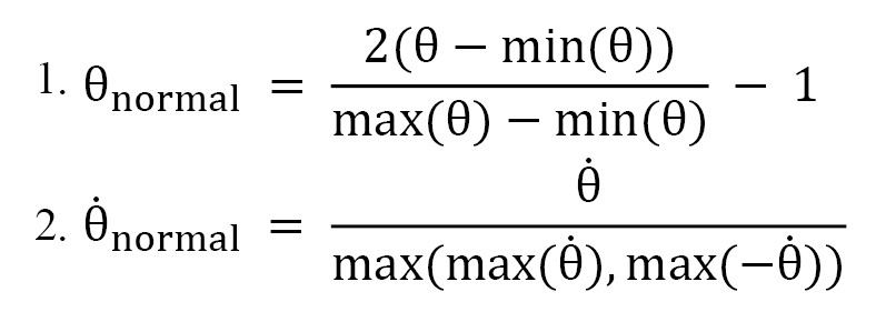
Lower limb joint kinematics were captured using eight motion analysis cameras (Oqus 5+, Qualisys, Sweden) at a frequency of 250 Hz. The data were filtered using a Butterworth low-pass filter with a 6 Hz cutoff frequency. The first 5 seconds of each trial were removed to minimize transient effects at the start of the movement. Twenty 14-mm reflective markers were affixed to anatomical landmarks according to the Visualize Three Dimensional (V3D) model. Visual 3D software, version 6 x64 was used to construct a biomechanical model comprising the pelvis, thigh, shank, and foot segments. This model was then used to calculate angular displacement, velocity, and acceleration of the ankle, knee, and hip joints within the sagittal plane. Angular jerk was computed using the three-point derivation method. To analyze non-sinusoidal signals, normalizing both the input signals’ displacement and their first derivative was necessary [7]. The objective was to transform the phase portraits in a manner that confines both displacement and its first derivative within the range of -1 to 1 [24]. This normalization allows for the comparability of data across different individuals with varying ranges. The signal’s displacement was normalized using Equation 1. The first derivative was normalized using Equation 2 [25]. In the phase planes, angular velocity versus angle, angular acceleration versus angular velocity, and angular jerk versus angular acceleration were considered as the input signals.
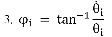
Following normalization, the phase angle of the signal at time ti is computed as the angle of the line between point i and the origin (0, 0) (Equation 3). The phase angle was calculated in all three phase planes.
Following the calculation of phase angles, CRP was computed between the hip-knee, hip-ankle, and knee-ankle in the sagittal plane (Equation 4). The average CRP curve across participant trials was then used as the ensemble CRP curve to simplify further analysis [26]. Synchronized joint movements were defined by in-phase (CRP=0°) and anti-phase (CRP=180°) patterns, representing movements in the same and opposite directions, respectively. Other CRP values indicate the phase deviation between the movements of the two joints. Finally, three variables were calculated from the CRP values for statistical analysis.

For statistical analysis of the CRP data, three variables were calculated. Firstly, the MARP, calculated as the mean of (CRP), was used to indicate the degree of coordination between two joints, representing the temporal relationship of their movements [27]. Secondly, the deviation phase (DP), determined as the standard deviation of the CRP, was calculated for both intra-individual (within each participant’s repetitions) and inter-individual (between the mean patterns of individuals within a group) cases [28, 29]. DP reflects the coordinative variability, indicating the extent of variability in a coordination pattern [30]. All calculations were performed using Circular Statistics [31]. Independent t-tests, with a significance level of α=0.05, were used for statistical analysis. Effect sizes (Cohen’s d) were calculated and interpreted using conventional thresholds: Small (≈0.2), medium (≈0.5), and large (≈0.8) [32]. MATLAB software, version R2019b and SPSS software, version 26 were used for calculations and statistical analysis, respectively.
Results
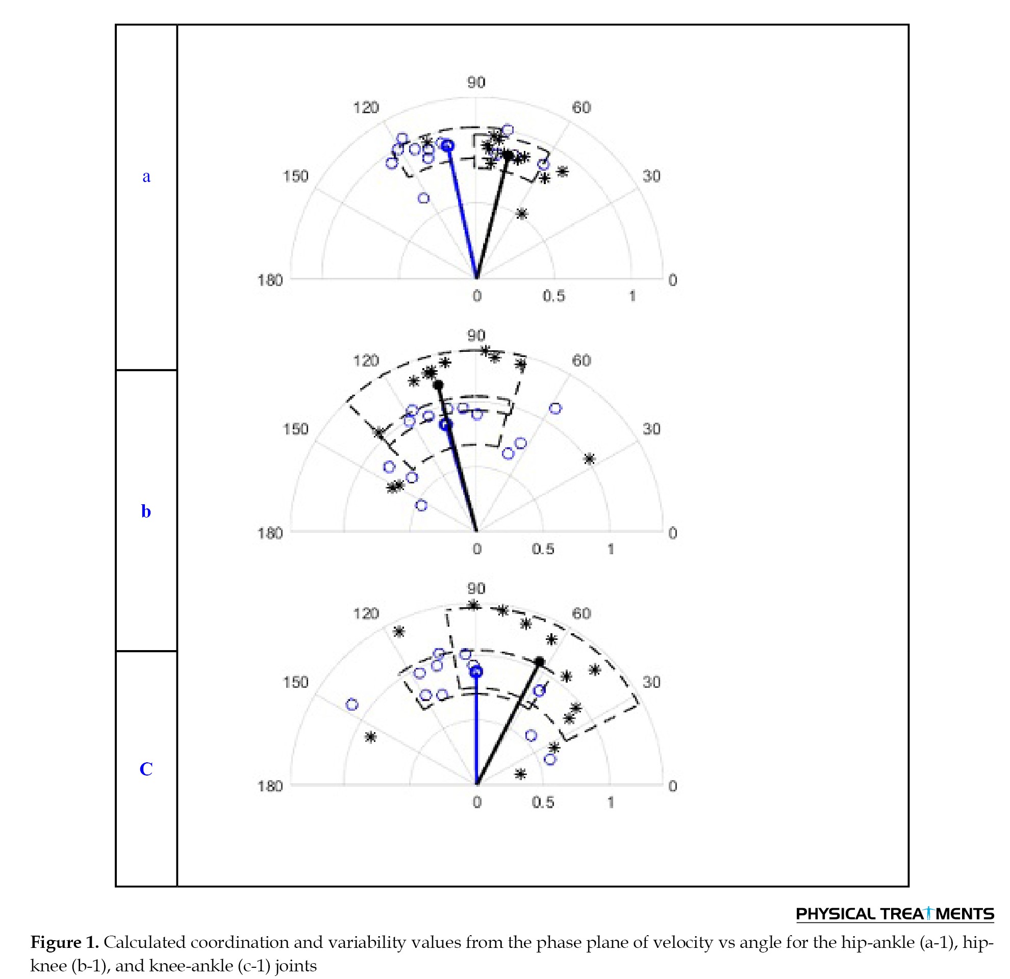
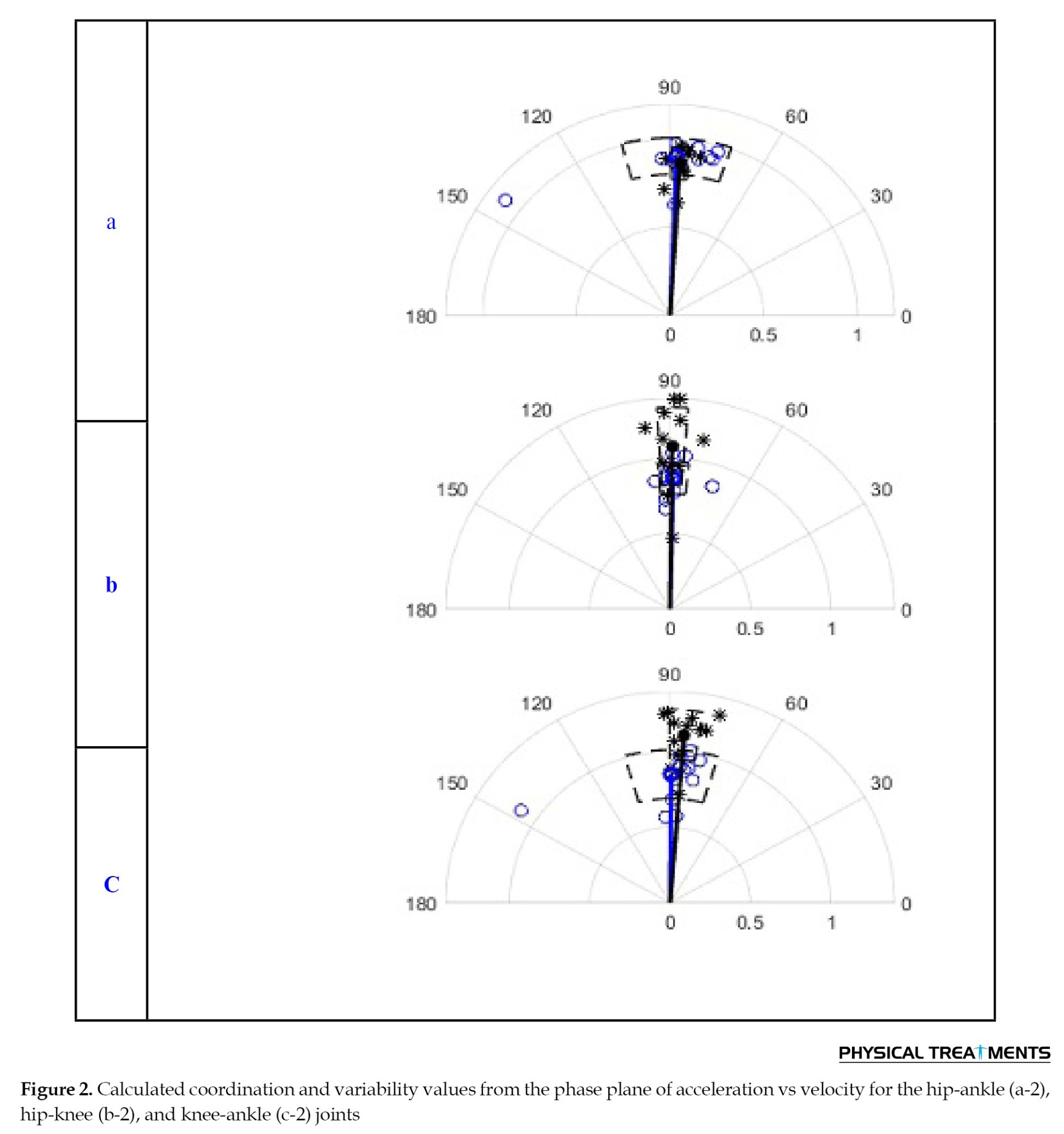
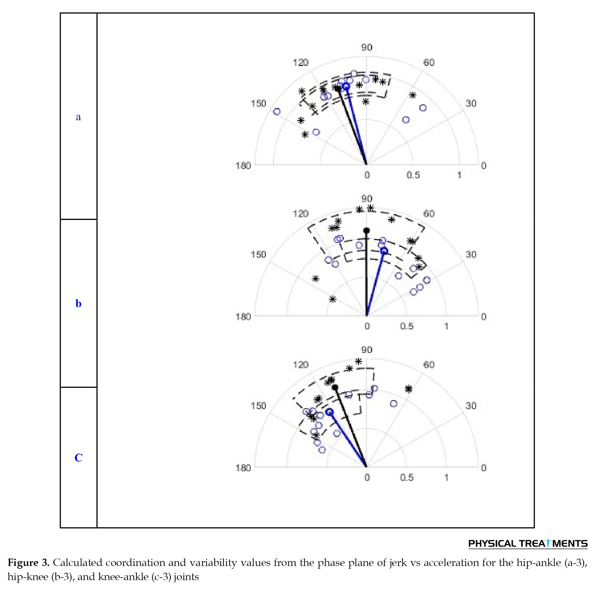
The coordination and coordination variability for both the control and experimental groups are illustrated in Figures 1, 2 and 3.
Knee joint stability is maintained through passive mechanisms, such as bone structure and ligamentous arrangements, and dynamic components, including muscular contractions and neuromuscular control [1]. Anterior cruciate ligament (ACL) rupture is common and can disrupt stability [1]. This leads to loss of mechanoreceptors, reduced afferent input to the sensorimotor system, compromised neuromuscular function, and subsequent deficits in knee functionality [2, 3]. ACL reconstruction (ACLR) is widely regarded as the standard treatment for restoring joint stability and function [4]. However, only about 65% of patients fully recover in terms of daily activities, and only 50% are able to return to their previous level of sports participation [5, 6], and they still face a high risk of re-injury [5].
Altered inter-joint coordination in the lower limbs is a common impairment following ACLR due to structural changes in the knee joint [6]. Movement coordination is defined as the selection and generation of movement, as well as the degrees of freedom available for a given task [7]. Coordination results from dynamic interactions between the musculoskeletal system, nervous system, and environment, with self-organized relationships among these components enabling flexibility in movement patterns [4]. Consequently, as a movement becomes more complex, the coordination between joints or the rhythm of the movement also becomes more intricate, increasing its significance [8, 9]. Research suggests that injuries to the ACL or other passive components of the knee joint are unlikely to result solely from dysfunction in a single joint [10]. When evaluating the effects of therapeutic interventions, it is essential to assess the function of the injured joint in relation to other joints within the kinetic chain [11]. Reduced coordination can lead to abnormal movement patterns, compensatory mechanisms, and an increased risk of injury [7]. These findings highlight the importance of understanding and improving inter-joint coordination in the lower limbs for effective rehabilitation after ACL reconstruction and injury prevention.
Coordination variability represents the dynamic balance between stability and adaptability in response to environmental demands [11, 12]. Diminished variability, which implies reduced flexibility, indicates a lowered ability to adapt to external perturbations [11]. Lower coordination variability may be linked to an increased occurrence of ACL injuries [11]. Several studies have analyzed inter-joint coordination and coordination variability in lower limb movements among ACLR individuals during various activities, such as walking [4, 5, 7, 13], running, and cutting maneuvers [5]. For instance, a study demonstrated a lower in-phase hip-knee coordination pattern in ACLR individuals during running and cutting [5]. In other cases, individuals with ACLR exhibited greater variability in hip-knee coordination during walking compared to healthy controls [4, 7]. Additionally, investigations into unipedal postural coordination and single-leg jumping have further shown altered coordination patterns in ACLR individuals, with reduced ankle-knee coordination but increased ankle-hip coordination variability compared to their non-injured counterparts [10, 11].
Clinically, it is crucial to assess the knee joint’s performance. Single-leg stance (SLS) postural control is a critical task in clinical assessments of ACLR individuals [13-16]. This task provides insight into the performance of the injured limb independently of the healthy limb. Researchers have specifically studied the center of pressure (COP) in patients with ACLR during SLS [17], as reduced somatosensory feedback and proprioceptive functionality following ACLR can lead to increased COP changes [18]. Investigating inter-joint coordination and its variability can discover abnormal movement patterns that elevate the risk of re-injury and help design suitable rehabilitation exercise programs [7, 19].
Previous studies have indicated that joint angular acceleration is a suitable kinematic variable for analyzing inter-joint coordination during a double quiet stance [20, 21]. Therefore, incorporating joint acceleration into continuous relative phase (CRP) calculations may help identify changes in inter-joint coordination in ACLR individuals during SLS. Currently, there are no existing reports on the inter-joint coordination patterns during SLS, prompting this study to analyze it using the CRP method. The analysis utilizes various phase planes, including angular velocity versus angle, angular acceleration versus angular velocity, and angular jerk versus angular acceleration. This study aimed to investigate the inter-joint coordination and coordination variability of the lower limb joints during SLS in individuals with ACLR. In this study, an effort was made to analyze the coordination in different phase planes to assess all potential changes and differences between the two evaluated groups.
Materials and Methods
This study involved two groups of subjects: 12 individuals who underwent ACL reconstruction and 12 healthy controls matched for age, weight, and height. The sample size is consistent with that used in previous ACL research [22, 23]. The participants were referred by the Bone, Joint, and Related Tissues Research Center at Shahid Beheshti University of Medical Sciences. An orthopedic physician evaluated the patients before their referral to the laboratory. The inclusion criteria included age between 20 and 37 years, right ACLR performed using a hamstring autograft, a timeframe of 12 to 16 months post-reconstruction, no pain or medication at the time of evaluation, completion of the same rehabilitation program, a normal musculoskeletal system (except the right knee) and an active lifestyle involving regular physical activity. The exclusion criteria included visual or vestibular disturbances and pain or deformities in the lower limbs. The control group consisted of active healthy individuals with no history of lower-limb injuries, surgeries, or musculoskeletal abnormalities; they were also neurologically sound. Ethical approval was obtained from the Institute of Physical Education and Sports Sciences, and all participants provided informed consent prior to data collection.
Balance was evaluated during an SLS, with each test lasting 35 seconds. Participants completed three trials of standing on their right leg, with a 2-minute rest interval between each attempt. For the duration of the tests, participants remained barefoot and kept their hands on their hips. They were instructed to bend the knee of the non-supporting leg to a 90-degree angle.
Lower limb joint kinematics were captured using eight motion analysis cameras (Oqus 5+, Qualisys, Sweden) at a frequency of 250 Hz. The data were filtered using a Butterworth low-pass filter with a 6 Hz cutoff frequency. The first 5 seconds of each trial were removed to minimize transient effects at the start of the movement. Twenty 14-mm reflective markers were affixed to anatomical landmarks according to the Visualize Three Dimensional (V3D) model. Visual 3D software, version 6 x64 was used to construct a biomechanical model comprising the pelvis, thigh, shank, and foot segments. This model was then used to calculate angular displacement, velocity, and acceleration of the ankle, knee, and hip joints within the sagittal plane. Angular jerk was computed using the three-point derivation method. To analyze non-sinusoidal signals, normalizing both the input signals’ displacement and their first derivative was necessary [7]. The objective was to transform the phase portraits in a manner that confines both displacement and its first derivative within the range of -1 to 1 [24]. This normalization allows for the comparability of data across different individuals with varying ranges. The signal’s displacement was normalized using Equation 1. The first derivative was normalized using Equation 2 [25]. In the phase planes, angular velocity versus angle, angular acceleration versus angular velocity, and angular jerk versus angular acceleration were considered as the input signals.
Following normalization, the phase angle of the signal at time ti is computed as the angle of the line between point i and the origin (0, 0) (Equation 3). The phase angle was calculated in all three phase planes.
Following the calculation of phase angles, CRP was computed between the hip-knee, hip-ankle, and knee-ankle in the sagittal plane (Equation 4). The average CRP curve across participant trials was then used as the ensemble CRP curve to simplify further analysis [26]. Synchronized joint movements were defined by in-phase (CRP=0°) and anti-phase (CRP=180°) patterns, representing movements in the same and opposite directions, respectively. Other CRP values indicate the phase deviation between the movements of the two joints. Finally, three variables were calculated from the CRP values for statistical analysis.
For statistical analysis of the CRP data, three variables were calculated. Firstly, the MARP, calculated as the mean of (CRP), was used to indicate the degree of coordination between two joints, representing the temporal relationship of their movements [27]. Secondly, the deviation phase (DP), determined as the standard deviation of the CRP, was calculated for both intra-individual (within each participant’s repetitions) and inter-individual (between the mean patterns of individuals within a group) cases [28, 29]. DP reflects the coordinative variability, indicating the extent of variability in a coordination pattern [30]. All calculations were performed using Circular Statistics [31]. Independent t-tests, with a significance level of α=0.05, were used for statistical analysis. Effect sizes (Cohen’s d) were calculated and interpreted using conventional thresholds: Small (≈0.2), medium (≈0.5), and large (≈0.8) [32]. MATLAB software, version R2019b and SPSS software, version 26 were used for calculations and statistical analysis, respectively.
Results
The coordination and coordination variability for both the control and experimental groups are illustrated in Figures 1, 2 and 3.
In these figures, the coordination with respect to the horizontal axis (zero origin) for the experimental group is represented by blue circles, while black stars indicate the control group. The radial distance from these points to the origin (0, 0) illustrates the variability for each participant’s repetitions. The angle between the blue line and the horizontal axis indicates the average coordination angle of the experimental group. In contrast, the angle between the black line and the horizontal axis represents that of the control group. The length of these lines indicates the average variability among participants in both groups. Specifically, in the graphical representation, the arc length corresponds to participants’ variability, while the chord length represents the standard deviation of the mean absolute relative phase (MARP). These lines are plotted for both groups.
Figure 1 shows coordination and variability values from the phase plane of angular velocity versus angle for hip-ankle, hip-knee and knee-ankle joints. The experimental group’s hip-ankle average coordination (101.7±22.2 degrees) was significantly greater than the control group (75.9±16.1 degrees), with a P=0.004 (d=0.41). However, no significant difference in coordination variability was observed between groups (experimental: 51.5±6.2; control: 48.2±6.7; P>0.05). The results show that hip-knee coordination was not significantly different between groups (control: 103.8±32.5; experimental: 105.5.5; P>0.05), but the average variability was significantly lower in the experimental group (49.2±11.2) than in the control group (66.7±13.9; P=0.003, d=0.66). Findings showed no significant differences in knee-ankle coordination between groups (experimental: 89.1±37.00; control: 66.00±40.3; P>0.05), nor in variability (experimental: 49.1±9.00; control: 60.4±18.7; P>0.05).
Figure 2 shows values from the phase plane of angular acceleration versus angular velocity for hip-ankle, hip-knee, and knee-ankle joints, revealing no significant differences in coordination values across all joint pairs (P>0.05). Coordination values for the experimental group were hip-ankle: 88.6±18.2, hip-knee: 88.7±6.00, knee-ankle: 90.3±18.1, compared to control values of hip-ankle: 86.5±3.3, hip-knee: 89.3±4.4, knee-ankle: 85.4±5.00. Variability Findings: The variability for hip-ankle (a-2) in the experimental and control groups was 51.9±6.2 and 49.8±5.7, respectively. No significant differences were observed (P>0.05). Significant differences were found for hip-knee (P=0.027, d=0.50) and knee-ankle (P=0.001, d=0.95) between groups.
Figure 3 shows coordination and variability values from the phase plane of angular jerk versus angular acceleration for hip-ankle, hip-knee, and knee-ankle joints, with no significant differences in coordination values between groups for any joint pairs (P>0.05). Coordination values for the experimental group were hip-ankle: 103.5±31.4, hip-knee: 75.1±36.7, knee-ankle: 122.3±29.2; control group values were hip-ankle: 109.8±28.8, hip-knee: 91.0±35.3. Knee-ankle: 110.5±27.2. Variability results showed no significant differences for hip-ankle joint movements between groups (experimental: 51.2±7.7; control: 51.3±5.6, P>0.05). The variability in the hip-knee joints was 49.7±7.7 for the experimental group and 62.8±15.2 for the control group. The knee-ankle joint values were 48.7±9.1 for the experimental group and 63.5±9.9 for the control group. The between-group differences in the variability of the hip-knee (P=0.015, d=0.54) and knee-ankle (P=0.001, d=0.99) joints were significant.
Discussion
This study aimed to investigate the impact of ACLR on the coordination and variability of lower limb joints using CRP analysis in the AP direction during balance control in the SLS position. The findings indicated that the average coordination (movement phase difference) for the hip-ankle joints in the velocity-angle phase plane was significantly higher in individuals with ACLR than in healthy controls, indicating a reduction in coordination between these joints. Additionally, the variability of coordination was lower in ACLR participants for hip-knee joints in all three phase planes, and knee-ankle joints in the acceleration-velocity and jerk-acceleration phase planes when compared to healthy individuals. Previous research has primarily focused on lower limb joint coordination during tasks, such as single-leg jumps and rhythmic coordination, while studies specifically examining SLS remain limited. Most findings indicate reduced coordination of hip-ankle joints in the velocity-angle phase plane during these tasks [33, 34].
The lower extremities act as linked chains [11]. The unique biomechanical composition of the lower limbs and the sequential interactions among the ankle, knee, and hip joints suggest a coordinated system [11]. Consequently, this interconnectedness suggests that an injury, such as one impacting the knee joint or ACL, seldom leads to dysfunction limited to only one joint [11]. Kiefer et al. (2013) indicated that in individuals who have undergone ACLR, the absence of functional mechanoreceptors in the grafted tissue can disturb the coordination of the ankle-hip joints [14]. Alterations in coordination and movement patterns elevate the risk of recurrent injuries and osteoarthritis in individuals with ACLR [35].
The current study’s findings on hip-knee joint coordination variability align with Park et al. (2021) [5] but contradict the results of Shi et al. (2021) [11], Blache et al. (2017) [23], and Srinivasan et al. (2018) [36]. These studies examined coordination variability of the hip-knee joint across tasks, such as 60-degree cutting, mid-stance gait, maximal vertical single-leg jumps, and one-leg hopping. Coordination variability is essential for adapting to environmental changes and accommodating shifting movement patterns [11].
Although ACLR surgery is generally effective in restoring mechanical stability of the knee, there is conflicting evidence regarding the restoration of sensory function post-ACLR [1, 37]. The absence of proper neural feedback mechanisms may result in alterations in coordination variability [36]. This study indicated reduced joint coordination variability in ACLR individuals, suggesting possible neuromuscular sensorimotor deficits related to neuromuscular control [4]. Diminished joint variability suggests a more rigid system, potentially leading to decreased adaptability to external perturbations [11, 38]. This lowered coordination variability could be interpreted pathologically as a cautious action strategy due to fear or movement discomfort [5], potentially increasing the risk of non-contact injuries in a less flexible system.
This study emphasizes the importance of the knee in coordination and coordination variability during single-leg balance control. The central nervous system (CNS) likely employs various performance criteria to plan movements, such as minimum jerk, minimum torque change, or minimum angle jerk, which are models proposed to describe the smoothness of human movement [39]. Therefore, in this study, the level of coordination and coordination variability were calculated in the phase planes of angular velocity vs. angle, acceleration vs. velocity, and jerk vs. acceleration in three joint pairs. Considering acceleration and jerk variables in the current study can be viewed as a comprehensive assessment of joint performance in individuals with ACLR.
Although ACL reconstruction restores the mechanical stability of the joint, the restoration of sensory and motor function remains controversial [1]. Impaired motor functions, such as joint coordination and coordination variability in individuals with ACLR, elevate the risk of re-injury during various sports activities [4]. These findings indicate that mechanosensitive nerve endings of the ACL play a role in providing information to the central nervous system (CNS) about the position and movement of the knee joint. This is indirectly supported by the impairment in the ability to recognize the position of the knee in space, known as joint position sense, in individuals with ACLR [40, 41]. Individuals with ACLR may exhibit lower mean amplitude compensatory responses in muscles compared to healthy individuals due to altered regulation of joint and muscle stiffness as a result of abnormalities in the gamma muscle spindle system of patients with ACLR compared to healthy individuals. Abnormal gamma loop sensitivity has been reported after ACLR [42]. This may alter muscle spindle signals, which are known to play a major role in signaling information regarding limb position in terms of muscle length [43]. The capacity of a joint to remain stable in the face of disturbances appears to rely on its stiffness, which, in addition to other factors, is also affected by the stiffness of the surrounding muscles [44]. The stiffness of joints and muscles can be dynamically adjusted by changing the intensity of muscle activation [45]. Therefore, increased activation of the muscles surrounding a joint leads to increased joint stiffness, enhancing the joint’s ability to withstand external forces (resulting in greater dynamic stability). Conversely, lower levels of muscle contraction may reduce an individual’s capacity to resist perturbation [46, 47].
Therefore, designing optimal rehabilitation protocols to restore natural movement patterns and reduce the risk of injury in individuals with ACLR is essential. The goals of a rehabilitation program after ACLR in athletes are to restore the strength and function needed for daily activities, increase neuromuscular control of the lower extremities through basic movement patterns, and adapt prescribed weight training to facilitate a gradual return to team sports [48]. Paying attention to lower limb coordination after ACLR can be valuable for improving rehabilitation strategies [4]. Blache et al. (2016) suggested that to enhance motor control, rehabilitation protocols should incorporate multi-joint and multiplanar movements [23].
It is suggested that researchers in future studies investigate how treatments may modify the adaptability and motor coordination of the affected lower limb. They should focus on designing and implementing rehabilitation programs with optimal timing to restore natural movement patterns and reduce the risk of re-injury in individuals after ACLR.
Study limitations
The results of this study are limited to male ACLR participants, and the relatively small sample size may reduce the ability to detect smaller effects. To enhance the generalizability of the findings, future research should be conducted with larger and mixed-gender samples. Additionally, the current study’s focus solely on sagittal plane kinematics limits the understanding of multiplanar movement patterns. Research should be conducted alongside electromyography data to examine muscle activation patterns and their effects on joint coordination changes.
Conclusion
In conclusion, this study demonstrates that despite successful surgical reconstruction, individuals with ACLR exhibit altered inter-joint coordination and markedly reduced coordination variability across lower limb joints during SLS. The observed shift toward an out-of-phase proximal–distal strategy alongside kinematic rigidity—manifested across angular velocity, acceleration, and jerk phase planes—reflects persistent sensorimotor deficits and compensatory motor adaptations. While such constrained variability may serve as an immediate protective response to impaired knee proprioception, it fundamentally limits the neuromuscular system’s dynamic adaptability to external perturbations, thereby potentially elevating the risk of secondary injuries. Therefore, post-ACLR rehabilitation protocols should progress beyond localized joint stability to incorporate functional, multi-joint coordination exercises and dynamic perturbation training aimed at restoring natural movement patterns and motor flexibility.
Ethical Considerations
Compliance with ethical guidelines
Ethical approval was obtained from the Ethics Committee of the Sport Sciences Research Institute (SSRC), Tehran, Iran (Code: IR.SSRC.REC.1404.091).
Funding
This research did not receive any grant from funding agencies in the public, commercial, or non-profit sectors.
Authors' contributions
Study design, original draft, methodology, investigation supervision: Leila.Ghazaleh; Data collection, manuscript writing, review and editing: Banafsheh.Mohammadi; Methodology support, data analysis: Maede.Alavikiaa; Conceptualization, manuscript review: Peyman.Aghaei Ataabad; Methodology support, project administration: Seyyed Morteza Kazemi; Manuscript review and editing: Nader.Farahpour.
Conflict of interest
The authors declared no conflicts of interest.
Acknowledgments
Appreciation to all individuals who collaborated in this research.
Figure 1 shows coordination and variability values from the phase plane of angular velocity versus angle for hip-ankle, hip-knee and knee-ankle joints. The experimental group’s hip-ankle average coordination (101.7±22.2 degrees) was significantly greater than the control group (75.9±16.1 degrees), with a P=0.004 (d=0.41). However, no significant difference in coordination variability was observed between groups (experimental: 51.5±6.2; control: 48.2±6.7; P>0.05). The results show that hip-knee coordination was not significantly different between groups (control: 103.8±32.5; experimental: 105.5.5; P>0.05), but the average variability was significantly lower in the experimental group (49.2±11.2) than in the control group (66.7±13.9; P=0.003, d=0.66). Findings showed no significant differences in knee-ankle coordination between groups (experimental: 89.1±37.00; control: 66.00±40.3; P>0.05), nor in variability (experimental: 49.1±9.00; control: 60.4±18.7; P>0.05).
Figure 2 shows values from the phase plane of angular acceleration versus angular velocity for hip-ankle, hip-knee, and knee-ankle joints, revealing no significant differences in coordination values across all joint pairs (P>0.05). Coordination values for the experimental group were hip-ankle: 88.6±18.2, hip-knee: 88.7±6.00, knee-ankle: 90.3±18.1, compared to control values of hip-ankle: 86.5±3.3, hip-knee: 89.3±4.4, knee-ankle: 85.4±5.00. Variability Findings: The variability for hip-ankle (a-2) in the experimental and control groups was 51.9±6.2 and 49.8±5.7, respectively. No significant differences were observed (P>0.05). Significant differences were found for hip-knee (P=0.027, d=0.50) and knee-ankle (P=0.001, d=0.95) between groups.
Figure 3 shows coordination and variability values from the phase plane of angular jerk versus angular acceleration for hip-ankle, hip-knee, and knee-ankle joints, with no significant differences in coordination values between groups for any joint pairs (P>0.05). Coordination values for the experimental group were hip-ankle: 103.5±31.4, hip-knee: 75.1±36.7, knee-ankle: 122.3±29.2; control group values were hip-ankle: 109.8±28.8, hip-knee: 91.0±35.3. Knee-ankle: 110.5±27.2. Variability results showed no significant differences for hip-ankle joint movements between groups (experimental: 51.2±7.7; control: 51.3±5.6, P>0.05). The variability in the hip-knee joints was 49.7±7.7 for the experimental group and 62.8±15.2 for the control group. The knee-ankle joint values were 48.7±9.1 for the experimental group and 63.5±9.9 for the control group. The between-group differences in the variability of the hip-knee (P=0.015, d=0.54) and knee-ankle (P=0.001, d=0.99) joints were significant.
Discussion
This study aimed to investigate the impact of ACLR on the coordination and variability of lower limb joints using CRP analysis in the AP direction during balance control in the SLS position. The findings indicated that the average coordination (movement phase difference) for the hip-ankle joints in the velocity-angle phase plane was significantly higher in individuals with ACLR than in healthy controls, indicating a reduction in coordination between these joints. Additionally, the variability of coordination was lower in ACLR participants for hip-knee joints in all three phase planes, and knee-ankle joints in the acceleration-velocity and jerk-acceleration phase planes when compared to healthy individuals. Previous research has primarily focused on lower limb joint coordination during tasks, such as single-leg jumps and rhythmic coordination, while studies specifically examining SLS remain limited. Most findings indicate reduced coordination of hip-ankle joints in the velocity-angle phase plane during these tasks [33, 34].
The lower extremities act as linked chains [11]. The unique biomechanical composition of the lower limbs and the sequential interactions among the ankle, knee, and hip joints suggest a coordinated system [11]. Consequently, this interconnectedness suggests that an injury, such as one impacting the knee joint or ACL, seldom leads to dysfunction limited to only one joint [11]. Kiefer et al. (2013) indicated that in individuals who have undergone ACLR, the absence of functional mechanoreceptors in the grafted tissue can disturb the coordination of the ankle-hip joints [14]. Alterations in coordination and movement patterns elevate the risk of recurrent injuries and osteoarthritis in individuals with ACLR [35].
The current study’s findings on hip-knee joint coordination variability align with Park et al. (2021) [5] but contradict the results of Shi et al. (2021) [11], Blache et al. (2017) [23], and Srinivasan et al. (2018) [36]. These studies examined coordination variability of the hip-knee joint across tasks, such as 60-degree cutting, mid-stance gait, maximal vertical single-leg jumps, and one-leg hopping. Coordination variability is essential for adapting to environmental changes and accommodating shifting movement patterns [11].
Although ACLR surgery is generally effective in restoring mechanical stability of the knee, there is conflicting evidence regarding the restoration of sensory function post-ACLR [1, 37]. The absence of proper neural feedback mechanisms may result in alterations in coordination variability [36]. This study indicated reduced joint coordination variability in ACLR individuals, suggesting possible neuromuscular sensorimotor deficits related to neuromuscular control [4]. Diminished joint variability suggests a more rigid system, potentially leading to decreased adaptability to external perturbations [11, 38]. This lowered coordination variability could be interpreted pathologically as a cautious action strategy due to fear or movement discomfort [5], potentially increasing the risk of non-contact injuries in a less flexible system.
This study emphasizes the importance of the knee in coordination and coordination variability during single-leg balance control. The central nervous system (CNS) likely employs various performance criteria to plan movements, such as minimum jerk, minimum torque change, or minimum angle jerk, which are models proposed to describe the smoothness of human movement [39]. Therefore, in this study, the level of coordination and coordination variability were calculated in the phase planes of angular velocity vs. angle, acceleration vs. velocity, and jerk vs. acceleration in three joint pairs. Considering acceleration and jerk variables in the current study can be viewed as a comprehensive assessment of joint performance in individuals with ACLR.
Although ACL reconstruction restores the mechanical stability of the joint, the restoration of sensory and motor function remains controversial [1]. Impaired motor functions, such as joint coordination and coordination variability in individuals with ACLR, elevate the risk of re-injury during various sports activities [4]. These findings indicate that mechanosensitive nerve endings of the ACL play a role in providing information to the central nervous system (CNS) about the position and movement of the knee joint. This is indirectly supported by the impairment in the ability to recognize the position of the knee in space, known as joint position sense, in individuals with ACLR [40, 41]. Individuals with ACLR may exhibit lower mean amplitude compensatory responses in muscles compared to healthy individuals due to altered regulation of joint and muscle stiffness as a result of abnormalities in the gamma muscle spindle system of patients with ACLR compared to healthy individuals. Abnormal gamma loop sensitivity has been reported after ACLR [42]. This may alter muscle spindle signals, which are known to play a major role in signaling information regarding limb position in terms of muscle length [43]. The capacity of a joint to remain stable in the face of disturbances appears to rely on its stiffness, which, in addition to other factors, is also affected by the stiffness of the surrounding muscles [44]. The stiffness of joints and muscles can be dynamically adjusted by changing the intensity of muscle activation [45]. Therefore, increased activation of the muscles surrounding a joint leads to increased joint stiffness, enhancing the joint’s ability to withstand external forces (resulting in greater dynamic stability). Conversely, lower levels of muscle contraction may reduce an individual’s capacity to resist perturbation [46, 47].
Therefore, designing optimal rehabilitation protocols to restore natural movement patterns and reduce the risk of injury in individuals with ACLR is essential. The goals of a rehabilitation program after ACLR in athletes are to restore the strength and function needed for daily activities, increase neuromuscular control of the lower extremities through basic movement patterns, and adapt prescribed weight training to facilitate a gradual return to team sports [48]. Paying attention to lower limb coordination after ACLR can be valuable for improving rehabilitation strategies [4]. Blache et al. (2016) suggested that to enhance motor control, rehabilitation protocols should incorporate multi-joint and multiplanar movements [23].
It is suggested that researchers in future studies investigate how treatments may modify the adaptability and motor coordination of the affected lower limb. They should focus on designing and implementing rehabilitation programs with optimal timing to restore natural movement patterns and reduce the risk of re-injury in individuals after ACLR.
Study limitations
The results of this study are limited to male ACLR participants, and the relatively small sample size may reduce the ability to detect smaller effects. To enhance the generalizability of the findings, future research should be conducted with larger and mixed-gender samples. Additionally, the current study’s focus solely on sagittal plane kinematics limits the understanding of multiplanar movement patterns. Research should be conducted alongside electromyography data to examine muscle activation patterns and their effects on joint coordination changes.
Conclusion
In conclusion, this study demonstrates that despite successful surgical reconstruction, individuals with ACLR exhibit altered inter-joint coordination and markedly reduced coordination variability across lower limb joints during SLS. The observed shift toward an out-of-phase proximal–distal strategy alongside kinematic rigidity—manifested across angular velocity, acceleration, and jerk phase planes—reflects persistent sensorimotor deficits and compensatory motor adaptations. While such constrained variability may serve as an immediate protective response to impaired knee proprioception, it fundamentally limits the neuromuscular system’s dynamic adaptability to external perturbations, thereby potentially elevating the risk of secondary injuries. Therefore, post-ACLR rehabilitation protocols should progress beyond localized joint stability to incorporate functional, multi-joint coordination exercises and dynamic perturbation training aimed at restoring natural movement patterns and motor flexibility.
Ethical Considerations
Compliance with ethical guidelines
Ethical approval was obtained from the Ethics Committee of the Sport Sciences Research Institute (SSRC), Tehran, Iran (Code: IR.SSRC.REC.1404.091).
Funding
This research did not receive any grant from funding agencies in the public, commercial, or non-profit sectors.
Authors' contributions
Study design, original draft, methodology, investigation supervision: Leila.Ghazaleh; Data collection, manuscript writing, review and editing: Banafsheh.Mohammadi; Methodology support, data analysis: Maede.Alavikiaa; Conceptualization, manuscript review: Peyman.Aghaei Ataabad; Methodology support, project administration: Seyyed Morteza Kazemi; Manuscript review and editing: Nader.Farahpour.
Conflict of interest
The authors declared no conflicts of interest.
Acknowledgments
Appreciation to all individuals who collaborated in this research.
References
- Lestari RD, Tinduh D, Pawana IPA, Utomo DN. Postural balance differences in athletes post anterior ligament cruciate reconstruction. Paper presented at: Proceedings of the International Meeting on Regenerative Medicine. 2017 August 15; Bergen, Norway. [DOI:10.5220/0007321503560358]
- Furlanetto TS, Peyré-Tartaruga LA, Pinho ASd, Bernardes EdS, Zaro MA. Proprioception, body balance and functionality in individuals with ACL reconstruction. Acta Ortopedica Brasileira. 2016; 24:67-72. [DOI:10.1590/1413-785220162402108949] [PMID] [PMCID]
- Lehmann T, Büchel D, Mouton C, Gokeler A, Seil R, Baumeister J. Functional cortical connectivity related to postural control in patients six weeks after anterior cruciate ligament reconstruction. Frontiers in Human Neuroscience. 2021; 15:655116. [DOI:10.3389/fnhum.2021.655116] [PMID] [PMCID]
- Davis K, Williams JL, Sanford BA, Zucker-Levin A. Assessing lower extremity coordination and coordination variability in individuals with anterior cruciate ligament reconstruction during walking. Gait & Posture. 2019; 67:154-9. [DOI:10.1016/j.gaitpost.2018.10.010] [PMID]
- Park S, Yoon S. Quantifying coordination and variability in the lower extremities after anterior cruciate ligament reconstruction. Sensors. 2021; 21(2):652. [DOI:10.3390/s21020652] [PMID] [PMCID]
- Gribbin TC, Slater LV, Herb CC, Hart JM, Chapman RM, Hertel J, et al. Differences in hip–knee joint coupling during gait after anterior cruciate ligament reconstruction. Clinical Biomechanics. 2016; 32:64-71. [DOI:10.1016/j.clinbiomech.2016.01.006]
- Hamill J, Palmer C, Van Emmerik RE. Coordinative variability and overuse injury. Sports Medicine, Arthroscopy, Rehabilitation, Therapy & Technology. 2012; 4(1):1-9. [DOI:10.1186/1758-2555-4-45] [PMID] [PMCID]
- Kim S, Qu F, Wang Y, Lam WK. Examining the joint coordination during dynamic balance learning using vector coding and statistical parametric mapping analyses. Scientific Reports. 2024; 14(1):1724. [DOI:10.1038/s41598-023-44216-1] [PMID] [PMCID]
- Cordo PJ, Gurfinkel VS. Motor coordination can be fully understood only by studying complex movements. Progress in Brain Research. 2004; 143:29-38. [DOI:10.1016/S0079-6123(03)43003-3] [PMID]
- Kacprzak B, Stańczak M, Surmacz J, Hagner-Derengowska M. Biophysics of ACL injuries. Orthopedic Reviews. 2024; 16:126041. [DOI:10.20944/preprints202408.0988.v1]
- Shi H, Ren S, Miao X, Zhang H, Yu Y, Hu X, et al. The effect of cognitive loading on the lower extremity movement coordination variability in patients with anterior cruciate ligament reconstruction. Gait & Posture. 2021; 84:141-7. [DOI:10.1016/j.gaitpost.2020.10.028] [PMID]
- Hamill J, van Emmerik RE, Heiderscheit BC, Li L. A dynamical systems approach to lower extremity running injuries. Clinical Biomechanics. 1999; 14(5):297-308.[DOI:10.1016/S0268-0033(98)90092-4] [PMID]
- Jiganti MR, Meyer BC, Chang E, Romanelli DA, An YW. Altered cortical activation after anterior cruciate ligament reconstruction during single-leg balance task. Translational Sports Medicine. 2020; 3(5):496-503. [DOI:10.1002/tsm2.164]
- Kiefer AW, Ford KR, Paterno MV, Schmitt LC, Myer GD, Riley MA, et al. Inter-segmental postural coordination measures differentiate athletes with ACL reconstruction from uninjured athletes. Gait & Posture. 2013; 37(2):149-53. [DOI:10.1016/j.gaitpost.2012.05.005] [PMID] [PMCID]
- Kirsch AN, Bodkin SG, Saliba SA, Hart JM. Measures of agility and single-legged balance as clinical assessments in patients with anterior cruciate ligament reconstruction and healthy individuals. Journal of Athletic Training. 2019; 54(12):1260-8. [DOI:10.4085/1062-6050-266-18] [PMID] [PMCID]
- Kouvelioti V, Kellis E, Kofotolis N, Amiridis I. Reliability of single-leg and double-leg balance tests in subjects with anterior cruciate ligament reconstruction and controls. Research in Sports Medicine. 2015; 23(2):151-66. [DOI:10.1080/15438627.2015.1005292] [PMID]
- Lehmann T, Paschen L, Baumeister J. Single-leg assessment of postural stability after anterior cruciate ligament injury: A systematic review and meta-analysis. Sports Medicine-Open. 2017; 3(1):32. [DOI:10.1186/s40798-017-0100-5] [PMID] [PMCID]
- Fleming JD, Ritzmann R, Centner C. Effect of an anterior cruciate ligament rupture on knee proprioception within 2 years after conservative and operative treatment: A systematic review with meta-analysis. Sports Medicine. 2022; 52(5):1091-102. [DOI:10.1007/s40279-021-01600-z] [PMID] [PMCID]
- Paterno MV, Kiefer AW, Bonnette S, Riley MA, Schmitt LC, Ford KR, et al. Prospectively identified deficits in sagittal plane hip–ankle coordination in female athletes who sustain a second anterior cruciate ligament injury after anterior cruciate ligament reconstruction and return to sport. Clinical Biomechanics. 2015; 30(10):1094-101. [DOI:10.1016/j.clinbiomech.2015.08.019]
- Aramaki Y, Nozaki D, Masani K, Sato T, Nakazawa K, Yano H. Reciprocal angular acceleration of the ankle and hip joints during quiet standing in humans. Experimental Brain Research. 2001; 136(4):463-73. [DOI:10.1007/s002210000603] [PMID]
- Yamamoto A, Sasagawa S, Oba N, Nakazawa K. Behavioral effect of knee joint motion on body’s center of mass during human quiet standing. Gait & Posture. 2015; 41(1):291-4. [DOI:10.1016/j.gaitpost.2014.08.016] [PMID]
- Mir SM HM, Talebian S, Nasseri N. Functional assessment of knee joint position sense following anterior cruciate ligament reconstruction. British Journal of Sports Medicine. 2008; 42(4):300-3. [DOI:10.1136/bjsm.2007.044875] [PMID]
- Blache Y, Pairot de Fontenay B, Argaud S, Monteil K. Asymmetry of inter-joint coordination during single leg jump after anterior cruciate ligament reconstruction. International Journal of Sports Medicine. 2017; 38(2):159-67. [DOI:10.1055/s-0042-109976] [PMID]
- Lamb PF SM. On the use of continuous relative phase: Review of current approaches and outline for a new standard.Clinical Biomechanics. 2014; 29(5):484-93. [DOI:10.1016/j.clinbiomech.2014.03.008] [PMID]
- Zehr JD HS, Beach TA. Using relative phase analyses and vector coding to quantify Pelvis-Thorax coordination during lifting-A methodological investigation. Journal of Electromyography and Kinesiology. 2018; 39:104-13. [DOI:10.1016/j.jelekin.2018.02.004] [PMID]
- Gittoes MJ, Wilson C. Intralimb joint coordination patterns of the lower extremity in maximal velocity phase sprint running. Journal of Applied Biomechanics. 2010; 26(2):188-95. [DOI:10.1123/jab.26.2.188] [PMID]
- Stergiou N, Scholten SD, Jensen JL, Blanke D. Intralimb coordination following obstacle clearance during running: the effect of obstacle height. Gait & Posture. 2001; 13(3):210-20. [DOI:10.1016/S0966-6362(00)00101-6] [PMID]
- Lamoth C, Beek PJ, Meijer OG. Pelvis-thorax coordination in the transverse plane during gait. Gait & Posture. 2002; 16(2):101-14. [DOI:10.1016/S0966-6362(01)00146-1] [PMID]
- Seifert L, Lardy J, Bourbousson J, Adé D, Nordez A, Thouvarecq R, et al. Interpersonal coordination and individual organization combined with shared phenomenological experience in rowing performance: Two case studies. Frontiers in Psychology. 2017; 8:75. [DOI:10.3389/fpsyg.2017.00075] [PMID] [PMCID]
- Seay JF, Van Emmerik RE, Hamill J. Trunk bend and twist coordination is affected by low back pain status during running. European Journal of Sport Science. 2014; 14(6):563-8. [DOI:10.1080/17461391.2013.866167] [PMID]
- Cremers J, Klugkist I. One direction? A tutorial for circular data analysis using R with examples in cognitive psychology. Frontiers in Psychology. 2018; 9:2040. [DOI:10.3389/fpsyg.2018.02040] [PMID] [PMCID]
- Cohen J. Statistical power analysis for the behavioral sciences. Abingdon: Routledge; 2013. [Link]
- Dingenen B, Janssens L, Claes S, Bellemans J, Staes FF. Lower extremity muscle activation onset times during the transition from double-leg stance to single-leg stance in anterior cruciate ligament reconstructed subjects. Clinical Biomechanics. 2016; 35:116-23. [DOI:10.1016/j.clinbiomech.2016.04.014]
- Claus AP, Hides JA, Moseley GL, Hodges PW. Different ways to balance the spine in sitting: Muscle activity in specific postures differs between individuals with and without a history of back pain in sitting. Clinical Biomechanics. 2018; 52:25-32. [DOI:10.1016/j.clinbiomech.2018.01.003]
- Armitano CN, Morrison S, Russell DM. Coordination stability between the legs is reduced after anterior cruciate ligament reconstruction. Clinical Biomechanics. 2018; 58:28-33. [DOI:10.1016/j.clinbiomech.2018.07.003] [PMID]
- Srinivasan D, Tengman E, Hager CK. Increased movement variability in one-leg hops about 20 years after treatment of anterior cruciate ligament injury. Clinical Biomechanics. 2018; 53:37-45. [DOI:10.1016/j.clinbiomech.2018.02.003] [PMID]
- Howells BE, Ardern CL, Webster KE. Is postural control restored following anterior cruciate ligament reconstruction? A systematic review. Knee Surgery, Sports Traumatology, Arthroscopy. 2011; 19(7):1168-77. [DOI:10.1007/s00167-011-1444-x] [PMID]
- Pollard CD, Stearns KM, Hayes AT, Heiderscheit BC. Altered lower extremity movement variability in female soccer players during side-step cutting after anterior cruciate ligament reconstruction. The American Journal of Sports Medicine. 2015; 43(2):460-5. [DOI:10.1177/0363546514560153] [PMID]
- Emadi Andani M, Bahrami F. COMAP: A new computational interpretation of human movement planning level based on coordinated minimum angle jerk policies and six universal movement elements. Human Movement Science. 2012; 31(5):1037-55. [DOI:10.1016/j.humov.2012.01.001] [PMID]
- Skinner HB, Barrack RL. Joint position sense in the normal and pathologic knee joint. Journal of Electromyography and Kinesiology. 1991; 1(3):180-90. [DOI:10.1016/1050-6411(91)90033-2] [PMID]
- Carter N, Jenkinson T, Wilson D, Jones D, Torode A. Joint position sense and rehabilitation in the anterior cruciate ligament deficient knee. British journal of sports medicine. 1997; 31(3):209-12. [DOI:10.1136/bjsm.31.3.209] [PMID] [PMCID]
- Konishi Y, Fukubayashi T, Takeshita D. Mechanism of quadriceps femoris muscle weakness in patients with anterior cruciate ligament reconstruction. Scandinavian Journal of Medicine & Science in Sports. 2002; 12(6):371-5. [DOI:10.1034/j.1600-0838.2002.01293.x] [PMID]
- Proske U, Gandevia SC. The proprioceptive senses: Their roles in signaling body shape, body position and movement, and muscle force. Physiological reviews. 2012; 92(4):1651-97.[DOI:10.1152/physrev.00048.2011] [PMID]
- Obusek J, Holt KG, Rosenstein R. The hybrid mass-spring pendulum model of human leg swinging: Stiffness in the control of cycle period. Biological Cybernetics. 1995; 73(2):139-47. [DOI:10.1007/BF00204052] [PMID]
- Silva PL, Fonseca ST, Ocarino JM, Gonçalves GP, Mancini MC. Contributions of cocontraction and eccentric activity to stiffness regulation. Journal of Motor Behavior. 2009; 41(3):207-18. [DOI:10.3200/JMBR.41.3.207-218] [PMID]
- da Fonseca ST, Silva PL, Ocarino JM, Guimaraes RB, Oliveira MT, Lage CA. Analyses of dynamic co-contraction level in individuals with anterior cruciate ligament injury. Journal of Electromyography and Kinesiology. 2004; 14(2):239-47. [DOI:10.1016/j.jelekin.2003.09.003] [PMID]
- Labanca L, Laudani L, Casabona A, Menotti F, Mariani PP, Macaluso A. Early compensatory and anticipatory postural adjustments following anterior cruciate ligament reconstruction. European journal of Applied Physiology. 2015; 115(7):1441-51. [DOI:10.1007/s00421-015-3126-8] [PMID]
- Badawy CR, Jan K, Beck EC, Fleet N, Taylor J, Ford K, et al. Contemporary principles for postoperative rehabilitation and return to sport for athletes undergoing anterior cruciate ligament reconstruction. Arthroscopy, Sports Medicine, and Rehabilitation. 2022; 4(1):e103-e13. [DOI:10.1016/j.asmr.2021.11.002] [PMID] [PMCID]
Type of Study: Research |
Subject:
Sport injury and corrective exercises
Received: 2025/04/17 | Accepted: 2025/10/25 | Published: 2026/10/18
Received: 2025/04/17 | Accepted: 2025/10/25 | Published: 2026/10/18
Send email to the article author
| Rights and permissions | |
 |
This work is licensed under a Creative Commons Attribution-NonCommercial 4.0 International License. |

This is an open access article distributed under the terms of the Creative Commons Attribution-NonCommercial 4.0 (CC-By-NC 4.0), which permits use, distribution, and reproduction in any medium, provided the original work is properly cited and is not used for commercial purposes.
Contact Information