Thu, Oct 22, 2026
Volume 16, Issue 3 (Summer 2026)
PTJ 2026, 16(3): 345-358 |
Back to browse issues page



Download citation:
BibTeX | RIS | EndNote | Medlars | ProCite | Reference Manager | RefWorks
Send citation to:


BibTeX | RIS | EndNote | Medlars | ProCite | Reference Manager | RefWorks
Send citation to:
Mashhadi Alizadeh S, Khoshraftar Yazdi N, Alirezaei Noghondar F. The Effect of a Pilates Exercise Program on Kyphosis Angle and Electrical Activity of Muscles in Kyphotic Adolescent Women. PTJ 2026; 16 (3) :345-358
URL: http://ptj.uswr.ac.ir/article-1-711-en.html
URL: http://ptj.uswr.ac.ir/article-1-711-en.html

1- Department of Exercise Physiology and Sports Injuries and Corrective Exercise, Faculty of Sports Sciences, Ferdowsi University of Mashhad, Mashhad, Iran.
Full-Text [PDF 1249 kb]
(1135 Downloads)
| Abstract (HTML) (2478 Views)
Full-Text: (190 Views)
Introduction
Epidemiological research has indicated a significant occurrence of spinal postural deformities and musculoskeletal pain in adolescents [1]. Kyphosis also increases with age. Forty percent of people over 60 years of age and 55% of people over 70 years of age have kyphosis greater than 40° [2]. Hyperkyphosis is a prevalent postural deviation associated with forward head posture and can lead to increased lumbar lordosis in more severe cases [3]. Hyperkyphosis can cause various complications, including weakness of the respiratory muscles and undesirable pressure on the heart [4, 5]. Such conditions are believed to increase compressive forces on the vertebral joints and alter the lengths and strength of connective tissues, leading to musculoskeletal imbalances and pain [6].
Previous studies have indicated that a deviation in one segment and the resulting alterations in the joints and muscles of that area can trigger a chain reaction, influencing other parts of the body and affecting various joints and muscles [7, 8]. Vladimir Janda (1923–2002) suggested that upper crossed syndrome is an abnormal posture that specifically alters muscle activation patterns (especially in the neck, trunk, and scapular muscles) and movement patterns (scapular dyskinesis) along with postural deviations (forward head and shoulder posture and increased thoracic kyphosis) [8, 9]. This syndrome is characterized by tightness of the upper trapezius (UT) and levator scapula, weakness of the deep cervical flexors, and weakness of the lower trapezius (LT) and middle trapezius (MT) [10]. Hyperkyphosis can cause side effects, such as slow walking, decreased balance, increased body sway, and disrupted muscle activity patterns. These side effects raise the risk of falls and diminish quality of life, underscoring the need for interventions to improve muscle recruitment strategies [11].
Pilates has emerged as an effective method for addressing postural deviations by focusing on core strength, body awareness, and flexibility [12, 13]. Research indicates that many individuals suffer from postural issues due to sedentary lifestyles, muscle imbalances, and improper movement patterns. Pilates specifically targets these areas, providing a comprehensive approach to improving posture [14]. Pilates is an exercise method that positively influences posture by emphasizing control of the pelvis and trunk through specific muscle recruitment strategies [15]. Additionally, Pilates exercises aim to correct posture while strengthening and lengthening the muscles, promoting a neutral pelvis and lumbar lordosis [15]. Pilates exercises emphasize core stability and breath control, which promote the activation of the diaphragm, transversus abdominis, multifidus, and pelvic floor muscles. Previous research has examined the impact of Pilates training on biomechanical characteristics, primarily concentrating on spinal and lower-limb alignment. González-Gálvez et al. (2023) showed that adolescents with thoracic hyperkyphosis exhibited reduced thoracic kyphosis while standing relaxed, along with enhanced hamstring flexibility [13]. Pilates training improves the pelvis and spinal stabilization, flexibility, power, posture, and coordination between movement and breathing [16]; therefore, it may reduce curvature of kyphosis and change the muscle electrical activity pattern.
It has been shown that 30 weeks of Pilates training improves posture and flexibility in women with hyperkyphosis [17]. Kim et al. (2014) showed that after three months of training, muscle electrical activity increased in healthy individuals [18]. Electrical muscle activity can provide useful information about exercise effectiveness and identify the most active muscle during exercise. Previous studies have examined the effects of Pilates exercises on posture [18]. According to this study, the effects of these exercises on muscle activity have not been examined so far.
According to previous studies, an exercise program should emphasize chain-reaction muscles to achieve optimal results in postural correction. No study appears to have examined the effects of Pilates training on local and global muscle function in people with kyphosis.
Therefore, the research hypothesis is whether Pilates exercises can affect the activity of the trapezius and erector spinae muscles, ultimately leading to positive changes in the kyphosis angle.
Materials and Methods
Study design
This study was quasi-experimental, with a pre-test-post-test design. The participants completed and signed the informed consent form. The study was carried out in accordance with the Declaration of Helsinki, and approval was obtained from the local committee for human research.
Participants
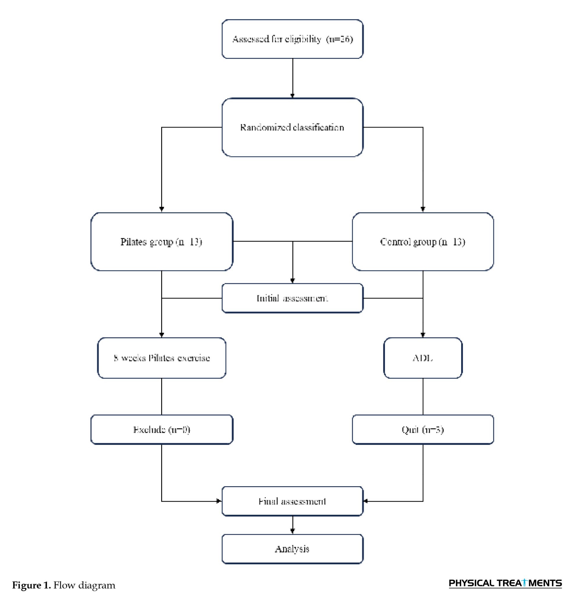
Twenty-three women diagnosed with functional kyphosis (Mean±SD age of 39.71±3.9 years, height of 160.34±6 cm, and mass of 65.43±8.2 kg) who met the inclusion criteria were included in this study. Participants were recruited using convenience sampling and then randomly allocated to either the control group (CG, n=10) or the Pilates group (PG, n=13). The sample size was estimated using G*Power software with effect size (ES)=0.35, α=0.05, and power=0.9. The inclusion criteria were (Figure 1):
Epidemiological research has indicated a significant occurrence of spinal postural deformities and musculoskeletal pain in adolescents [1]. Kyphosis also increases with age. Forty percent of people over 60 years of age and 55% of people over 70 years of age have kyphosis greater than 40° [2]. Hyperkyphosis is a prevalent postural deviation associated with forward head posture and can lead to increased lumbar lordosis in more severe cases [3]. Hyperkyphosis can cause various complications, including weakness of the respiratory muscles and undesirable pressure on the heart [4, 5]. Such conditions are believed to increase compressive forces on the vertebral joints and alter the lengths and strength of connective tissues, leading to musculoskeletal imbalances and pain [6].
Previous studies have indicated that a deviation in one segment and the resulting alterations in the joints and muscles of that area can trigger a chain reaction, influencing other parts of the body and affecting various joints and muscles [7, 8]. Vladimir Janda (1923–2002) suggested that upper crossed syndrome is an abnormal posture that specifically alters muscle activation patterns (especially in the neck, trunk, and scapular muscles) and movement patterns (scapular dyskinesis) along with postural deviations (forward head and shoulder posture and increased thoracic kyphosis) [8, 9]. This syndrome is characterized by tightness of the upper trapezius (UT) and levator scapula, weakness of the deep cervical flexors, and weakness of the lower trapezius (LT) and middle trapezius (MT) [10]. Hyperkyphosis can cause side effects, such as slow walking, decreased balance, increased body sway, and disrupted muscle activity patterns. These side effects raise the risk of falls and diminish quality of life, underscoring the need for interventions to improve muscle recruitment strategies [11].
Pilates has emerged as an effective method for addressing postural deviations by focusing on core strength, body awareness, and flexibility [12, 13]. Research indicates that many individuals suffer from postural issues due to sedentary lifestyles, muscle imbalances, and improper movement patterns. Pilates specifically targets these areas, providing a comprehensive approach to improving posture [14]. Pilates is an exercise method that positively influences posture by emphasizing control of the pelvis and trunk through specific muscle recruitment strategies [15]. Additionally, Pilates exercises aim to correct posture while strengthening and lengthening the muscles, promoting a neutral pelvis and lumbar lordosis [15]. Pilates exercises emphasize core stability and breath control, which promote the activation of the diaphragm, transversus abdominis, multifidus, and pelvic floor muscles. Previous research has examined the impact of Pilates training on biomechanical characteristics, primarily concentrating on spinal and lower-limb alignment. González-Gálvez et al. (2023) showed that adolescents with thoracic hyperkyphosis exhibited reduced thoracic kyphosis while standing relaxed, along with enhanced hamstring flexibility [13]. Pilates training improves the pelvis and spinal stabilization, flexibility, power, posture, and coordination between movement and breathing [16]; therefore, it may reduce curvature of kyphosis and change the muscle electrical activity pattern.
It has been shown that 30 weeks of Pilates training improves posture and flexibility in women with hyperkyphosis [17]. Kim et al. (2014) showed that after three months of training, muscle electrical activity increased in healthy individuals [18]. Electrical muscle activity can provide useful information about exercise effectiveness and identify the most active muscle during exercise. Previous studies have examined the effects of Pilates exercises on posture [18]. According to this study, the effects of these exercises on muscle activity have not been examined so far.
According to previous studies, an exercise program should emphasize chain-reaction muscles to achieve optimal results in postural correction. No study appears to have examined the effects of Pilates training on local and global muscle function in people with kyphosis.
Therefore, the research hypothesis is whether Pilates exercises can affect the activity of the trapezius and erector spinae muscles, ultimately leading to positive changes in the kyphosis angle.
Materials and Methods
Study design
This study was quasi-experimental, with a pre-test-post-test design. The participants completed and signed the informed consent form. The study was carried out in accordance with the Declaration of Helsinki, and approval was obtained from the local committee for human research.
Participants
Twenty-three women diagnosed with functional kyphosis (Mean±SD age of 39.71±3.9 years, height of 160.34±6 cm, and mass of 65.43±8.2 kg) who met the inclusion criteria were included in this study. Participants were recruited using convenience sampling and then randomly allocated to either the control group (CG, n=10) or the Pilates group (PG, n=13). The sample size was estimated using G*Power software with effect size (ES)=0.35, α=0.05, and power=0.9. The inclusion criteria were (Figure 1):
1) Having no history of trauma, injury, or surgery, spinal fracture, or any other disease in the last 6 months; 2) Having had no regular exercise and Pilates training for the past two months; 3) Having postural changes, such as functional thoracic kyphosis greater than 42 degrees (According to medical history and functional testing of forward and lateral bending)
The kyphosis angle and muscle activity of the selected muscles were measured at baseline. The PG received the Pilates training program, while the CG received no intervention. All assessments were repeated after six weeks. The PG performed Pilates exercise interventions for 6 weeks, three sessions per week. The training sessions lasted 35 minutes, gradually increasing to 60 minutes, and included warm-up, Pilates exercises, and cool-down. The principles of Pilates and how to perform the exercises in one session were explained to the participants.
Kyphosis angle
The inclinometer was used to measure the thoracic kyphosis angle, a valid and reliable tool [19]. This tool was chosen over other methods due to its ease of use, ease of analysis, and improved access to the inclinometer. The measurement method was as follows: the participant was asked to stand in her anatomical position. On the spinous process, the T1_T2 and T12_L1 vertebrae were marked. Then, the bases of the inclinometer, which are adjusted based on the distance between the two spinal processes of the two vertebrae, were placed on the spinous processes of T1 and T2. T12, L1, and the intervertebral angle were also measured. Measurements were performed with an inclinometer 3 times, and the average between the obtained angle and the kyphosis angle was calculated [19].
Electrical activity of muscles
Electrical activity was measured using surface electromyography (sEMG) of selected muscles, including the UT, MT, LT, and erector spine in L1 and L2 vertebrae, during maximal voluntary isometric contraction (MVIC). Participants applied their maximum force against the assessor’s fixed arm.
For the UT muscle, the electrodes were positioned along a line 1.2 cm from the acromion to the C7 vertebra. For the MT muscle, the electrodes were placed along a line 1.2 cm from the inner border of the scapula to the spine, at the same level as the T3 vertebra. For the LT muscle, the electrodes were positioned at an angle two-thirds of the way from the scapular root to the T8 vertebra. To measure the electrical activity of the erector spinae muscles, the participant was placed in a supine position, and the electrodes were positioned six centimeters lateral to the spinous processes of the L1 and L2 vertebrae [19].
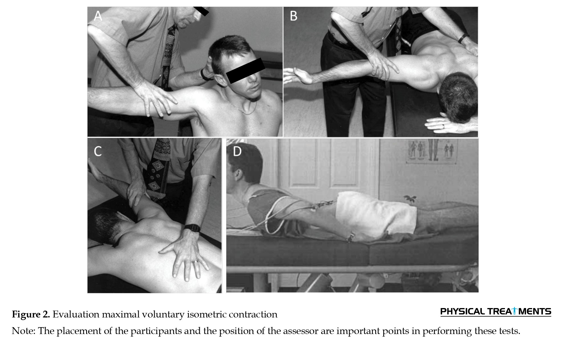
To assess the MVIC of the UT muscle (Figure 2A), the participant was seated perpendicular to the chair, at some distance from the back.
The kyphosis angle and muscle activity of the selected muscles were measured at baseline. The PG received the Pilates training program, while the CG received no intervention. All assessments were repeated after six weeks. The PG performed Pilates exercise interventions for 6 weeks, three sessions per week. The training sessions lasted 35 minutes, gradually increasing to 60 minutes, and included warm-up, Pilates exercises, and cool-down. The principles of Pilates and how to perform the exercises in one session were explained to the participants.
Kyphosis angle
The inclinometer was used to measure the thoracic kyphosis angle, a valid and reliable tool [19]. This tool was chosen over other methods due to its ease of use, ease of analysis, and improved access to the inclinometer. The measurement method was as follows: the participant was asked to stand in her anatomical position. On the spinous process, the T1_T2 and T12_L1 vertebrae were marked. Then, the bases of the inclinometer, which are adjusted based on the distance between the two spinal processes of the two vertebrae, were placed on the spinous processes of T1 and T2. T12, L1, and the intervertebral angle were also measured. Measurements were performed with an inclinometer 3 times, and the average between the obtained angle and the kyphosis angle was calculated [19].
Electrical activity of muscles
Electrical activity was measured using surface electromyography (sEMG) of selected muscles, including the UT, MT, LT, and erector spine in L1 and L2 vertebrae, during maximal voluntary isometric contraction (MVIC). Participants applied their maximum force against the assessor’s fixed arm.
For the UT muscle, the electrodes were positioned along a line 1.2 cm from the acromion to the C7 vertebra. For the MT muscle, the electrodes were placed along a line 1.2 cm from the inner border of the scapula to the spine, at the same level as the T3 vertebra. For the LT muscle, the electrodes were positioned at an angle two-thirds of the way from the scapular root to the T8 vertebra. To measure the electrical activity of the erector spinae muscles, the participant was placed in a supine position, and the electrodes were positioned six centimeters lateral to the spinous processes of the L1 and L2 vertebrae [19].
To assess the MVIC of the UT muscle (Figure 2A), the participant was seated perpendicular to the chair, at some distance from the back.
She raised her arm to a 90° abduction angle while flexing and rotating her neck outward. The tester applied resistance to prevent head extension and excessive arm abduction [20, 21]. For the MT muscle (Figure 2B), the participant was positioned supine with the shoulder in horizontal abduction and external rotation, close to the head in alignment with the MT muscle fibers. Resistance was applied to the arm [22]. To measure the MVIC of the LT muscle (Figure 2C), the participant remained supine while raising the arm toward the LT muscle fibers, with resistance applied to the arm [21]. The participant was placed supine for the erector spinae muscles (Figure 2D), with the pelvic, knee, and ankle areas secured. The participant performed trunk extension to the maximum degree facilitated by the applied resistance [22]. One person performed this for all participants in two assessment stages.
Bipolar silver/silver chloride electrodes with an active area of approximately 1 cm were positioned with a center-to-center spacing of 2 cm along the muscle fibers. The location of the electrode on the examined muscles was determined and marked by the researcher according to the recommendations of Surface ElectroMyoGraphy for the non-invasive assessment of muscles instruction [19]. By carefully removing the hair, abrading, and cleansing the skin, low impedance was achieved at the interface between the skin and electrode. Other electrical devices were kept away from the measuring device to reduce noise. Electrical muscle activity was recorded at a sampling frequency of 1000 Hz during maximal voluntary muscle contraction. The EMG signals were automatically amplified and recorded utilizing the Biopac system (Biopac, Santa Barbara, CA). To provide a clear picture of the EMG data and to compare and interpret the differences, the maximum voluntary contraction of individuals (calculated by following the curve) was compared in three attempts. After each test, the person rested in a neutral position for 30 seconds [21]. EMG signals collected during MVIC tests were carefully examined for noise, and the first and the last few seconds of each attempt, which were less stable, were removed and then processed with a second-order low-pass Butterworth filter (20-500 Hz) and full-wave rectified. Subsequently, the root mean square was calculated, and its maximum value was extracted from the desired range.
Pilates exercise
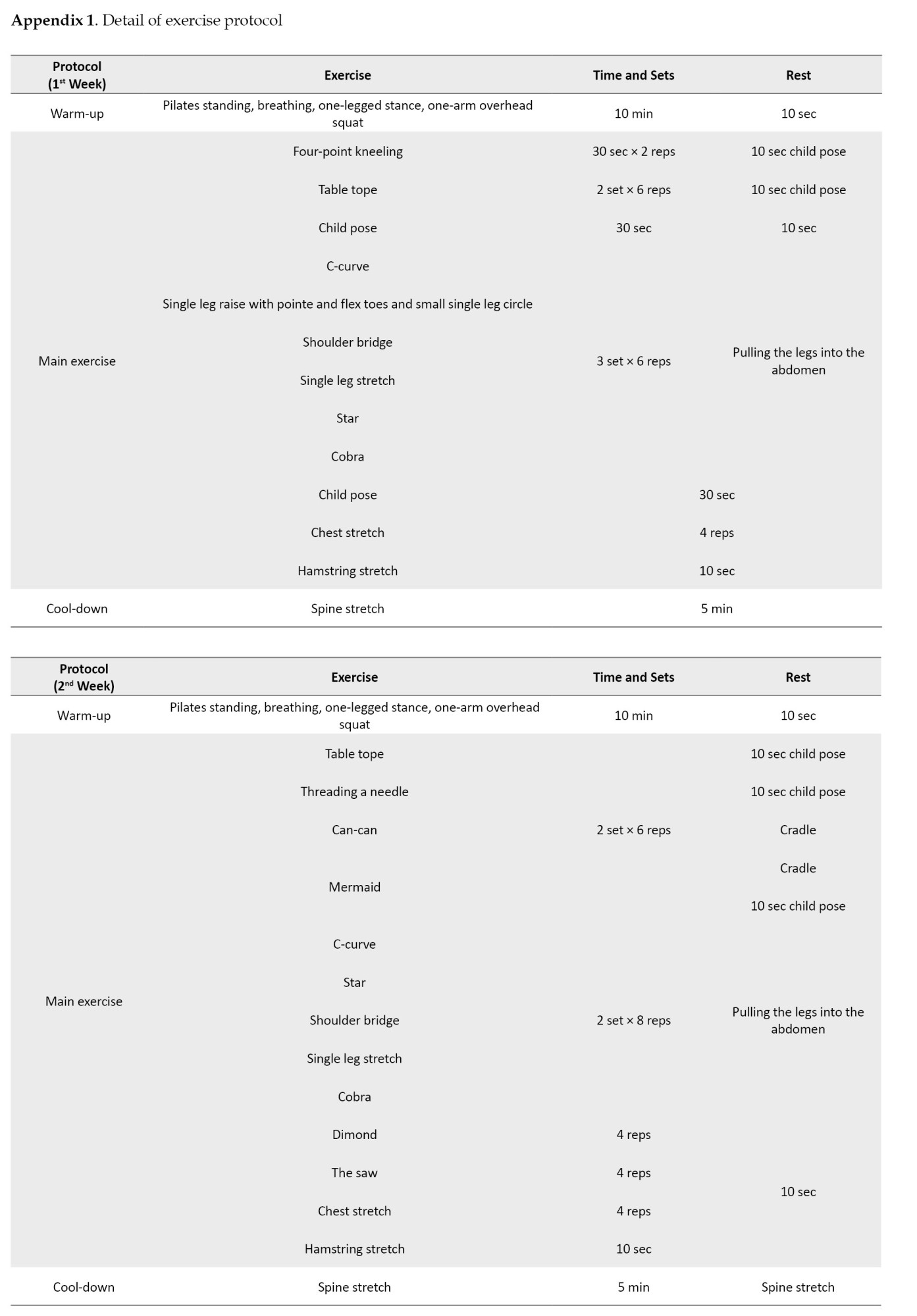
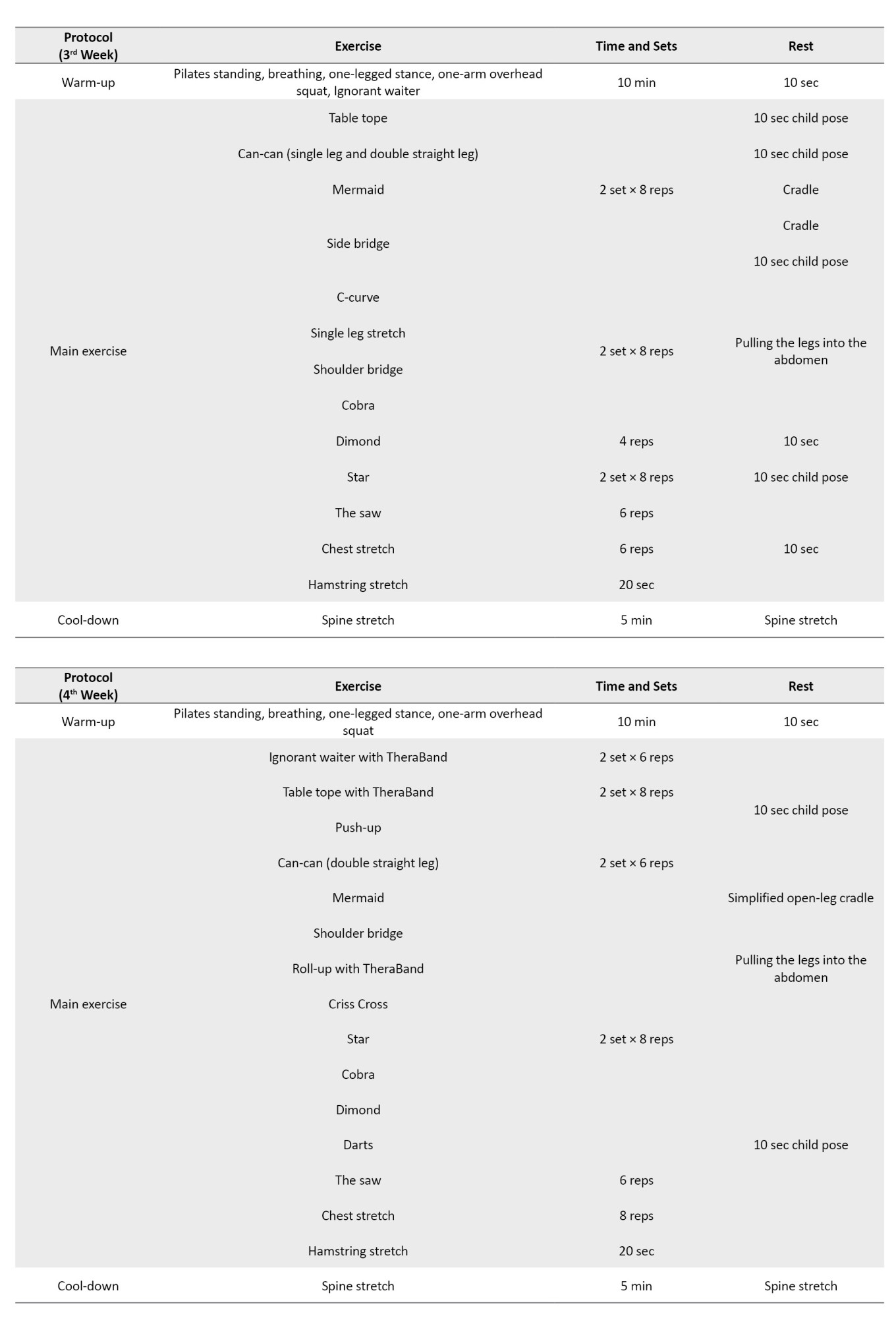
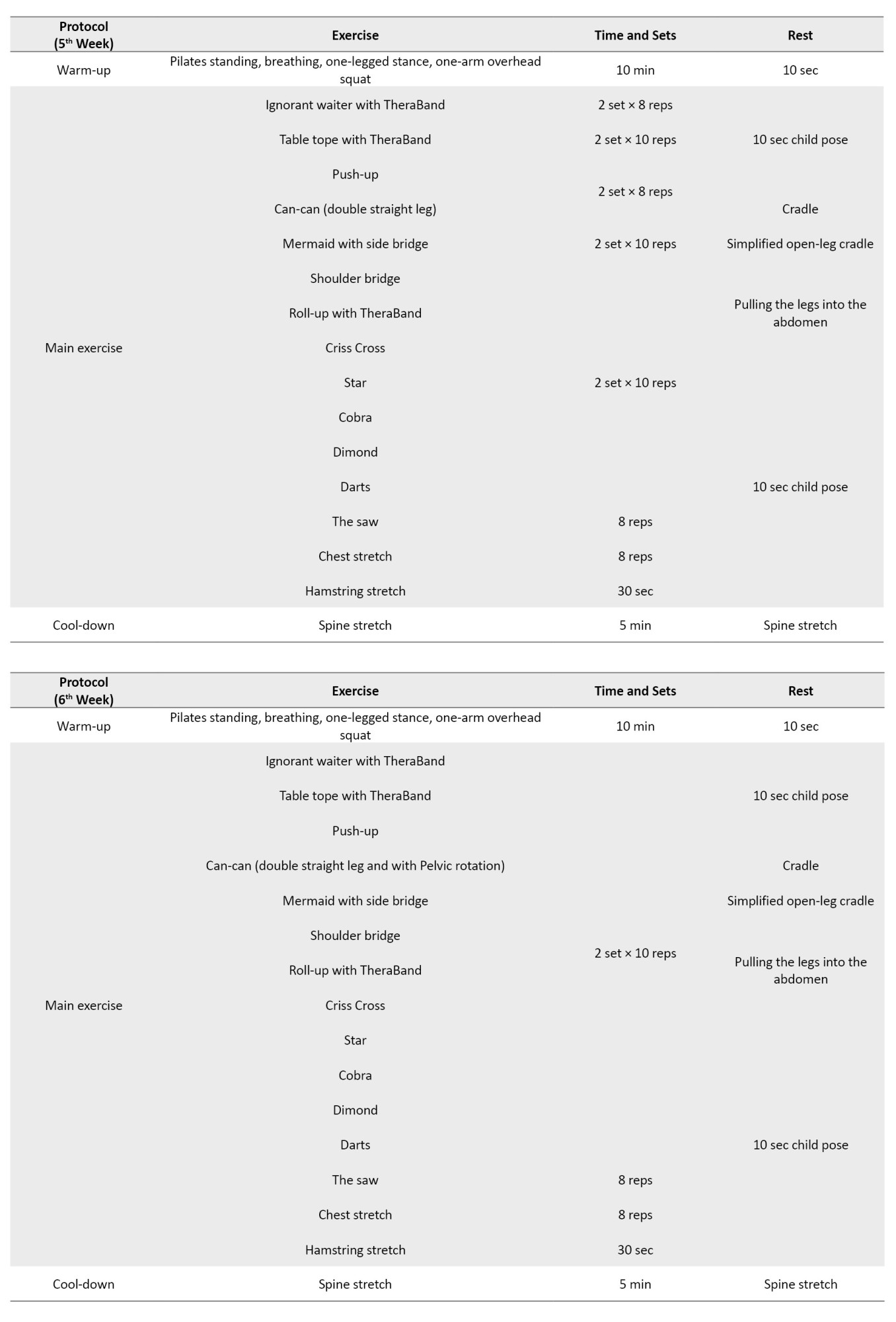
The PG participated in Pilates exercises for 6 weeks, attending three sessions each week. Each training session began at 35 minutes and gradually extended to 60 minutes, including a warm-up, Pilates exercises, and a cool-down period. Participants were educated on the principles of Pilates and how to properly execute the exercises during each session. Initially, participants performed two sets of six repetitions, which progressed to 2 sets of 10 repetitions over six weeks. The detailed exercise protocol is listed in the Appendix 1.
Data analysis
Raw data were processed using MATLAB software, version R2018b (IBM Inc., Chicago, USA). The mean value of three attempts was calculated and represented for further analysis.
Statistical analysis
The Shapiro-Wilk test was employed to assess the normality of the distributions of all variables. The independent-samples t-test was utilized to identify initial differences between groups. Paired sample t-tests were used to compare within groups. An analysis of covariance (ANCOVA) was used to compare post-training values between PG and CG, using baseline values as covariates. Statistical analysis was conducted using SPSS software, version 27 for Windows (SPSS Inc., Chicago, IL, USA) at a significance level of P<0.05.
Results
Table 1 presents the baseline demographic data and characteristics of all variables for each group.
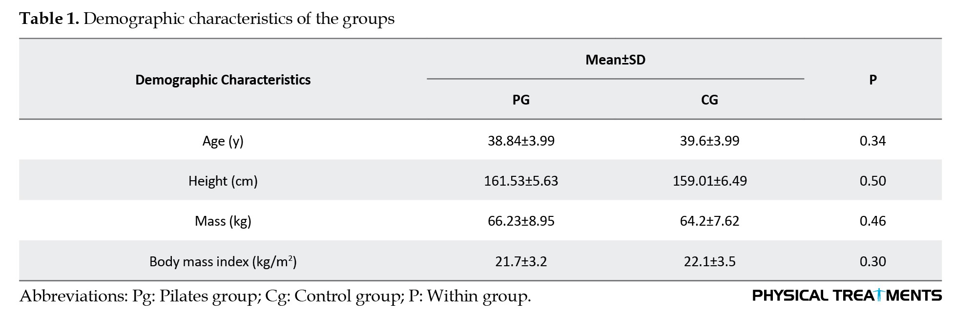
No significant difference was observed in any demographic or outcome variables between groups.
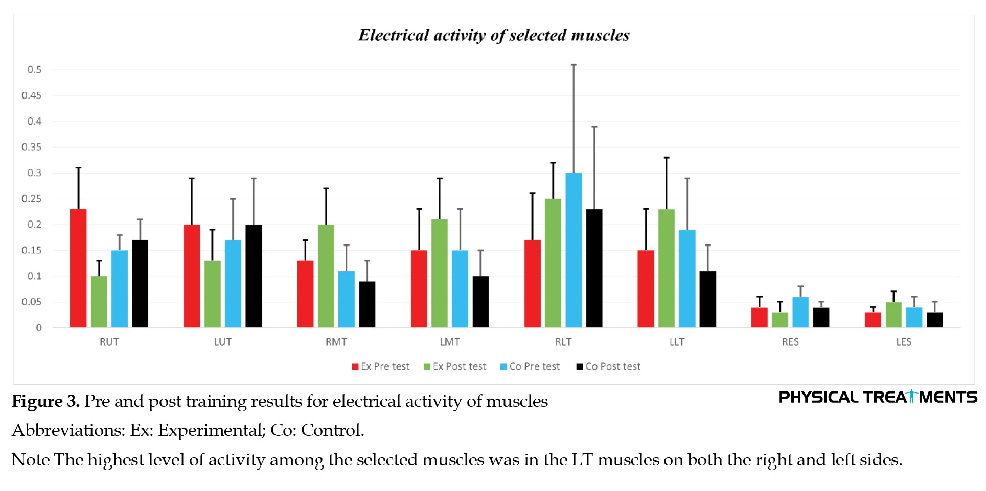
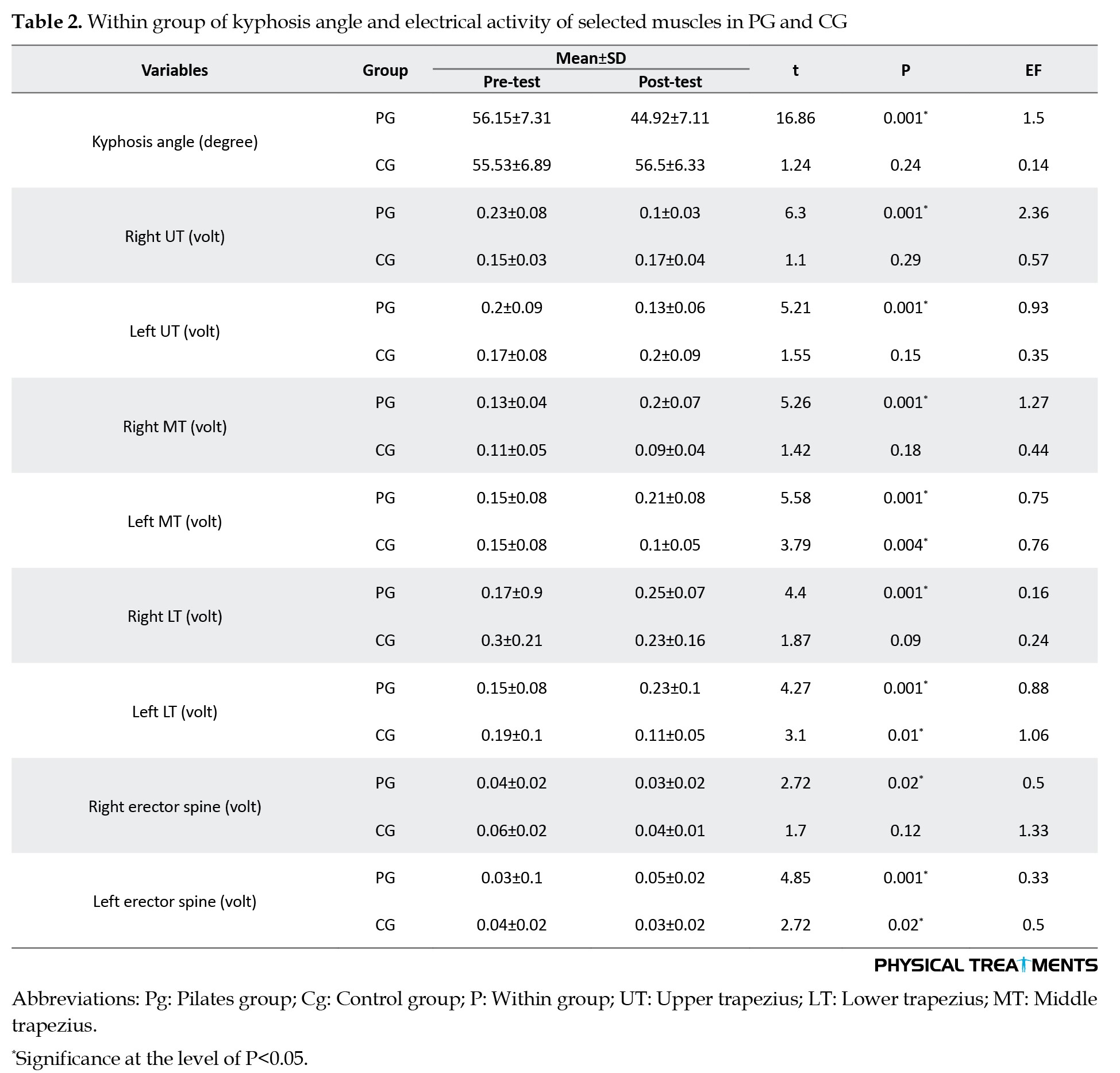
Table 2 and Figure 3 show the pre- and post-training results for kyphosis angle and muscle electrical activity.
Bipolar silver/silver chloride electrodes with an active area of approximately 1 cm were positioned with a center-to-center spacing of 2 cm along the muscle fibers. The location of the electrode on the examined muscles was determined and marked by the researcher according to the recommendations of Surface ElectroMyoGraphy for the non-invasive assessment of muscles instruction [19]. By carefully removing the hair, abrading, and cleansing the skin, low impedance was achieved at the interface between the skin and electrode. Other electrical devices were kept away from the measuring device to reduce noise. Electrical muscle activity was recorded at a sampling frequency of 1000 Hz during maximal voluntary muscle contraction. The EMG signals were automatically amplified and recorded utilizing the Biopac system (Biopac, Santa Barbara, CA). To provide a clear picture of the EMG data and to compare and interpret the differences, the maximum voluntary contraction of individuals (calculated by following the curve) was compared in three attempts. After each test, the person rested in a neutral position for 30 seconds [21]. EMG signals collected during MVIC tests were carefully examined for noise, and the first and the last few seconds of each attempt, which were less stable, were removed and then processed with a second-order low-pass Butterworth filter (20-500 Hz) and full-wave rectified. Subsequently, the root mean square was calculated, and its maximum value was extracted from the desired range.
Pilates exercise
The PG participated in Pilates exercises for 6 weeks, attending three sessions each week. Each training session began at 35 minutes and gradually extended to 60 minutes, including a warm-up, Pilates exercises, and a cool-down period. Participants were educated on the principles of Pilates and how to properly execute the exercises during each session. Initially, participants performed two sets of six repetitions, which progressed to 2 sets of 10 repetitions over six weeks. The detailed exercise protocol is listed in the Appendix 1.
Data analysis
Raw data were processed using MATLAB software, version R2018b (IBM Inc., Chicago, USA). The mean value of three attempts was calculated and represented for further analysis.
Statistical analysis
The Shapiro-Wilk test was employed to assess the normality of the distributions of all variables. The independent-samples t-test was utilized to identify initial differences between groups. Paired sample t-tests were used to compare within groups. An analysis of covariance (ANCOVA) was used to compare post-training values between PG and CG, using baseline values as covariates. Statistical analysis was conducted using SPSS software, version 27 for Windows (SPSS Inc., Chicago, IL, USA) at a significance level of P<0.05.
Results
Table 1 presents the baseline demographic data and characteristics of all variables for each group.
No significant difference was observed in any demographic or outcome variables between groups.
Table 2 and Figure 3 show the pre- and post-training results for kyphosis angle and muscle electrical activity.
Significant decreases were observed in kyphosis angle for PG (P=0.001) but not for CG. Significant differences were observed in electrical activities of the left and right UT, MT, and LT muscles (P=0.001) and erector spine muscles (P<0.05) in within-group comparison in PG (P<0.05). In addition, there were significant differences in the electrical activity of the left MT and LT muscles (P<0.05) and right erector spine muscle in the CG (P=0.02).
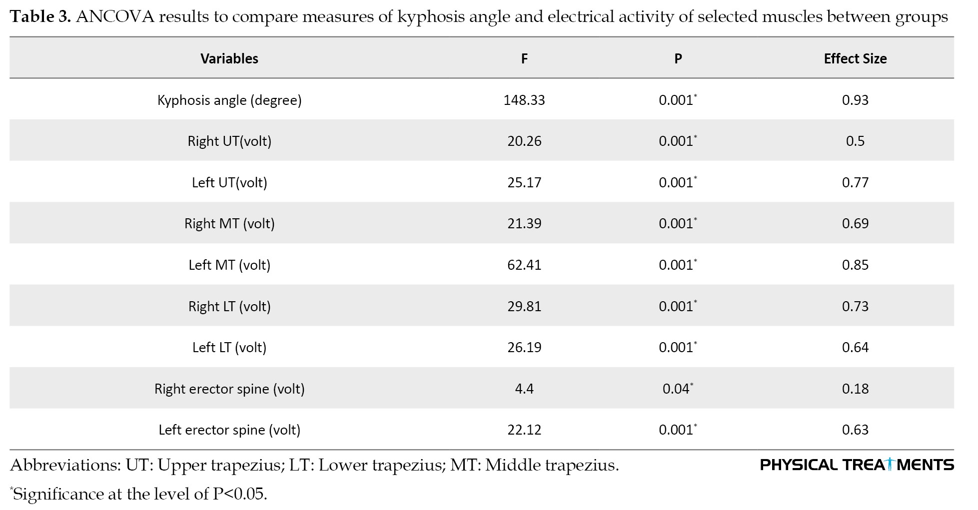
ANCOVA results reveal significant differences in the post-training values of PG and CG (P<0.05) (Table 3).
Additionally, these differences were significant in the PG compared to the CG (P<0.05).
Discussion
This study assessed the effectiveness of Pilates exercises compared to a CG regarding the kyphosis angle and the muscle activation of key scapular stabilizers and erector spinae in women with hyperkyphotic posture. The mean EMG amplitudes of the UT, MT, and LT, as well as the kyphosis angle, were measured. According to the results of this study, the thoracic kyphosis angle decreased significantly in the PG group but not in the CG group. These results pointed out that the Pilates method positively affected the kyphosis angle. It has been shown that corrective exercises or interventions aimed at correcting the kyphosis angle should emphasize stretching exercises for the pectoral muscles and strengthening of the core extensor muscles [4]. Pilates exercises emphasize correcting posture, strengthening and lengthening muscles, enhancing core stability, and improving breath control, all aimed at achieving a neutral pelvis alignment and lumbar lordosis [15].
Pilates plays a crucial role in strengthening the abdominal and lumbar muscles. These muscles provide stability and support to the spine, influencing overall body movements [23]. A strong core supports proper posture by aligning the spine and reducing strain on surrounding muscles. Pilates encourages awareness of body alignment, helping practitioners learn to maintain good posture in various positions, whether sitting, standing, or during physical activity. An improved posture can alleviate back pain and discomfort associated with muscle imbalances [14]. The results showed a reduction in the electrical activity of the trapezius and erector spinae muscles in PG. One possible reason for the reduction in electrical activity in the UT muscles may be a decrease in muscle tension. Individuals with kyphosis, due to the shoulders being positioned away from the body’s midline, may experience tension in these muscles [24]. This excessive tension increases the motor unit requirements, resulting in increased muscle activity [25]. Therefore, following the reduction of the kyphosis angle, the tension of the UT muscle may be reduced, and the electrical activity of this muscle may be lowered.
In addition, the mean electrical activity of the right and left MT and right and left LT muscles increased in PG. The reason for the changes in the electrical activity of the MT and LT muscles after 6 weeks of Pilates exercise may be explained as follows: In kyphosis, upper rotation and scapular abduction occur, which may be due to the weakness of the lower and MT muscles. As a result, less electrical activity was observed on the pre-test due to muscle weakness. Weakness leads to muscle fatigue, which reduces the speed of afferent message transmission. It also affects the presynaptic-postsynaptic mechanism and the potential action position at the environmental level. This causes an inability to transmit nerve signals or to respond to nerve stimulation, and changes the muscle’s pattern of electrical activity. Therefore, the increase in electrical activity in the MT and LT muscles is likely due to muscle strengthening and the correction of the abnormality after 6 weeks of Pilates exercises in PG. The electrical activity of the left MT muscle was in the CG after 6 weeks. The electrical activity of the lower right trapezius muscle in the post-test was not significantly different from that of the pre-test in the CG. This is probably due to a lack of exercise and physical activity in the control group or the superiority of the subjects’ right limb. Lack of training in this muscle causes weakness, and the use of the superior limbs makes the muscles on the opposite side of the body less active. These factors might cause a reduction in the electrical activity of the other side.
The average electrical activity of the right and left erector spine increased in PG. The reason for the changes in the electrical activity of the erector spine muscles is that these muscles are the backbone of the spine and central stabilizers of the body. These muscles become stretched and weak due to hyperkyphosis. This weakness impairs the transmission of nerve signals to the muscles, leading to a change in the strategy for using the muscle to stabilize the joints, followed by a change in the movement program [18]. The reason for the increase in muscle electrical activity is that straightening the spine after 6 weeks of Pilates exercise strengthens these muscles and returns them to normal alignment, thereby changing the muscle response to nerve signals. According to the principle of centrality in Pilates, movements are designed so that with every performance, the central parts of the body, including the transverse abdominal muscles, the internal and external obliques, and the erector spine, which are the body’s central stabilizers and mobilizers, get involved [25].
The positive effect of Pilates on muscle electrical activity can be explained by the fact that, according to scientific findings, muscle tone or tension depends on two factors: the basic viscoelastic properties of the muscle soft tissue and the degree of activation of muscle contraction network [26].
Viscoelastic properties include shortness, stiffness, and length loss, while the contractile network includes increased contractile activity. The muscles may shorten or tighten to compensate for the shortening of contractile fibers, the retraction of extra-muscular connective tissue, or the adjacent fascia. The combination of these two muscle properties causes a relaxed muscle tone.
Stiff muscles have a higher muscle tone and a lower irritability threshold. This means that these muscles are readily recruited for movement [27]. Due to an abnormality, the neural control unit may temporarily change the strategy for recruiting muscles for joint stability. This change in activation patterns affects muscle balance and movement patterns, ultimately altering the motor program and leading to reciprocal inhibition of the muscles. Reciprocal inhibition is a neuromuscular condition that occurs when a muscle becomes more active than normal, and to cause stimulation, the nerve signal increases [8]. This reduces stimulus and increases the inhibitory signal to the antagonist’s muscle. In addition, habitual postures can stretch the muscles for a considerable amount of time, resulting in postural weakness. Postural weakness might be due to inhibition caused by muscle stiffness.
These exercises emphasize slow, controlled movements that require focus and balance and train the body to respond more effectively to weight shifts or changes in direction. This is supported by timely muscle activation [12]. Delaying muscle activation in different situations has different effects, including decreased balance. Pilates exercises can change muscle timing, and neuromuscular adaptations occur with continued practice [12, 28].
Muscle weakness caused by excessive traction also inhibits spindle activation and can increase the number of sarcomeres. These changes can affect the pattern of electrical activity in the muscle [29]. According to Pilates principles that affect posture and centrality, one should always keep the transverse abdominal muscle in voluntary contraction while performing these exercises. This increases intra-abdominal pressure and tension in the thoracolumbar fascia, providing strong support for muscle contraction before limb movements [30]. Contraction of other abdominal muscles stabilizes the spine and provides suitable conditions in the joint movement chain for proper limb movement [31]. Therefore, it is possible to focus on the imbalance of the target muscles in kyphosis instability and restore muscle balance with the least amount of energy.
Pilates is an effective way to develop body awareness and postural control [28]. Research has shown that Pilates exercises lead to more activation of deep abdominal and core stabilizer muscles [31]. Proper distribution of forces and reduction of compressive forces in the joints of the motor chain are created following the stability in the body’s center. Dysfunction in this area can affect other parts of the motor chain, cause the deviation of the spine from its regular position, and lead to injuries and abnormalities in this area.
The limitations of this study include the small sample size; employing larger samples would facilitate more robust and accurate generalizations of the results. Additionally, the study lacked a follow-up period; future research should include a follow-up assessment to evaluate the persistence of the effects of these exercises. Furthermore, implementing blinding for the evaluator could enhance the accuracy and reliability of the results.
Conclusion
Pilates exercises in the motor chain create equilibrium, and the muscles are in the correct position regarding strength and flexibility. These factors can affect electrical activity, which is the result we are aiming for. Six weeks of Pilates exercise emphasizing core stabilizers improved the severity of kyphosis and muscle activity. The reduction of UT muscle activity and erector spine, along with the increased activity of the MT and LT muscles, is related to a reduction in the kyphosis angle. It can effectively reduce the kyphotic angle and restore balance to weakened, shortened muscles. Future studies could explore the long-term effects of Pilates on kyphosis, particularly with extended follow-up periods to assess the sustainability of improvements in muscle activity and spinal alignment, and incorporate Pilates into preventive health strategies to benefit individuals at risk of developing kyphosis, especially among populations engaged in activities that lead to postural imbalances.
Ethical Considerations
Compliance with ethical guidelines
There were no ethical considerations to be considered in this research.
Funding
This research did not receive any grant from funding agencies in the public, commercial, or non-profit sectors.
Authors' contributions
All authors contributed equally to the conception and design of the study, data collection and analysis, interpretation of the results, and drafting of the manuscript. Each author approved the final version of the manuscript for submission.
Conflict of interest
The authors declared no conflict of interest.
ANCOVA results reveal significant differences in the post-training values of PG and CG (P<0.05) (Table 3).
Additionally, these differences were significant in the PG compared to the CG (P<0.05).
Discussion
This study assessed the effectiveness of Pilates exercises compared to a CG regarding the kyphosis angle and the muscle activation of key scapular stabilizers and erector spinae in women with hyperkyphotic posture. The mean EMG amplitudes of the UT, MT, and LT, as well as the kyphosis angle, were measured. According to the results of this study, the thoracic kyphosis angle decreased significantly in the PG group but not in the CG group. These results pointed out that the Pilates method positively affected the kyphosis angle. It has been shown that corrective exercises or interventions aimed at correcting the kyphosis angle should emphasize stretching exercises for the pectoral muscles and strengthening of the core extensor muscles [4]. Pilates exercises emphasize correcting posture, strengthening and lengthening muscles, enhancing core stability, and improving breath control, all aimed at achieving a neutral pelvis alignment and lumbar lordosis [15].
Pilates plays a crucial role in strengthening the abdominal and lumbar muscles. These muscles provide stability and support to the spine, influencing overall body movements [23]. A strong core supports proper posture by aligning the spine and reducing strain on surrounding muscles. Pilates encourages awareness of body alignment, helping practitioners learn to maintain good posture in various positions, whether sitting, standing, or during physical activity. An improved posture can alleviate back pain and discomfort associated with muscle imbalances [14]. The results showed a reduction in the electrical activity of the trapezius and erector spinae muscles in PG. One possible reason for the reduction in electrical activity in the UT muscles may be a decrease in muscle tension. Individuals with kyphosis, due to the shoulders being positioned away from the body’s midline, may experience tension in these muscles [24]. This excessive tension increases the motor unit requirements, resulting in increased muscle activity [25]. Therefore, following the reduction of the kyphosis angle, the tension of the UT muscle may be reduced, and the electrical activity of this muscle may be lowered.
In addition, the mean electrical activity of the right and left MT and right and left LT muscles increased in PG. The reason for the changes in the electrical activity of the MT and LT muscles after 6 weeks of Pilates exercise may be explained as follows: In kyphosis, upper rotation and scapular abduction occur, which may be due to the weakness of the lower and MT muscles. As a result, less electrical activity was observed on the pre-test due to muscle weakness. Weakness leads to muscle fatigue, which reduces the speed of afferent message transmission. It also affects the presynaptic-postsynaptic mechanism and the potential action position at the environmental level. This causes an inability to transmit nerve signals or to respond to nerve stimulation, and changes the muscle’s pattern of electrical activity. Therefore, the increase in electrical activity in the MT and LT muscles is likely due to muscle strengthening and the correction of the abnormality after 6 weeks of Pilates exercises in PG. The electrical activity of the left MT muscle was in the CG after 6 weeks. The electrical activity of the lower right trapezius muscle in the post-test was not significantly different from that of the pre-test in the CG. This is probably due to a lack of exercise and physical activity in the control group or the superiority of the subjects’ right limb. Lack of training in this muscle causes weakness, and the use of the superior limbs makes the muscles on the opposite side of the body less active. These factors might cause a reduction in the electrical activity of the other side.
The average electrical activity of the right and left erector spine increased in PG. The reason for the changes in the electrical activity of the erector spine muscles is that these muscles are the backbone of the spine and central stabilizers of the body. These muscles become stretched and weak due to hyperkyphosis. This weakness impairs the transmission of nerve signals to the muscles, leading to a change in the strategy for using the muscle to stabilize the joints, followed by a change in the movement program [18]. The reason for the increase in muscle electrical activity is that straightening the spine after 6 weeks of Pilates exercise strengthens these muscles and returns them to normal alignment, thereby changing the muscle response to nerve signals. According to the principle of centrality in Pilates, movements are designed so that with every performance, the central parts of the body, including the transverse abdominal muscles, the internal and external obliques, and the erector spine, which are the body’s central stabilizers and mobilizers, get involved [25].
The positive effect of Pilates on muscle electrical activity can be explained by the fact that, according to scientific findings, muscle tone or tension depends on two factors: the basic viscoelastic properties of the muscle soft tissue and the degree of activation of muscle contraction network [26].
Viscoelastic properties include shortness, stiffness, and length loss, while the contractile network includes increased contractile activity. The muscles may shorten or tighten to compensate for the shortening of contractile fibers, the retraction of extra-muscular connective tissue, or the adjacent fascia. The combination of these two muscle properties causes a relaxed muscle tone.
Stiff muscles have a higher muscle tone and a lower irritability threshold. This means that these muscles are readily recruited for movement [27]. Due to an abnormality, the neural control unit may temporarily change the strategy for recruiting muscles for joint stability. This change in activation patterns affects muscle balance and movement patterns, ultimately altering the motor program and leading to reciprocal inhibition of the muscles. Reciprocal inhibition is a neuromuscular condition that occurs when a muscle becomes more active than normal, and to cause stimulation, the nerve signal increases [8]. This reduces stimulus and increases the inhibitory signal to the antagonist’s muscle. In addition, habitual postures can stretch the muscles for a considerable amount of time, resulting in postural weakness. Postural weakness might be due to inhibition caused by muscle stiffness.
These exercises emphasize slow, controlled movements that require focus and balance and train the body to respond more effectively to weight shifts or changes in direction. This is supported by timely muscle activation [12]. Delaying muscle activation in different situations has different effects, including decreased balance. Pilates exercises can change muscle timing, and neuromuscular adaptations occur with continued practice [12, 28].
Muscle weakness caused by excessive traction also inhibits spindle activation and can increase the number of sarcomeres. These changes can affect the pattern of electrical activity in the muscle [29]. According to Pilates principles that affect posture and centrality, one should always keep the transverse abdominal muscle in voluntary contraction while performing these exercises. This increases intra-abdominal pressure and tension in the thoracolumbar fascia, providing strong support for muscle contraction before limb movements [30]. Contraction of other abdominal muscles stabilizes the spine and provides suitable conditions in the joint movement chain for proper limb movement [31]. Therefore, it is possible to focus on the imbalance of the target muscles in kyphosis instability and restore muscle balance with the least amount of energy.
Pilates is an effective way to develop body awareness and postural control [28]. Research has shown that Pilates exercises lead to more activation of deep abdominal and core stabilizer muscles [31]. Proper distribution of forces and reduction of compressive forces in the joints of the motor chain are created following the stability in the body’s center. Dysfunction in this area can affect other parts of the motor chain, cause the deviation of the spine from its regular position, and lead to injuries and abnormalities in this area.
The limitations of this study include the small sample size; employing larger samples would facilitate more robust and accurate generalizations of the results. Additionally, the study lacked a follow-up period; future research should include a follow-up assessment to evaluate the persistence of the effects of these exercises. Furthermore, implementing blinding for the evaluator could enhance the accuracy and reliability of the results.
Conclusion
Pilates exercises in the motor chain create equilibrium, and the muscles are in the correct position regarding strength and flexibility. These factors can affect electrical activity, which is the result we are aiming for. Six weeks of Pilates exercise emphasizing core stabilizers improved the severity of kyphosis and muscle activity. The reduction of UT muscle activity and erector spine, along with the increased activity of the MT and LT muscles, is related to a reduction in the kyphosis angle. It can effectively reduce the kyphotic angle and restore balance to weakened, shortened muscles. Future studies could explore the long-term effects of Pilates on kyphosis, particularly with extended follow-up periods to assess the sustainability of improvements in muscle activity and spinal alignment, and incorporate Pilates into preventive health strategies to benefit individuals at risk of developing kyphosis, especially among populations engaged in activities that lead to postural imbalances.
Ethical Considerations
Compliance with ethical guidelines
There were no ethical considerations to be considered in this research.
Funding
This research did not receive any grant from funding agencies in the public, commercial, or non-profit sectors.
Authors' contributions
All authors contributed equally to the conception and design of the study, data collection and analysis, interpretation of the results, and drafting of the manuscript. Each author approved the final version of the manuscript for submission.
Conflict of interest
The authors declared no conflict of interest.
References
- Golalizadeh D, Toopchizadeh V, Fasaie N, Dolatkhah N. Body composition indices in a sample of female adolescents with postural deformity: A case control study. BMC Research Notes. 2019; 12(1):754. [DOI:10.1186/s13104-019-4794-y] [PMID] [PMCID]
- Koelé M, Lems W, Willems H. The clinical relevance of hyperkyphosis: A narrative review. Frontiers in Endocrinology. 2020; 11:5. [DOI:10.3389/fendo.2020.00005] [PMID] [PMCID]
- Seidi F, Bayattork M, Minoonejad H, Andersen LL, Page P. Comprehensive corrective exercise program improves alignment, muscle activation and movement pattern of men with upper crossed syndrome: Randomized controlled trial. Scientific Reports. 2020; 10(1):20688. [DOI:10.1038/s41598-020-77571-4] [PMID] [PMCID]
- Vaughn DW, Brown EW. The influence of an in-home based therapeutic exercise program on thoracic kyphosis angles. Journal of Back and Musculoskeletal Rehabilitation. 2007; 20(4):155-65. [DOI:10.3233/BMR-2007-20404]
- Azizi A, Mahdavinejhad R, Taheri-tizani A, Jafarzadeh T, Rezaeinasab A. [The effect of 8 weeks specific aquatic therapy on kyphosis angle and some pulmonary indices in male university students with kyphosis (Persian)]. Journal of Kerman University of Medical Sciences. 2012; 19(2):440-50. [Link]
- Silva AG, Punt TD, Sharples P, Vilas-Boas JP, Johnson MI. Head posture and neck pain of chronic nontraumatic origin: A comparison between patients and pain-free persons. Archives of Physical Medicine and Rehabilitation. 2009; 90(4):669-74. [DOI:10.1016/j.apmr.2008.10.018] [PMID]
- Altan L, Korkmaz N, Bingol Ü, Gunay B. Effect of pilates training on people with fibromyalgia syndrome: A pilot study. Archives of Physical Medicine and Rehabilitation. 2009; 90(12):1983-8. [DOI:10.1016/j.apmr.2009.06.021] [PMID]
- Page P, Frank CC, Lardner R. Assessment and treatment of muscle imbalance: The Janda Approach. Champaign, IL Human Kinetics; 2010. [DOI:10.5040/9781718211445]
- Morris CE, Greenman PE, Bullock MI, Basmajian JV, Kobesova A. Vladimir Janda, MD, DSc: Tribute to a master of rehabilitation. Spine. 2006; 31(9):1060-4. [DOI:10.1097/01.brs.0000214879.93102.4e] [PMID]
- Sinaki M, Brey RH, Hughes CA, Larson DR, Kaufman KR. Balance disorder and increased risk of falls in osteoporosis and kyphosis: significance of kyphotic posture and muscle strength. Osteoporosis International. 2005; 16(8):1004-10. [DOI:10.1007/s00198-004-1791-2] [PMID]
- Paillard T. Effects of general and local fatigue on postural control: A review. Neuroscience & Biobehavioral Reviews. 2012; 36(1):162-76. [DOI:10.1016/j.neubiorev.2011.05.009] [PMID]
- Ko HS, Jung HU, Park TY, Song JK, Wang J, Jung HC. Comparisons of functional movements and core muscle activity in women according to Pilates proficiency. Frontiers in Physiology. 2024; 15:1435671. [DOI:10.3389/fphys.2024.1435671] [PMID] [PMCID]
- González-Gálvez N, Marcos-Pardo PJ, Albaladejo-Saura M, López-Vivancos A, Vaquero-Cristóbal R. Effects of a Pilates programme in spinal curvatures and hamstring extensibility in adolescents with thoracic hyperkyphosis: A randomised controlled trial. Postgraduate Medical Journal. 2023; 99(1171):433-41. [DOI:10.1136/postgradmedj-2021-140901] [PMID]
- Eliks M, Zgorzalewicz-Stachowiak M, Zeńczak-Praga K. Application of Pilates-based exercises in the treatment of chronic non-specific low back pain: State of the art. Postgraduate Medical Journal. 2019; 95(1119):41-5. [DOI:10.1136/postgradmedj-2018-135920] [PMID] [PMCID]
- Lyon Jr D, Arbizu WA. The complete book of Pilates for men: The lifetime plan for strength, power, and peak performance. New York: HarperCollins Publishers; 2005. [Link]
- Segal NA, Hein J, Basford JR. The effects of Pilates training on flexibility and body composition: An observational study. Archives of Physical Medicine and Rehabilitation. 2004; 85(12):1977-81. [DOI:10.1016/j.apmr.2004.01.036] [PMID]
- Junges S, Gottlieb MG, Baptista RR, Quadros CBd, Resende TdL, Gomes I. Effectiveness of pilates method for the posture and flexibility of women with hyperkyphosis. Revista Brasileira de Ciência e Movimento. 2012; 20(1):21-33. [Link]
- Kim BI, Jung JH, Shim J, Kwon HY, Kim H. An analysis of muscle activities of healthy women during Pilates exercises in a prone position. Journal of Physical Therapy Science. 2014; 26(1):77-9. [DOI:10.1589/jpts.26.77] [PMID] [PMCID]
- Lewis JS, Valentine RE. Clinical measurement of the thoracic kyphosis. A study of the intra-rater reliability in subjects with and without shoulder pain. BMC Musculoskeletal Disorders. 2010; 11:39. [DOI:10.1186/1471-2474-11-39] [PMID] [PMCID]
- Sekendiz B, Cug M, Korkusuz F. Effects of Swiss-ball core strength training on strength, endurance, flexibility, and balance in sedentary women. The Journal of Strength & Conditioning Research. 2010; 24(11):3032-40. [DOI:10.1519/JSC.0b013e3181d82e70] [PMID]
- Ekstrom RA, Donatelli RA, Soderberg GL. Surface electromyographic analysis of exercises for the trapezius and serratus anterior muscles. Journal of Orthopaedic & Sports Physical Therapy. 2003; 33(5):247-58. [DOI:10.2519/jospt.2003.33.5.247] [PMID]
- Keller TS, Colloca CJ. Mechanical force spinal manipulation increases trunk muscle strength assessed by electromyography: A comparative clinical trial. Journal of Manipulative and Physiological Therapeutics. 2000; 23(9):585-95. [DOI:10.1067/mmt.2000.110947] [PMID]
- Gholamalishahi S, Backhaus I, Cilindro C, Masala D, La Torre G. Pilates-based exercise in the reduction of the low back pain: An overview of reviews. European Review for Medical & Pharmacological Sciences. 2022; 26(13):4557-63. [DOI:10.26355/eurrev_202207_29176] [PMID]
- Scheuermann H. Kyphosis dorsalis juvenilis. Ugeskr laeger. 1920; 82:385. [Link]
- Tecco S, Mummolo S, Marchetti E, Tetè S, Campanella V, Gatto R, et al. sEMG activity of masticatory, neck, and trunk muscles during the treatment of scoliosis with functional braces. A longitudinal controlled study. Journal of Electromyography and Kinesiology. 2011; 21(6):885-92. [DOI:10.1016/j.jelekin.2011.08.004] [PMID]
- Wolkodoff N, Peterson S, Miller J. The fitness effects of a combined aerobic and pilates program an eight-week Study using the stamina aeropilates pro XP555. AeroPilates Pro XP. 2008; 555. [Link]
- Quek J, Pua YH, Clark RA, Bryant AL. Effects of thoracic kyphosis and forward head posture on cervical range of motion in older adults. Manual Therapy. 2013; 18(1):65-71. [DOI:10.1016/j.math.2012.07.005] [PMID]
- Kamali A, Mahdavi Nezhad R, Norouzi K. [The effect of selected pilates exercises on thigh muscle strength and depression in elderly women (Persian)]. Journal of Paramedical Sciences & Rehabilitation. 2016; 5(2):67-75. [DOI:10.22038/jpsr.2016.6915]
- Clark M, Lucett S. NASM essentials of corrective exercise training. Philadelphia: Lippincott Williams & Wilkins; 2010. [Link]
- Gabriel DA, Kamen G, Frost G. Neural adaptations to resistive exercise: Mechanisms and recommendations for training practices. Sports Medicine. 2006; 36(2):133-49.[DOI:10.2165/00007256-200636020-00004] [PMID]
- Mousavi SH, Minoonejad H, Rajabi R, Seidi F. Follow-up comparison of the effect of eight-weeks corrective exercises with and without myofascial release on postural kyphosis deformity. Sport Sciences and Health Research. 2019; 11(1):1-12. [DOI:10.32598/JESM.11.1.1]
Type of Study: Research |
Subject:
General
Received: 2024/12/20 | Accepted: 2025/05/31 | Published: 2026/07/1
Received: 2024/12/20 | Accepted: 2025/05/31 | Published: 2026/07/1
Send email to the article author
| Rights and permissions | |
 |
This work is licensed under a Creative Commons Attribution-NonCommercial 4.0 International License. |

This is an open access article distributed under the terms of the Creative Commons Attribution-NonCommercial 4.0 (CC-By-NC 4.0), which permits use, distribution, and reproduction in any medium, provided the original work is properly cited and is not used for commercial purposes.
Contact Information