Tue, Sep 29, 2026
Volume 16 - Special Issue(Exercise Therapy in Action)
PTJ 2026, 16 - Special Issue(Exercise Therapy in Action): 401-412 |
Back to browse issues page



Download citation:
BibTeX | RIS | EndNote | Medlars | ProCite | Reference Manager | RefWorks
Send citation to:


BibTeX | RIS | EndNote | Medlars | ProCite | Reference Manager | RefWorks
Send citation to:
Imanibrouj S, Jafarnezhadgero A, Sheikhalizade H, Mousavi S H. Effect of Sand Surface Training on Knee and Ankle Co-contraction in Individuals With Pronated Feet and ACL Reconstruction. PTJ 2026; 16 (S1) :401-412
URL: http://ptj.uswr.ac.ir/article-1-671-en.html
URL: http://ptj.uswr.ac.ir/article-1-671-en.html

1- Department of Sports Biomechanics, Faculty of Educational Sciences and Psychology, University of Mohaghegh Ardabili, Ardabil, Iran.
2- Department of Health and Sports Medicine, Faculty of Physical Education and Sport Sciences, University of Tehran, Tehran, Iran.
2- Department of Health and Sports Medicine, Faculty of Physical Education and Sport Sciences, University of Tehran, Tehran, Iran.
Full-Text [PDF 594 kb]
(1097 Downloads)
| Abstract (HTML) (1914 Views)
Full-Text: (14 Views)
Introduction
Anterior cruciate ligament (ACL) tears are very common [1]. The human skeletal-muscular system operates as an interconnected framework, meaning changes in one part can influence other areas and potentially disrupt fundamental motor skills, such as walking [2, 3]. Walking is a primary function of the lower body, involving tasks, such as absorbing the impact forces from foot strikes, maintaining stability, and generating forward propulsion. These actions are crucial for forming a coordinated and efficient walking pattern [3, 4].
Research indicates that natural walking relies on neural control, muscle force production, and an adequate range of motion. Disruptions in any of these factors can lead to abnormal gait patterns [5]. Abnormal gait can cause various issues in the lower limbs, with foot pronation being one potential factor that increases the risk of walking-related injuries. ACL reconstruction (ACLR) is commonly required for ACL injuries, with over 175,000 procedures performed annually in the U.S. The incidence of ACLR has increased from 32.94 per 100,000 person-years in 1994 to 43.48 per 100,000 by 2006. Similarly, annual rates of ACLR from 2004 to 2007 in Scandinavian countries were 32, 34, and 38 per 100,000 person-years for Sweden, Norway, and Denmark, respectively. Despite advancements in surgical techniques that effectively restore knee function, ACLR patients face an elevated risk of developing early-onset knee osteoarthritis (OA) [6, 7].
The precise mechanisms contributing to the elevated susceptibility to OA in individuals who have undergone ACLR remain uncertain. Nevertheless, the increased risk of knee OA in this group may be attributed to an abnormal elevation in joint compressive force resulting from altered neuromuscular strategies. Studies have documented specific adaptations in gait mechanics following ACLR, including reduced internal knee extensor moments [8, 9] and increased internal hip extensor moments [4, 10]. These biomechanical modifications may represent compensatory movement strategies employed to safeguard the reconstructed knee joints. Alterations in neuromuscular activity patterns may accompany these adaptations. Individuals who undergo ACLR demonstrate increased hamstring activity and concurrent activation of both quadriceps and hamstring muscles during various functional movements, including running, jumping, and walking [11-13]. Muscle co-contraction augmentation has been shown to increase tibiofemoral compressive force in a simulated knee model with ACL deficiency [14]. This muscle recruitment pattern—frequently observed in individuals who have undergone ACLR—is thought to enhance joint stability and lessen anterior shear forces on the knee by creating a posterior shear force through the hamstrings [15]. Thus, one possible explanation for the increased compressive pressure at the tibiofemoral joint is the greater co-contraction of muscles observed in ACLR patients [15].
Foot overpronation is considered a risk factor for ACL injuries and is common in the general population [16, 17]. Beckett et al. (2018) established a direct correlation between ACL tears and excessive pronation of the subtalar joint [18]. There is clear evidence that excessive internal rotation of the tibia and overpronation of the feet can generate twisting forces, increasing forces on the knee [19].
According to a recent comprehensive review and meta-analysis, gait retraining shows promise as a treatment for decreasing foot pronation [20]. Research suggests that walking barefoot activates plantar cutaneous mechanoreceptors, especially on uneven ground, such as sand [21]. Our results showed that, in people with ACLR and pronated feet, exercise [22]. The current study’s researchers did not find any existing research examining the impact of walking on a sand surface on muscle co-contraction in individuals with pronated feet and ACLR. Therefore, it is important to identify effective therapeutic modalities to enhance muscular co-contraction in people with ACLR and pronated feet. Therefore, the purpose of this study was to evaluate how sand-surface training affects co-contraction of the knee and ankle muscles in people with pronated feet and ACLR while walking. We hypothesized that sand training could decrease directed ankle and knee joint co-contraction at the loading phase in both sagittal and frontal planes.
Materials and Methods
Study design
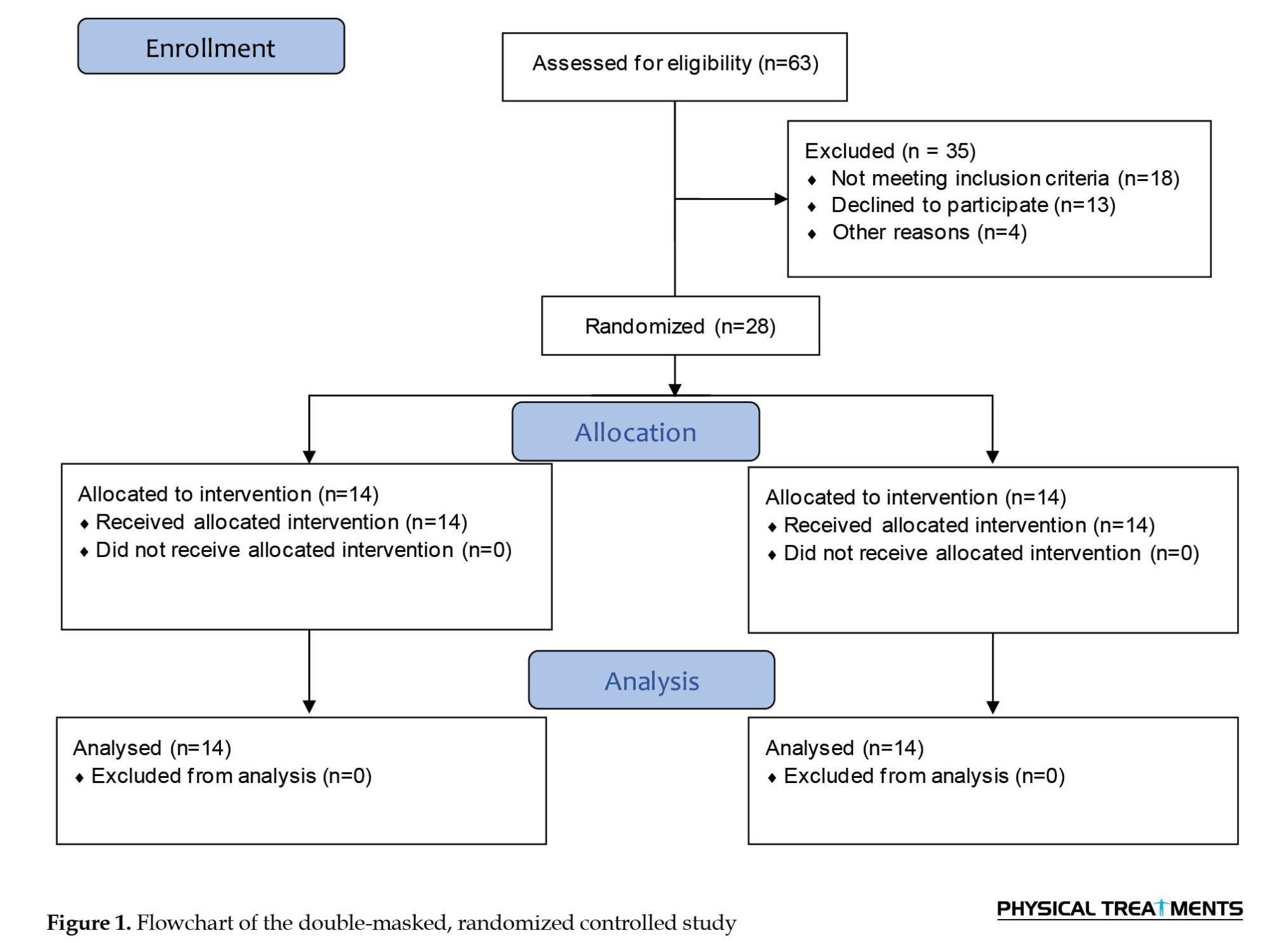
A randomized controlled trial was used in this investigation (Figure 1).
Anterior cruciate ligament (ACL) tears are very common [1]. The human skeletal-muscular system operates as an interconnected framework, meaning changes in one part can influence other areas and potentially disrupt fundamental motor skills, such as walking [2, 3]. Walking is a primary function of the lower body, involving tasks, such as absorbing the impact forces from foot strikes, maintaining stability, and generating forward propulsion. These actions are crucial for forming a coordinated and efficient walking pattern [3, 4].
Research indicates that natural walking relies on neural control, muscle force production, and an adequate range of motion. Disruptions in any of these factors can lead to abnormal gait patterns [5]. Abnormal gait can cause various issues in the lower limbs, with foot pronation being one potential factor that increases the risk of walking-related injuries. ACL reconstruction (ACLR) is commonly required for ACL injuries, with over 175,000 procedures performed annually in the U.S. The incidence of ACLR has increased from 32.94 per 100,000 person-years in 1994 to 43.48 per 100,000 by 2006. Similarly, annual rates of ACLR from 2004 to 2007 in Scandinavian countries were 32, 34, and 38 per 100,000 person-years for Sweden, Norway, and Denmark, respectively. Despite advancements in surgical techniques that effectively restore knee function, ACLR patients face an elevated risk of developing early-onset knee osteoarthritis (OA) [6, 7].
The precise mechanisms contributing to the elevated susceptibility to OA in individuals who have undergone ACLR remain uncertain. Nevertheless, the increased risk of knee OA in this group may be attributed to an abnormal elevation in joint compressive force resulting from altered neuromuscular strategies. Studies have documented specific adaptations in gait mechanics following ACLR, including reduced internal knee extensor moments [8, 9] and increased internal hip extensor moments [4, 10]. These biomechanical modifications may represent compensatory movement strategies employed to safeguard the reconstructed knee joints. Alterations in neuromuscular activity patterns may accompany these adaptations. Individuals who undergo ACLR demonstrate increased hamstring activity and concurrent activation of both quadriceps and hamstring muscles during various functional movements, including running, jumping, and walking [11-13]. Muscle co-contraction augmentation has been shown to increase tibiofemoral compressive force in a simulated knee model with ACL deficiency [14]. This muscle recruitment pattern—frequently observed in individuals who have undergone ACLR—is thought to enhance joint stability and lessen anterior shear forces on the knee by creating a posterior shear force through the hamstrings [15]. Thus, one possible explanation for the increased compressive pressure at the tibiofemoral joint is the greater co-contraction of muscles observed in ACLR patients [15].
Foot overpronation is considered a risk factor for ACL injuries and is common in the general population [16, 17]. Beckett et al. (2018) established a direct correlation between ACL tears and excessive pronation of the subtalar joint [18]. There is clear evidence that excessive internal rotation of the tibia and overpronation of the feet can generate twisting forces, increasing forces on the knee [19].
According to a recent comprehensive review and meta-analysis, gait retraining shows promise as a treatment for decreasing foot pronation [20]. Research suggests that walking barefoot activates plantar cutaneous mechanoreceptors, especially on uneven ground, such as sand [21]. Our results showed that, in people with ACLR and pronated feet, exercise [22]. The current study’s researchers did not find any existing research examining the impact of walking on a sand surface on muscle co-contraction in individuals with pronated feet and ACLR. Therefore, it is important to identify effective therapeutic modalities to enhance muscular co-contraction in people with ACLR and pronated feet. Therefore, the purpose of this study was to evaluate how sand-surface training affects co-contraction of the knee and ankle muscles in people with pronated feet and ACLR while walking. We hypothesized that sand training could decrease directed ankle and knee joint co-contraction at the loading phase in both sagittal and frontal planes.
Materials and Methods
Study design
A randomized controlled trial was used in this investigation (Figure 1).
A one-tailed preliminary power analysis was performed using the free G*Power software. For the power analysis, the F-test family—particularly the repeated measures analysis of variance (ANOVA) within-between interactions—was employed. The foundation of this analysis was a related study [23]. The power analysis demonstrated that at least 28 samples were needed for this study design. Due to differences in walking biomechanical characteristics in accordance with gender, only male participants were used in this study. A total of 28 male individuals aged 22-25 with a history of ACLR volunteered to participate. A random assignment was employed to distribute the participants into two groups:
An intervention group (IG) (n=14), a control group (CG) (n=14), and a CG consisting of 14 individuals. The study participants were divided into these experimental groups using the block randomization approach with a block size of 4. The participants were blinded to their group allocation, and the examiners responsible for data collection were unaware of the group assignments.
The study received ethical approval from the local Ethics Committee prior to its commencement. Moreover, the Iranian Registry of Clinical Trials (IRCT) registered the study. Before participating in the trial, all subjects provided written informed consent. The research followed the guidelines of the CONSORT statement.
Inclusion and exclusion criteria
The inclusion criteria were: a foot posture index score ranging from 6 to 12, prior ACLR, absence of ankle injury within the preceding six months, absence of ankle pain at the time of the study, and capacity to provide informed written consent. A total of 63 individuals met these criteria and were enrolled in the study after providing their informed written consent. The rights and well-being of all participants were duly protected throughout the study.
The exclusion criteria included the inability to walk independently without pain or assistive devices, as well as the presence of cardiac conditions, unstable hypertension, musculoskeletal disorders, or disabilities resulting from stroke, cerebral palsy, polio, rheumatoid arthritis, the use of prosthetic devices, or moderate to advanced OA. Additionally, individuals who had exercised regularly within the previous six months were not eligible.
Assessment of muscle activities
A wireless electromyography (EMG) system was used to quantify muscle activity in the right leg, specifically in the rectus femoris (RF), biceps femoris (BF), vastus lateralis (VL), vastus medialis (VM), and tibialis anterior (TA). These muscles were selected because they play a critical role in the stability and mobility of the lower-limb joints. This system used seven pairs of bipolar silver/silver chloride surface electrodes with a center-to-center distance of 25 mm, an input impedance of 100 MΩ, and a common-mode rejection ratio greater than 110 dB (EMG Preamplifier, Biometrics Ltd., Nine Mile Point Industrial Estate, Newport, UK) [24]. The electrodes were securely attached to the participants’ muscular bellies using a double-sided adhesive tape that included die-cutting and was appropriate for medical use. After being digitally transformed at 1000 Hz, the original EMG signals were Bluetooth-enabled and wirelessly transmitted to a computer for further analysis. Following the recommendations of SENIAM for surface EMG, the skin covering the affected muscles was carefully shaved and cleaned with a 70% ethanol (C2H5OH) solution [25]. According to Dugan et al. (2005), Jafarnezhadgero et al. (2019), Jafarnezhadgero et al., (2021), and Jafarnezhadgero et al. (2021), the walking stance phase has been divided into three separate sub-phases for EMG analyses: loading response occurs from 0–20% of the gait cycle, followed by the mid-stance phase from 20–47%, and the push-off phase, which spans from 47–70% of the gait cycle [23, 26-29]. To standardize the EMG results, the maximum voluntary isometric contraction (MVIC) of each muscle recorded during walking was measured using a handheld dynamometer. Participants were instructed to give their all on the tests [30]. Three test trials were performed, with 1-2 minutes of rest between trials. An isometric belt set at zero velocity and guaranteed to immobilize joints was used to test MVIC. For normalization, the test’s highest recorded MVIC value was used [31].
Biometrics DataLITE software was used to process the EMG data, applying a low-pass filter set to 10-500 Hz. The RMS value for each muscle was divided by the corresponding MVIC measurement, and this quotient was then multiplied by 100 to standardize the EMG signals. The following formulas were used to determine the values of general and directed co-contraction across different walking phases [32].
To determine directed co-contraction, if the mean EMG value of the agonist muscle is greater than that of the antagonist, then directed co-contraction is calculated as:
In directed muscle co-contraction, as the numerical value approaches zero, the level of co-contraction intensifies. Conversely, when the value approaches either 1 or -1, the degree of co-contraction diminishes [32].
Sand walking training protocol
The training regimen included a variety of barefoot workouts, including running nonstop, walking, running in place, galloping, and short-distance sprinting. A 50-minute training session began with a 5-minute warm-up and stretching routine, and each session ended with a 5-minute cool-down [33]. Throughout the sessions, a physiotherapist supervised to ensure proper technique execution and made necessary adjustments to meet the program requirements. The program was the same for both groups; however, the IG group executed it on sand, whereas the CG group performed the task on a stable ground surface. Six days following the last training session, post-tests were administered to participants to ensure they had fully recovered [34]. Throughout the intervention period, participants in both the IG and CG were instructed not to engage in any other forms of exercise. The training activities recommended for both groups included specific exercises to improve their fitness and performance, ensuring strict adherence to the prescribed regimen during the study [35].
Walking, jogging, striding, leaping, galloping, and brief sprints are among the workouts. Each exercise has training characteristics, such as duration, intensity, number of repetitions, distance traveled, and rest intervals.
Walking is performed for 5 minutes. During the first 4 weeks, the intensity is 1.2 m/s, which increases to 1.4 m/s for weeks 5 to 8. The distance covered is 50 meters, and no repetitions or rest periods are specified.
Jogging lasts 20 minutes at an intensity of 2.0 m/s during the first 4 weeks, then increases to 2.5 m/s in weeks 5-8. Similar to walking, the distance is 50 meters, and no repetitions or rest periods are specified.
Striding is performed for 3 minutes at an intensity of 3.5 m/s in the first 4 weeks and 4.5 m/s in the following 4 weeks. The exercise includes two repetitions for the first 4 weeks and 3 repetitions for weeks 5-8, with a 1-minute rest between repetitions. The distance covered per repetition is 50 meters.
Bounding is also a 3-minute exercise with the same intensity progression as striding (3.5 m/s to 4.5 m/s). Similarly, it includes two repetitions for the first 4 weeks and 3 for weeks 5-8, with 1 minute of rest after each. The distance covered is 30 meters per repetition.
Galloping follows the same structure as bounding, with a duration of 3 minutes, an increase in intensity from 3.5 m/s to 4.5 m/s, two repetitions for the first 4 weeks, and 3 for weeks 5-8. The distance covered per repetition is 30 meters, with a 1-minute rest period.
Short sprints last 6 minutes, with the participants running as fast as possible throughout both phases of the program. The first 4 weeks involve 3 repetitions, increasing to 4-5 repetitions for weeks 5-8. Each sprint covers 25 meters, with a 2-minute rest between repetitions.
The descriptions of the MVIC tests for TA, Gas-M, BF, ST, VL, VM, and RF muscles were as follows. Throughout the intervention period, participants in both the IG and the CG were instructed not to engage in any additional exercises. The test protocols for the muscle evaluations are as follows:
TA: Participants sat in a chair with a backrest, maintaining 90 degrees of flexion at the hip, knee, and ankle joints. They were instructed to exert maximal effort in activating the TA against resistance.
Gas-M: Subjects exerted maximal activation of their plantar flexor muscles against resistance while positioned seated on the examination table, with the hip flexed to a 90-degree angle and both the knee and ankle in a neutral alignment.
BF: The participants used their hamstrings at maximum effort against resistance while sitting on a chair, with their knees and hips extended to a 90-degree angle.
ST: Participants maximally engaged their knee flexors against resistance while sitting with their hips and knees flexed to 90 degrees.
VL, VM, and RF: Participants maximally engaged their knee extensors against resistance while sitting on a chair with their hips and knees flexed to a 90-degree angle.
Statistical analyses
We conducted a within-between repeated measures analysis of variance (ANOVA) to examine the effects of time (pre vs post) and group (CG vs IG) on outcomes. The Bonferroni test was used in the post hoc analysis. Eta-squared (η²p) was transformed into Cohen’s d to estimate effect sizes; values less than 0.5, 0.5–0.8, and d≥0.8 indicate minor effects, medium effects, and large effects, respectively. All analyses were conducted using SPSS software, version 26.0, with a significance threshold of P<0.05.
Results
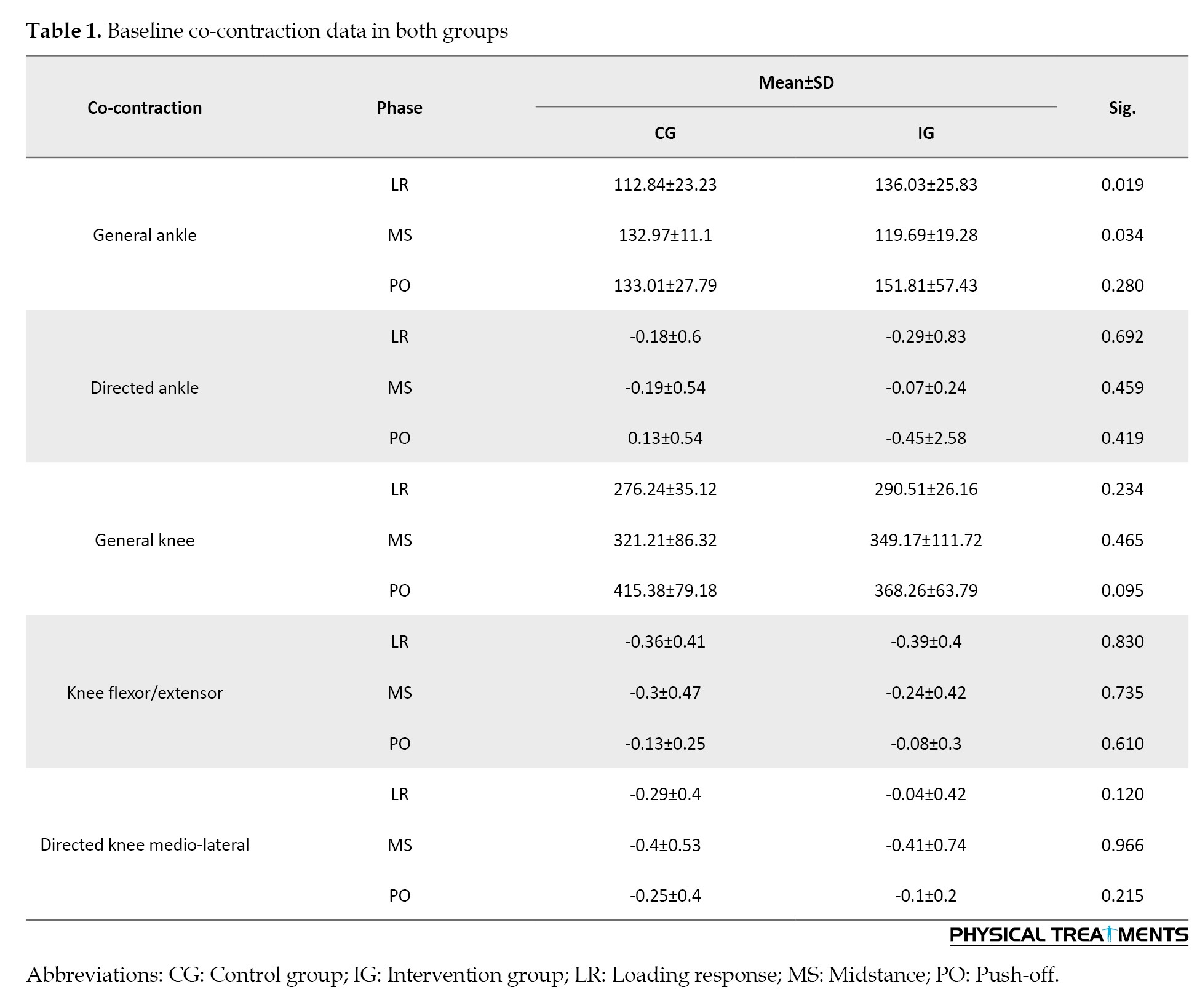
The general ankle co-contraction during the loading response (P=0.019) and mid-stance phases (P=0.034) at baseline showed significant differences, according to the results (Table 1).
The findings showed that “time” had a substantial impact on the overall muscle co-contraction of the knee muscles throughout the loading (P=0.003; ƞ2=0.298), mid-stance (P=0.002; ƞ2=0.305), and push-off (P<0.001; ƞ2=0.84) phases. Additionally, results indicated that “time” had a significant main influence on directed knee flexor/extensor muscle co-contraction during the propulsion phase (P=0.003, ƞ2=0.288) as well as directed knee mediolateral co-contraction during the loading (P=0.003, ƞ2=0.285) and mid-stance (P<0.001, ƞ2=0.46) phases (Table 2).
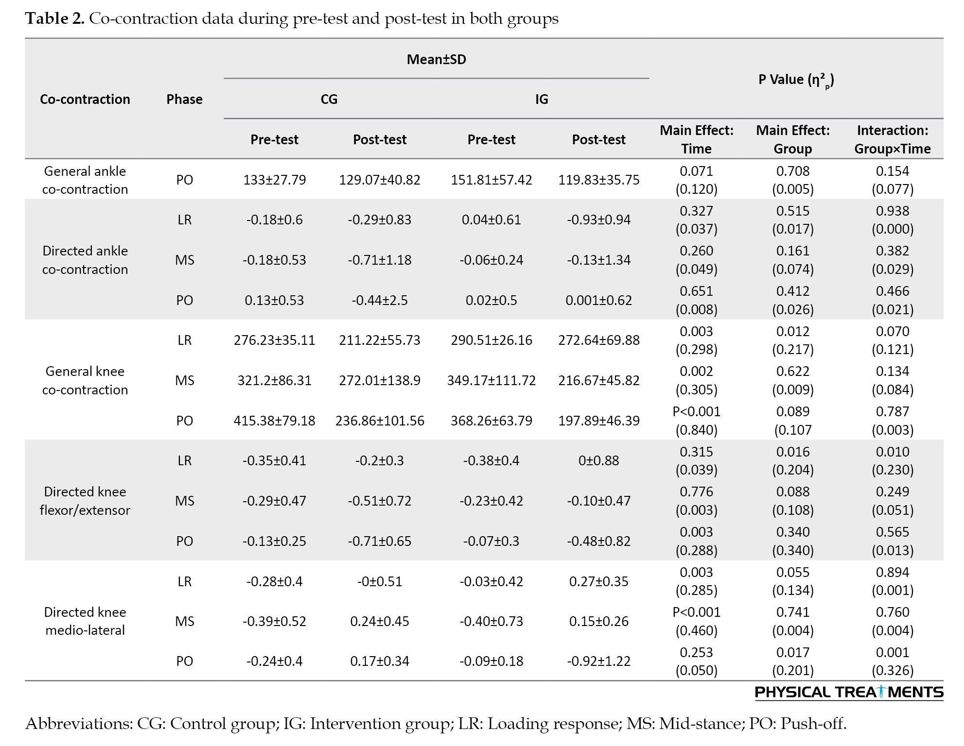
The results showed that “group” had a significant main influence on overall knee muscle co-contraction during the loading phase (P=0.012; ƞ2=0.217). Furthermore, directed knee flexor/extensor muscle co-contraction during the loading phase (P=0.016; ƞ2=0.204) and directed knee medio-lateral muscle co-contraction during push-off (P=0.017; ƞ2=0.201) showed significant main effects of “group” (Table 2).
Directed knee flexor/extensor muscle co-contraction during the loading phase (P=0.010; ƞ2=0.23) and directed knee mediolateral muscle co-contraction during the propulsion phase (P=0.001; ƞ2=0.326) both showed significant group-by-time interactions (Table 2). Post-hoc analysis revealed significantly greater directed knee flexor/extensor co-contraction during the loading period in the IG (but not in the CG). Furthermore, post-hoc analysis revealed a substantial decrease in directed knee mediolateral muscle co-contraction during the propulsion phase in the IG (but not in the CG).
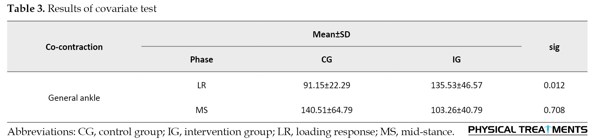
Covariate analysis showed greater general ankle co-contraction at post-test during the loading phase in IG than in CG (P=0.012) (Table 3).
Discussion
This study aimed to assess the effects of sand surface training on knee and ankle muscle co-contraction during gait in individuals with a pronated foot and ACLR. Overall, the findings demonstrated that: I) directed knee flexor/extensor co-contraction increased significantly during the loading phase in the IG; II) directed knee mediolateral co-contraction significantly decreased during the push-off phase in the IG; and III) the overall activation of ankle muscles during the loading phase was higher in the IG than in the CG after the post-test.
Our research results indicated that training on sand had no significant impact on overall knee co-contraction while walking. Knee muscle co-contraction serves to modify joint stability and articular loading [36]. There are two primary forms of knee muscle co-contraction: generalized and directed co-contraction [37]. In generalized co-contraction, both the agonist and antagonist muscles of the knee are activated equally, potentially impacting articular loading [38, 39]. Notably, our research demonstrated a significant increase in directed knee flexor/extensor co-contraction during the loading phase in the IG, while such an increase was not observed in the CG.
The results showed a significant reduction in mediolateral knee muscle co-contraction during the propulsion phase in the IG compared to the CG. This specific form of co-contraction involves the simultaneous activation of both medial agonist and antagonist muscles to assist the lateral muscles in generating adduction moments. Directed co-contraction is believed to help maintain the external moment, thereby preventing condylar lift-off and reducing the load concentration on the medial knee compartment [40]. Our findings indicate that training in sand may help decrease articular stress in the medial knee region. In line with our results, other researchers have suggested that exercises aimed at lowering co-contraction could be beneficial in reducing joint load, given its potential adverse effects on knee stress and disease progression [41, 42].
Muscle co-contraction increases the stiffness of the joint(s) around which the muscles act [43]. During novel situations in which the postural control system is challenged, increased co-contraction may be employed as a strategy to reduce the degrees of freedom the postural control system must organize [44], thereby stabilizing the body’s center of mass and increasing postural stability [45]. However, high levels of co-contraction require greater energy expenditure [46], reducing movement efficiency [47] and contributing to fatigue and potential injury [48]. In addition, increased joint rigidity resulting from co-contraction may hinder accurate balance reactions and/or the ability to update movement strategies quickly [49].
The findings revealed a greater general ankle co-contraction during the loading response in the post-test in the IG compared to the CG. TA and GL are key muscles involved in lifting and lowering the foot during the gait cycle [50, 51]. In line with previous studies, TA and GL exhibited significant variability in activation patterns while walking, including the frequency of each activation mode. Both TA and GL use different activation modalities from one stride to the next. During mid-stance, the ankle’s antagonistic pair does not function in their usual opposing manner; rather than acting solely as an ankle dorsiflexor, research indicates that TA behaves more like an inverter of the foot [52]. The TA and GL muscles collaborate synergistically to regulate the movement of the tibia over the talus bone in the ankle joint. During walking, when the contralateral limb is swinging forward, the TA and GL help decelerate the lower limb’s displacement and maintain balance [53]. Our findings demonstrated that running sand training increased general ankle co-contraction during the loading response at post-test due to training-induced adaptation. Our study results are consistent with earlier research, which found that the short-foot exercise—acting through mechanisms similar to sand training in activating foot muscles—was more effective than traditional methods [54]. The intrinsic muscles of the lower limb play an important role in energy transfer and force production during dynamic activities. These small but powerful muscles act across the joints of the foot and ankle to provide stabilization and precise movement. Thus, sand training, which strengthens the intrinsic muscles, not only improves medial longitudinal arch support and stability but also enhances energy transfer throughout the lower limb [55].
This study has certain limitations that warrant attention. We examined the long-term effects of walking on sand in males with pronated feet and patients who had undergone ACLR. Therefore, it is not possible to extrapolate our findings to females. Future studies are required to determine whether sand walking is a preventive or therapeutic strategy for females with pronated feet. The current study did not investigate walking kinematics. Consequently, it is recommended that future studies investigate how sand-based training influences lower-limb movement patterns during walking.
Conclusion
Based on our findings, we can conclude that sand training may influence the knee joint co-contraction pattern in adult males with pronated feet and those who have undergone. Overall, this study highlights the potential benefits of sand surface training in modulating muscle co-contraction in individuals with pronated feet and ACLR during gait. The findings demonstrate that sand training significantly increased directed knee flexor/extensor co-contraction during the loading phase and reduced mediolateral knee co-contraction during the push-off phase, potentially decreasing medial knee compartment stress. Additionally, sand training enhanced general ankle muscle co-contraction during the loading response, suggesting improved stability and energy transfer within the lower limb. These outcomes emphasize the role of sand training in strengthening intrinsic foot and ankle muscles, thereby contributing to joint stability and efficient dynamic movement.
Ethical Considerations
Compliance with ethical guidelines
This study was approved by the Research Ethics Committee of University of Mohaghegh Ardabili, Ardabil, Iran (Code: IR.UMA.REC.1403.063). Moreover, this study was vregistered by the Iranian Clinical Trial Organization (IRCT), Tehran, Iran (Code: IRCT20170806035517N6). Before participating in the trial, all subjects provided written informed consent.
Funding
This research did not receive any grant from funding agencies in the public, commercial, or non-profit sectors.
Authors' contributions
All authors equally contributed to preparing this article.
Conflict of interest
The authors declared no conflict of interest.
Acknowledgments
The authors express their gratitude to all participants who volunteered for this study.
An intervention group (IG) (n=14), a control group (CG) (n=14), and a CG consisting of 14 individuals. The study participants were divided into these experimental groups using the block randomization approach with a block size of 4. The participants were blinded to their group allocation, and the examiners responsible for data collection were unaware of the group assignments.
The study received ethical approval from the local Ethics Committee prior to its commencement. Moreover, the Iranian Registry of Clinical Trials (IRCT) registered the study. Before participating in the trial, all subjects provided written informed consent. The research followed the guidelines of the CONSORT statement.
Inclusion and exclusion criteria
The inclusion criteria were: a foot posture index score ranging from 6 to 12, prior ACLR, absence of ankle injury within the preceding six months, absence of ankle pain at the time of the study, and capacity to provide informed written consent. A total of 63 individuals met these criteria and were enrolled in the study after providing their informed written consent. The rights and well-being of all participants were duly protected throughout the study.
The exclusion criteria included the inability to walk independently without pain or assistive devices, as well as the presence of cardiac conditions, unstable hypertension, musculoskeletal disorders, or disabilities resulting from stroke, cerebral palsy, polio, rheumatoid arthritis, the use of prosthetic devices, or moderate to advanced OA. Additionally, individuals who had exercised regularly within the previous six months were not eligible.
Assessment of muscle activities
A wireless electromyography (EMG) system was used to quantify muscle activity in the right leg, specifically in the rectus femoris (RF), biceps femoris (BF), vastus lateralis (VL), vastus medialis (VM), and tibialis anterior (TA). These muscles were selected because they play a critical role in the stability and mobility of the lower-limb joints. This system used seven pairs of bipolar silver/silver chloride surface electrodes with a center-to-center distance of 25 mm, an input impedance of 100 MΩ, and a common-mode rejection ratio greater than 110 dB (EMG Preamplifier, Biometrics Ltd., Nine Mile Point Industrial Estate, Newport, UK) [24]. The electrodes were securely attached to the participants’ muscular bellies using a double-sided adhesive tape that included die-cutting and was appropriate for medical use. After being digitally transformed at 1000 Hz, the original EMG signals were Bluetooth-enabled and wirelessly transmitted to a computer for further analysis. Following the recommendations of SENIAM for surface EMG, the skin covering the affected muscles was carefully shaved and cleaned with a 70% ethanol (C2H5OH) solution [25]. According to Dugan et al. (2005), Jafarnezhadgero et al. (2019), Jafarnezhadgero et al., (2021), and Jafarnezhadgero et al. (2021), the walking stance phase has been divided into three separate sub-phases for EMG analyses: loading response occurs from 0–20% of the gait cycle, followed by the mid-stance phase from 20–47%, and the push-off phase, which spans from 47–70% of the gait cycle [23, 26-29]. To standardize the EMG results, the maximum voluntary isometric contraction (MVIC) of each muscle recorded during walking was measured using a handheld dynamometer. Participants were instructed to give their all on the tests [30]. Three test trials were performed, with 1-2 minutes of rest between trials. An isometric belt set at zero velocity and guaranteed to immobilize joints was used to test MVIC. For normalization, the test’s highest recorded MVIC value was used [31].
Biometrics DataLITE software was used to process the EMG data, applying a low-pass filter set to 10-500 Hz. The RMS value for each muscle was divided by the corresponding MVIC measurement, and this quotient was then multiplied by 100 to standardize the EMG signals. The following formulas were used to determine the values of general and directed co-contraction across different walking phases [32].
To determine directed co-contraction, if the mean EMG value of the agonist muscle is greater than that of the antagonist, then directed co-contraction is calculated as:
In directed muscle co-contraction, as the numerical value approaches zero, the level of co-contraction intensifies. Conversely, when the value approaches either 1 or -1, the degree of co-contraction diminishes [32].
Sand walking training protocol
The training regimen included a variety of barefoot workouts, including running nonstop, walking, running in place, galloping, and short-distance sprinting. A 50-minute training session began with a 5-minute warm-up and stretching routine, and each session ended with a 5-minute cool-down [33]. Throughout the sessions, a physiotherapist supervised to ensure proper technique execution and made necessary adjustments to meet the program requirements. The program was the same for both groups; however, the IG group executed it on sand, whereas the CG group performed the task on a stable ground surface. Six days following the last training session, post-tests were administered to participants to ensure they had fully recovered [34]. Throughout the intervention period, participants in both the IG and CG were instructed not to engage in any other forms of exercise. The training activities recommended for both groups included specific exercises to improve their fitness and performance, ensuring strict adherence to the prescribed regimen during the study [35].
Walking, jogging, striding, leaping, galloping, and brief sprints are among the workouts. Each exercise has training characteristics, such as duration, intensity, number of repetitions, distance traveled, and rest intervals.
Walking is performed for 5 minutes. During the first 4 weeks, the intensity is 1.2 m/s, which increases to 1.4 m/s for weeks 5 to 8. The distance covered is 50 meters, and no repetitions or rest periods are specified.
Jogging lasts 20 minutes at an intensity of 2.0 m/s during the first 4 weeks, then increases to 2.5 m/s in weeks 5-8. Similar to walking, the distance is 50 meters, and no repetitions or rest periods are specified.
Striding is performed for 3 minutes at an intensity of 3.5 m/s in the first 4 weeks and 4.5 m/s in the following 4 weeks. The exercise includes two repetitions for the first 4 weeks and 3 repetitions for weeks 5-8, with a 1-minute rest between repetitions. The distance covered per repetition is 50 meters.
Bounding is also a 3-minute exercise with the same intensity progression as striding (3.5 m/s to 4.5 m/s). Similarly, it includes two repetitions for the first 4 weeks and 3 for weeks 5-8, with 1 minute of rest after each. The distance covered is 30 meters per repetition.
Galloping follows the same structure as bounding, with a duration of 3 minutes, an increase in intensity from 3.5 m/s to 4.5 m/s, two repetitions for the first 4 weeks, and 3 for weeks 5-8. The distance covered per repetition is 30 meters, with a 1-minute rest period.
Short sprints last 6 minutes, with the participants running as fast as possible throughout both phases of the program. The first 4 weeks involve 3 repetitions, increasing to 4-5 repetitions for weeks 5-8. Each sprint covers 25 meters, with a 2-minute rest between repetitions.
The descriptions of the MVIC tests for TA, Gas-M, BF, ST, VL, VM, and RF muscles were as follows. Throughout the intervention period, participants in both the IG and the CG were instructed not to engage in any additional exercises. The test protocols for the muscle evaluations are as follows:
TA: Participants sat in a chair with a backrest, maintaining 90 degrees of flexion at the hip, knee, and ankle joints. They were instructed to exert maximal effort in activating the TA against resistance.
Gas-M: Subjects exerted maximal activation of their plantar flexor muscles against resistance while positioned seated on the examination table, with the hip flexed to a 90-degree angle and both the knee and ankle in a neutral alignment.
BF: The participants used their hamstrings at maximum effort against resistance while sitting on a chair, with their knees and hips extended to a 90-degree angle.
ST: Participants maximally engaged their knee flexors against resistance while sitting with their hips and knees flexed to 90 degrees.
VL, VM, and RF: Participants maximally engaged their knee extensors against resistance while sitting on a chair with their hips and knees flexed to a 90-degree angle.
Statistical analyses
We conducted a within-between repeated measures analysis of variance (ANOVA) to examine the effects of time (pre vs post) and group (CG vs IG) on outcomes. The Bonferroni test was used in the post hoc analysis. Eta-squared (η²p) was transformed into Cohen’s d to estimate effect sizes; values less than 0.5, 0.5–0.8, and d≥0.8 indicate minor effects, medium effects, and large effects, respectively. All analyses were conducted using SPSS software, version 26.0, with a significance threshold of P<0.05.
Results
The general ankle co-contraction during the loading response (P=0.019) and mid-stance phases (P=0.034) at baseline showed significant differences, according to the results (Table 1).
The findings showed that “time” had a substantial impact on the overall muscle co-contraction of the knee muscles throughout the loading (P=0.003; ƞ2=0.298), mid-stance (P=0.002; ƞ2=0.305), and push-off (P<0.001; ƞ2=0.84) phases. Additionally, results indicated that “time” had a significant main influence on directed knee flexor/extensor muscle co-contraction during the propulsion phase (P=0.003, ƞ2=0.288) as well as directed knee mediolateral co-contraction during the loading (P=0.003, ƞ2=0.285) and mid-stance (P<0.001, ƞ2=0.46) phases (Table 2).
The results showed that “group” had a significant main influence on overall knee muscle co-contraction during the loading phase (P=0.012; ƞ2=0.217). Furthermore, directed knee flexor/extensor muscle co-contraction during the loading phase (P=0.016; ƞ2=0.204) and directed knee medio-lateral muscle co-contraction during push-off (P=0.017; ƞ2=0.201) showed significant main effects of “group” (Table 2).
Directed knee flexor/extensor muscle co-contraction during the loading phase (P=0.010; ƞ2=0.23) and directed knee mediolateral muscle co-contraction during the propulsion phase (P=0.001; ƞ2=0.326) both showed significant group-by-time interactions (Table 2). Post-hoc analysis revealed significantly greater directed knee flexor/extensor co-contraction during the loading period in the IG (but not in the CG). Furthermore, post-hoc analysis revealed a substantial decrease in directed knee mediolateral muscle co-contraction during the propulsion phase in the IG (but not in the CG).
Covariate analysis showed greater general ankle co-contraction at post-test during the loading phase in IG than in CG (P=0.012) (Table 3).
Discussion
This study aimed to assess the effects of sand surface training on knee and ankle muscle co-contraction during gait in individuals with a pronated foot and ACLR. Overall, the findings demonstrated that: I) directed knee flexor/extensor co-contraction increased significantly during the loading phase in the IG; II) directed knee mediolateral co-contraction significantly decreased during the push-off phase in the IG; and III) the overall activation of ankle muscles during the loading phase was higher in the IG than in the CG after the post-test.
Our research results indicated that training on sand had no significant impact on overall knee co-contraction while walking. Knee muscle co-contraction serves to modify joint stability and articular loading [36]. There are two primary forms of knee muscle co-contraction: generalized and directed co-contraction [37]. In generalized co-contraction, both the agonist and antagonist muscles of the knee are activated equally, potentially impacting articular loading [38, 39]. Notably, our research demonstrated a significant increase in directed knee flexor/extensor co-contraction during the loading phase in the IG, while such an increase was not observed in the CG.
The results showed a significant reduction in mediolateral knee muscle co-contraction during the propulsion phase in the IG compared to the CG. This specific form of co-contraction involves the simultaneous activation of both medial agonist and antagonist muscles to assist the lateral muscles in generating adduction moments. Directed co-contraction is believed to help maintain the external moment, thereby preventing condylar lift-off and reducing the load concentration on the medial knee compartment [40]. Our findings indicate that training in sand may help decrease articular stress in the medial knee region. In line with our results, other researchers have suggested that exercises aimed at lowering co-contraction could be beneficial in reducing joint load, given its potential adverse effects on knee stress and disease progression [41, 42].
Muscle co-contraction increases the stiffness of the joint(s) around which the muscles act [43]. During novel situations in which the postural control system is challenged, increased co-contraction may be employed as a strategy to reduce the degrees of freedom the postural control system must organize [44], thereby stabilizing the body’s center of mass and increasing postural stability [45]. However, high levels of co-contraction require greater energy expenditure [46], reducing movement efficiency [47] and contributing to fatigue and potential injury [48]. In addition, increased joint rigidity resulting from co-contraction may hinder accurate balance reactions and/or the ability to update movement strategies quickly [49].
The findings revealed a greater general ankle co-contraction during the loading response in the post-test in the IG compared to the CG. TA and GL are key muscles involved in lifting and lowering the foot during the gait cycle [50, 51]. In line with previous studies, TA and GL exhibited significant variability in activation patterns while walking, including the frequency of each activation mode. Both TA and GL use different activation modalities from one stride to the next. During mid-stance, the ankle’s antagonistic pair does not function in their usual opposing manner; rather than acting solely as an ankle dorsiflexor, research indicates that TA behaves more like an inverter of the foot [52]. The TA and GL muscles collaborate synergistically to regulate the movement of the tibia over the talus bone in the ankle joint. During walking, when the contralateral limb is swinging forward, the TA and GL help decelerate the lower limb’s displacement and maintain balance [53]. Our findings demonstrated that running sand training increased general ankle co-contraction during the loading response at post-test due to training-induced adaptation. Our study results are consistent with earlier research, which found that the short-foot exercise—acting through mechanisms similar to sand training in activating foot muscles—was more effective than traditional methods [54]. The intrinsic muscles of the lower limb play an important role in energy transfer and force production during dynamic activities. These small but powerful muscles act across the joints of the foot and ankle to provide stabilization and precise movement. Thus, sand training, which strengthens the intrinsic muscles, not only improves medial longitudinal arch support and stability but also enhances energy transfer throughout the lower limb [55].
This study has certain limitations that warrant attention. We examined the long-term effects of walking on sand in males with pronated feet and patients who had undergone ACLR. Therefore, it is not possible to extrapolate our findings to females. Future studies are required to determine whether sand walking is a preventive or therapeutic strategy for females with pronated feet. The current study did not investigate walking kinematics. Consequently, it is recommended that future studies investigate how sand-based training influences lower-limb movement patterns during walking.
Conclusion
Based on our findings, we can conclude that sand training may influence the knee joint co-contraction pattern in adult males with pronated feet and those who have undergone. Overall, this study highlights the potential benefits of sand surface training in modulating muscle co-contraction in individuals with pronated feet and ACLR during gait. The findings demonstrate that sand training significantly increased directed knee flexor/extensor co-contraction during the loading phase and reduced mediolateral knee co-contraction during the push-off phase, potentially decreasing medial knee compartment stress. Additionally, sand training enhanced general ankle muscle co-contraction during the loading response, suggesting improved stability and energy transfer within the lower limb. These outcomes emphasize the role of sand training in strengthening intrinsic foot and ankle muscles, thereby contributing to joint stability and efficient dynamic movement.
Ethical Considerations
Compliance with ethical guidelines
This study was approved by the Research Ethics Committee of University of Mohaghegh Ardabili, Ardabil, Iran (Code: IR.UMA.REC.1403.063). Moreover, this study was vregistered by the Iranian Clinical Trial Organization (IRCT), Tehran, Iran (Code: IRCT20170806035517N6). Before participating in the trial, all subjects provided written informed consent.
Funding
This research did not receive any grant from funding agencies in the public, commercial, or non-profit sectors.
Authors' contributions
All authors equally contributed to preparing this article.
Conflict of interest
The authors declared no conflict of interest.
Acknowledgments
The authors express their gratitude to all participants who volunteered for this study.
References
- Tsai LC, McLean S, Colletti PM, Powers CM. Greater muscle co‐contraction results in increased tibiofemoral compressive forces in females who have undergone anterior cruciate ligament reconstruction. Journal of Orthopaedic Research. 2012; 30(12):2007-14. [DOI:10.1002/jor.22176] [PMID]
- Ledoux WR, Hillstrom HJ. The distributed plantar vertical force of neutrally aligned and pes planus feet. Gait & Posture. 2002; 15(1):1-9. [DOI:10.1016/S0966-6362(01)00165-5] [PMID]
- Tajdini kakavandi H, Sadeghi H, Abasi A. [Investigating ground reaction forces and electromyography muscle activity of lower extremity during the stance phase of walking in active male with and without genu varum deformity in Kharazmi University in 2017: A descriptive study (Persian)]. Journal of Rafsanjan University of Medical Sciences. 2019; 18(7):675-88. [Link]
- Rethlefsen SA, Blumstein G, Kay RM, Dorey F, Wren TA. Prevalence of specific gait abnormalities in children with cerebral palsy revisited: Influence of age, prior surgery, and Gross Motor Function Classification System level. Developmental Medicine & Child Neurology. 2017; 59(1):79-88. [DOI:10.1111/dmcn.13205] [PMID]
- Fasihi A, Siahkouhian M, Sheikhalizade H, Jafarnezhadgero A. [Effect of fatigue at the heart rate deflection point on the frequency components of ground reaction forces during walking in individuals with pronated feet (Persian)]. The Scientific Journal of Rehabilitation Medicine. 2020; 9(2):228-38. [Link]
- Pinczewski LA, Lyman J, Salmon LJ, Russell VJ, Roe J, Linklater J. A 10-year comparison of anterior cruciate ligament reconstructions with hamstring tendon and patellar tendon autograft: A controlled, prospective trial. The American Journal of Sports Medicine. 2007; 35(4):564-74. [DOI:10.1177/0363546506296042] [PMID]
- Sajovic M, Vengust V, Komadina R, Tavcar R, Skaza K. A prospective, randomized comparison of semitendinosus and gracilis tendon versus patellar tendon autografts for anterior cruciate ligament reconstruction: Five-year follow-up. The American Journal of Sports Medicine. 2006; 34(12):1933-40. [DOI:10.1177/0363546506290726] [PMID]
- Bush-Joseph CA, Hurwitz DE, Patel RR, Bahrani Y, Garretson R, Bach Jr BR, et al. Dynamic function after anterior cruciate ligament reconstruction with autologous patellar tendon. The American Journal of Sports Medicine. 2001; 29(1):36-41. [DOI:10.1177/03635465010290011101] [PMID]
- Hall M, Stevermer CA, Gillette JC. Gait analysis post anterior cruciate ligament reconstruction: Knee osteoarthritis perspective. Gait & posture. 2012; 36(1):56-60. [DOI:10.1016/j.gaitpost.2012.01.003] [PMID]
- Nyland J, Klein S, Caborn DN. Lower extremity compensatory neuromuscular and biomechanical adaptations 2 to 11 years after anterior cruciate ligament reconstruction. Arthroscopy: The Journal of Arthroscopic & Related Surgery. 2010; 26(9):1212-25. [DOI:10.1016/j.arthro.2010.01.003] [PMID]
- Ferber R, Osternig LR, Woollacott MH, Wasielewski NJ, Lee JH. Gait mechanics in chronic ACL deficiency and subsequent repair. Clinical Biomechanics. 2002; 17(4):274-85. [DOI:10.1016/S0268-0033(02)00016-5] [PMID]
- Ortiz A, Olson S, Libby CL, Trudelle-Jackson E, Kwon YH, Etnyre B, et al. Landing mechanics between noninjured women and women with anterior cruciate ligament reconstruction during 2 jump tasks. The American Journal of Sports Medicine. 2008; 36(1):149-57. [DOI:10.1177/0363546507307758] [PMID]
- Vairo GL, Myers JB, Sell TC, Fu FH, Harner CD, Lephart SM. Neuromuscular and biomechanical landing performance subsequent to ipsilateral semitendinosus and gracilis autograft anterior cruciate ligament reconstruction. Knee Surgery, Sports Traumatology, Arthroscopy. 2008; 16(1):2-14. [DOI:10.1007/s00167-007-0427-4] [PMID]
- Liu W, Maitland ME. The effect of hamstring muscle compensation for anterior laxity in the ACL-deficient knee during gait. Journal of Biomechanics. 2000; 33(7):871-9. [DOI:10.1016/S0021-9290(00)00047-6] [PMID]
- Drechsler WI, Cramp MC, Scott OM. Changes in muscle strength and EMG median frequency after anterior cruciate ligament reconstruction. European Journal of Applied Physiology. 2006; 98(6):613-23. [DOI:10.1007/s00421-006-0311-9] [PMID]
- Smith HC, Vacek P, Johnson RJ, Slauterbeck JR, Hashemi J, Shultz S, et al. Risk factors for anterior cruciate ligament injury: A review of the literature - part 1: Neuromuscular and anatomic risk. Sports Health. 2012; 4(1):69-78. [DOI:10.1177/1941738111428281] [PMID]
- Aenumulapalli A, Kulkarni MM, Gandotra AR. Prevalence of Flexible Flat Foot in Adults: A Cross-sectional Study. Journal of Clinical and Diagnostic Research. 2017; 11(6):Ac17-20. [DOI:10.7860/JCDR/2017/26566.10059] [PMID]
- Beckett ME, Massie DL, Bowers KD, Stoll DA. Incidence of hyperpronation in the ACL Injured knee: A clinical perspective. Journal of Athletic Training. 1992; 27(1):58-62. [PMID]
- Mokhtari Malekabadi A, Jafarnezhadgero A, Gghorbanlou F. The effects of changing footstrike pattern on the amplitude and frequency spectrum of ground reaction forces during running in individuals with pronated feet. Physical Treatments-Specific Physical Therapy Journal. 2020; 10(4):205-12. [DOI:10.32598/ptj.10.4.348.2]
- Mousavi SH, Khorramroo F, Jafarnezhadgero A. Gait retraining targeting foot pronation: A systematic review and meta-analysis. Plos One. 2024; 19(3):e0298646. [DOI:10.1371/journal.pone.0298646] [PMID]
- Sheikhalizade H, Jafarnezhadgero A, Imanibrouj S, Mafi M, Asheghan M. Effect of Sand Training on Walking Mechanics in Men With Anterior Cruciate Ligament Reconstruction and Pronated Feet. Physical Treatments - Specific Physical Therapy. 2024; 14(3):241-52. [DOI:10.32598/ptj.14.3.591.1]
- de Lacey J, Brughelli M, McGuigan M, Hansen K, Samozino P, Morin JB. The effects of tapering on power-force-velocity profiling and jump performance in professional rugby league players. The Journal of Strength & Conditioning Research. 2014; 28(12):3567-70. [DOI:10.1519/JSC.0000000000000572] [PMID]
- Jafarnezhadgero A, Fatollahi A, Sheykholeslami A, Dionisio VC, Akrami M. Long-term training on sand changes lower limb muscle activities during running in runners with over-pronated feet. Biomedical Engineering Online 2021; 20(1):118. [DOI:10.1186/s12938-021-00955-8] [PMID]
- Sheikhalizade H, Imani F, Jafarnezhadgero A, Imani Brouj S. Effect of exercises on the sand on the frequency content of the lower limb muscles in individuals with anterior cruciate ligament reconstruction and pronated feet during walking. Physical Treatments - Specific Physical Therapy Journal. 2024; 14(2):93-100. [DOI:10.32598/ptj.14.2.348.9]
- Mafi M, Sheikhalizade H, Jafarnezhadgero A, Asheghan M. Investigating the effect of sand training on running mechanics in individuals with anterior cruciate ligament reconstruction and pronated feet. Gait & Posture. 2023; 104:129-34. [DOI:10.1016/j.gaitpost.2023.06.009] [PMID]
- Jafarnezhadgero A, Ghorbanloo F, Fatollahi A, Dionisio VC, Granacher U. Effects of an elastic resistance band exercise program on kinetics and muscle activities during walking in young adults with genu valgus: A double-blinded randomized controlled trial. Clinical Biomechanics. 2021; 81:105215. [DOI:10.1016/j.clinbiomech.2020.105215] [PMID]
- Whittle MW. Gait analysis: An introduction. Oxford: Butterworth-Heinemann; 2014. [Link]
- Dugan SA, Bhat KP. Biomechanics and analysis of running gait. Physical Medicine and Rehabilitation Clinics of North America. 2005; 16(3):603-21. [DOI:10.1016/j.pmr.2005.02.007] [PMID]
- Jafarnezhadgero A, Fatollahi A, Amirzadeh N, Siahkouhian M, Granacher U. Ground reaction forces and muscle activity while walking on sand versus stable ground in individuals with pronated feet compared with healthy controls. Plos One. 2019; 14(9):e0223219. [DOI:10.1371/journal.pone.0223219] [PMID]
- Jafarnezhadgero A, Sheikhalizadeh H, Salahi Movasagh S. [Running ground reaction force characteristics in children with forward head posture compared to healthy control ones (Persian)]. The Scientific Journal of Rehabilitation Medicine. 2018; 7(4):217-26. [Link]
- Jafarnezhadgero AA, Dehghani M, Abdollahpourdarvishani M, Sheikhalizadeh H, Akrami M. Effect of textured foot orthoses on walking plantar pressure variables in children with autism spectrum disorders. Journal of Biomechanics. 2021; 129:110775. [DOI:10.1016/j.jbiomech.2021.110775] [PMID]
- Heiden TL, Lloyd DG, Ackland TR. Knee joint kinematics, kinetics and muscle co-contraction in knee osteoarthritis patient gait. Clinical Biomechanics. 2009; 24(10):833-41. [DOI:10.1016/j.clinbiomech.2009.08.005] [PMID]
- Kortge CS. Healing walks for hard times: quiet your mind, strengthen your body, and get your life back. Colorado: Shambhala Publications; 2010. [Link]
- Jafarnezhadgero AA, Sorkhe E, Oliveira AS. Motion-control shoes help maintaining low loading rate levels during fatiguing running in pronated female runners. Gait & Posture. 2019; 73:65-70. [DOI:10.1016/j.gaitpost.2019.07.133] [PMID]
- Anderson M, Barnum M. Foundations of athletic training: Prevention, assessment, and management. Netherlands: Wolters Kluwer Health; 2021. [Link]
- Hubley-Kozey CL, Deluzio KJ, Landry SC, McNutt JS, Stanish WD. Neuromuscular alterations during walking in persons with moderate knee osteoarthritis. Journal of Electromyography and Kinesiology. 2006; 16(4):365-78. [DOI:10.1016/j.jelekin.2005.07.014] [PMID]
- Lloyd DG, Besier TF, Winby CR, Buchanan TS. Neuromusculoskeletal modelling and simulation of tissue load in the lower extremities. Handbook of Biomechanics and Human Movement Science. New York: Routledge; 2008. [Link]
- Andriacchi TP, Ogle JA, Galante JO. Walking speed as a basis for normal and abnormal gait measurements. Journal of Biomechanics. 1977; 10(4):261-8. [DOI:10.1016/0021-9290(77)90049-5] [PMID]
- Zhang LQ, Xu D, Wang G, Hendrix RW. Muscle strength in knee varus and valgus. Medicine & Science in Sports & Exercise. 2001; 33(7):1194-9. [DOI:10.1097/00005768-200107000-00018] [PMID]
- Schipplein OD, Andriacchi TP. Interaction between active and passive knee stabilizers during level walking. Journal of Orthopaedic Research. 1991; 9(1):113-9. [DOI:10.1002/jor.1100090114] [PMID]
- Hortobágyi T, Westerkamp L, Beam S, Moody J, Garry J, Holbert D, et al. Altered hamstring-quadriceps muscle balance in patients with knee osteoarthritis. Clinical Biomechanics. 2005; 20(1):97-104. [DOI:10.1016/j.clinbiomech.2004.08.004] [PMID]
- Lewek MD, Rudolph KS, Snyder-Mackler L. Control of frontal plane knee laxity during gait in patients with medial compartment knee osteoarthritis. Osteoarthritis and Cartilage. 2004; 12(9):745-51. [DOI:10.1016/j.joca.2004.05.005] [PMID]
- Granata K, Wilson S, Massimini A, Gabriel R. Active stiffness of the ankle in response to inertial and elastic loads. Journal of Electromyography and Kinesiology. 2004; 14(5):599-609. [DOI:10.1016/j.jelekin.2004.03.005] [PMID]
- Tucker MG, Kavanagh JJ, Barrett RS, Morrison S. Age-related differences in postural reaction time and coordination during voluntary sway movements. Human Movement Science. 2008; 27(5):728-37. [DOI:10.1016/j.humov.2008.03.002] [PMID]
- Nagai K, Yamada M, Uemura K, Yamada Y, Ichihashi N, Tsuboyama T. Differences in muscle coactivation during postural control between healthy older and young adults. Archives of Gerontology and Geriatrics. 2011; 53(3):338-43. [DOI:10.1016/j.archger.2011.01.003] [PMID]
- Cholewicki J, McGill SM. Mechanical stability of the in vivo lumbar spine: Implications for injury and chronic low back pain. Clinical Biomechanics. 1996; 11(1):1-15. [DOI:10.1016/0268-0033(95)00035-6] [PMID]
- Gregory DE, Narula S, Howarth SJ, Russell C, Callaghan JP. The effect of fatigue on trunk muscle activation patterns and spine postures during simulated firefighting tasks. Ergonomics. 2008; 51(7):1032-41. [DOI:10.1080/00140130701813178] [PMID]
- Kumar S. Theories of musculoskeletal injury causation. Ergonomics. 2001; 44(1):17-47. [DOI:10.1080/00140130120716] [PMID]
- Freyler K, Krause A, Gollhofer A, Ritzmann R. Specific stimuli induce specific adaptations: sensorimotor training vs. reactive balance training. PLoS One. 2016; 11(12):e0167557. [DOI:10.1371/journal.pone.0167557] [PMID]
- Winter DA. Biomechanics and motor control of human movement. Hoboken: John Wiley & Sons; 2009. [DOI:10.1002/9780470549148]
- Di Nardo F, Fioretti S. EMG-based analysis of treadmill and ground walking in distal leg muscles. In: Roa Romero L, editor. XIII Mediterranean Conference on Medical and Biological Engineering and Computing 2013. IFMBE Proceedings, vol 41. Cham: Springer; 2014. [DOI:10.1007/978-3-319-00846-2_151]
- Di Nardo F, Mengarelli A, Maranesi E, Burattini L, Fioretti S. Assessment of the ankle muscle co-contraction during normal gait: A surface electromyography study. Journal of Electromyography and Kinesiology. 2015; 25(2):347-54. [DOI:10.1016/j.jelekin.2014.10.016] [PMID]
- Mengarelli A, Maranesi E, Burattini L, Fioretti S, Di Nardo F. Co-contraction activity of ankle muscles during walking: A gender comparison. Biomedical Signal Processing and Control. 2017; 33:1-9. [DOI:10.1016/j.bspc.2016.11.010]
- Jung DY, Kim MH, Koh EK, Kwon OY, Cynn HS, Lee WH. A comparison in the muscle activity of the abductor hallucis and the medial longitudinal arch angle during toe curl and short foot exercises. Physical Therapy in Sport. 2011; 12(1):30-5. [DOI:10.1016/j.ptsp.2010.08.001] [PMID]
- Sulowska I, Mika A, Oleksy Ł, Stolarczyk A. The influence of plantar short foot muscle exercises on the lower extremity muscle strength and power in proximal segments of the kinematic chain in long-distance runners. BioMed Research International. 2019; 2019:6947273. [DOI:10.1155/2019/6947273] [PMID]
Type of Study: Research |
Subject:
General
Received: 2024/08/21 | Accepted: 2024/12/22 | Published: 2026/06/23
Received: 2024/08/21 | Accepted: 2024/12/22 | Published: 2026/06/23
Send email to the article author
| Rights and permissions | |
 |
This work is licensed under a Creative Commons Attribution-NonCommercial 4.0 International License. |

This is an open access article distributed under the terms of the Creative Commons Attribution-NonCommercial 4.0 (CC-By-NC 4.0), which permits use, distribution, and reproduction in any medium, provided the original work is properly cited and is not used for commercial purposes.
Contact Information