Thu, Oct 22, 2026
Volume 16, Issue 3 (Summer 2026)
PTJ 2026, 16(3): 283-294 |
Back to browse issues page



Download citation:
BibTeX | RIS | EndNote | Medlars | ProCite | Reference Manager | RefWorks
Send citation to:


BibTeX | RIS | EndNote | Medlars | ProCite | Reference Manager | RefWorks
Send citation to:
Moghadas Tabrizi Y, Kateb M Y, Rezaei Z. A Single Session of Transcranial Direct Current Stimulation-imagery for Balance in Athletes With Chronic Ankle Instability. PTJ 2026; 16 (3) :283-294
URL: http://ptj.uswr.ac.ir/article-1-698-en.html
URL: http://ptj.uswr.ac.ir/article-1-698-en.html
1- Department of Sport Injuries and Corrective Exercise, Faculty of Sports and Health Sciences, University of Tehran, Tehran, Iran.
2- Department of Behavioral and Cognitive Sciences, Faculty of Sports and Health Sciences, University of Tehran, Tehran, Iran.
2- Department of Behavioral and Cognitive Sciences, Faculty of Sports and Health Sciences, University of Tehran, Tehran, Iran.
Keywords: Balance, Chronic ankle instability (CAI), Motor imagery practice (MIP), Postural control, Transcranial direct current stimulation (tDCS)
Full-Text [PDF 570 kb]
(886 Downloads)
| Abstract (HTML) (2578 Views)
Full-Text: (103 Views)
Introduction
An acute ankle sprain represents one of the most prevalent musculoskeletal injuries in physically active populations. Approximately one-third of those who sustain such sprains may develop chronic ankle instability (CAI), characterized by recurrent ankle sprains and a subjective sensation of the joint “giving way” [1]. This condition is commonly associated with functional deficits, including impaired balance, diminished proprioceptive acuity, and altered neuromuscular control [2]. A previous study identified static and dynamic balance as key variables commonly assessed and targeted in CAI rehabilitation [3]. The somatosensory system is critical in postural and balance control by integrating sensory input with the central nervous system (CNS). Mechanoreceptors and joint sensory information, transmitted by peripheral nerves to the CNS, are essential for maintaining balance [4]. CAI alters sensory receptors and proprioception, leading to impaired transmission of information to the cerebral cortex and resulting in balance deficits [5]. These impairments in postural control can reduce functional independence, limit daily activities, and negatively affect quality of life [6]. Therefore, restoring postural function is a central goal in rehabilitation programs for individuals with significant motor impairments [7]. Conventional rehabilitation for CAI primarily targets peripheral neuromuscular elements [8]. However, research has also linked reduced cortical activation to biomechanical impairments and a heightened likelihood of recurrent ankle sprains [9]. Integrating approaches that stimulate central neural pathways alongside peripheral neuromuscular interventions may enhance recovery outcomes in individuals with CAI. Techniques such as action observation and motor imagery (MI) have shown promise in promoting balance and improving postural control [10, 11].
MI, mental rehearsal of movements without physical execution, is an effective intervention for motor skill development and rehabilitation. Extensive research indicates repeated motor imagery practice (MIP) significantly benefits athletic performance and physical rehabilitation [12]. For instance, MIP has been shown to improve motor function in postural control tasks, such as weight shifting, by reducing the time required to execute the movement [13]. While physical training typically yields more pronounced improvements, MIP has proven effective at facilitating the acquisition of postural control skills and improving dynamic balance in unpredictable environments [14]. Additionally, MIP activates brain regions involved in gait planning, making it particularly valuable for populations with balance deficits, such as older adults and stroke survivors [15]. MIP enhances dynamic postural control and stabilizes movement by engaging neural pathways similar to those activated during physical exercise, particularly in the primary motor cortex (M1). Neuroimaging studies have demonstrated that MIP promotes activity-dependent plasticity, which is essential for motor learning, as it parallels the increases in neural excitability observed during physical training [16].
Transcranial direct current stimulation (tDCS) is another noteworthy non-invasive brain stimulation method that alters cortical excitability and facilitates motor learning. Anodal tDCS increases neural excitability and enhances interneuronal communication during motor tasks, facilitating learning. This stimulation improves performance on sequential and visuomotor tasks, reduces reaction times, and enhances functional abilities [17, 18]. Prior studies have shown that tDCS can augment motor learning, particularly in populations requiring rehabilitative care or skill acquisition. Similar improvements in balance and gait performance have been observed with tDCS interventions, comparable to those observed with physical exercises [19]. Recently, tDCS has been combined with various therapeutic modalities, such as physical therapy, virtual reality, and mindfulness, to optimize functional outcomes by facilitating neuroplasticity [20, 21].
Using anodal tDCS over the right dorsolateral prefrontal cortex (DLPFC) during MI has enhanced Mu desynchronization, a neural marker of motor execution and imagery [22]. This suggests that anodal tDCS can augment the neuroplastic effects of MIP, thereby enhancing performance. For instance, Yavari et al. reported that the simultaneous application of tDCS and mindfulness training led to significant improvements in self-control and emotion regulation among skilled badminton players, with the combined approach proving more effective than mindfulness alone [23]. Similarly, Matsumoto et al. found greater performance improvements in sequential tasks when MIP was paired with anodal tDCS than either intervention alone [24]. While prior research has explored the individual effects of MIP and tDCS on motor performance, the integrated application of these two approaches—especially with stimulation targeting the DLPFC—remains underexplored in the context of balance regulation among athletes experiencing CAI. Given the DLPFC’s involvement in executive functioning, attentional processes, and motor coordination, it is a potentially valuable neural target for enhancing MIP outcomes via tDCS-induced modulation [25, 26]. Moreover, effective balance regulation is a key component in the rehabilitation of individuals with CAI. Nonetheless, the neural mechanisms underlying cognitive-motor integration achieved through the combined use of MIP and DLPFC-targeted stimulation remain insufficiently understood. To address this, the present study investigates whether a single session of anodal tDCS over the right DLPFC can amplify the effects of MIP on both static and dynamic balance in physically active individuals with CAI.
Materials and Methods
The current study utilized a double-blind, randomized clinical trial with a pre-test-post-test design and a control (sham) group. Fourteen right-handed athletes (mean age: 24.5±2.62 years; seven females) who had sustained an ankle sprain more than a year prior and reported experiencing at least two episodes of ankle “giving way” on the same side were recruited for the study [27], and physician-diagnosed CAI were randomly selected based on predefined inclusion criteria. Participants were recruited from the Faculty of Sport Sciences and Health at the University of Tehran. Before initiating the intervention, all subjects completed written informed consent for voluntary participation and submitted demographic, sports, and medical history data. Participants were excluded if they had a history of seizures, any neurological or psychiatric conditions, previous lower limb surgery or fractures, or if they were concurrently involved in other intervention programs. The Ethics Committee of the Faculty of Sport Sciences and Health at the University of Tehran approved the study protocol. Baseline assessments were conducted using the Biodex Balance System (BBS), in which participants performed both static and dynamic balance tests to establish pre-intervention balance metrics. Participants were then randomly assigned to one of two groups: The anodal stimulation group (MI + AtDCS) or the imagery sham stimulation group (MI + StDCS), each consisting of seven individuals. The intervention included an MI session and tDCS targeting the DLPFC. All experimental procedures, including the intervention and subsequent assessments, were conducted at the laboratory of the Faculty of Sport Sciences and Health. Participants were randomly allocated to the experimental or control group using a computer-generated random number sequence. The study was double-blind, with both the participants and the assessors unaware of group assignments. After the intervention, post-test assessments were performed to evaluate dynamic and static balance using the BBS. The BBS was selected due to its high reliability and validity in assessing both static and dynamic balance. Given the study’s focus on balance performance in athletes with CAI and to ensure methodological clarity and control, a single outcome measure was used. This allowed for a focused evaluation of the intervention’s effectiveness without introducing confounding variability from multiple assessment tools.
Study design
This experiment employed a randomized, double-blind design with placebo control involving sham and anodal stimulation. Participants were randomly allocated into two groups: imagery anodal stimulation (MI-AtDCS) and imagery sham stimulation (MI-StDCS), with seven participants in each group.
tDCS
A tDCS device (ActivaDose) was used to administer the stimulation protocol. For electrode placement, a 5×5 cm saline-soaked sponge served as the anodal electrode and was positioned over the left DLPFC (F3), The cathodal electrode, identical in size, was positioned on the right forehead. The stimulation protocol involved delivering a direct current of 1.5 mA for a single 15-minute session [23]. For the sham condition, the device delivered the current for 60 seconds before turning off to mimic stimulation without maintaining active current flow, thereby ensuring participant blinding to the condition.
MI
The MI exercise program consisted of a guided imagery procedure tailored to balance tasks. Participants were first instructed to visualize, step by step, the process of maintaining static balance as assessed during the pre-test using the BBS, while focusing on executing the task positively. This visualization lasted approximately two minutes, followed by a one-minute rest period, and was repeated twice. After completing the static balance imagery phase, the participants engaged in the dynamic balance imagery procedure, which followed the same protocol as the static balance exercise. The total imaging and rest duration for static and dynamic balance exercises were 12 minutes [13]. Before the guided imagery exercises began, participants received tDCS stimulation for an initial three-minute period without performing any specific activity, allowing them to adapt to the stimulation protocol.
Assessments
The BBS is reliable for evaluating static and dynamic balance in clinical and athletic populations. The BBS measures participants’ ability to maintain stability on a fixed platform during static balance assessments. Dynamic balance assessments require participants to respond to multi-directional platform tilts, thereby evaluating functional stability and proprioception [28]. These metrics are essential for identifying balance deficits that may increase injury risk and monitoring improvements following interventions. The BBS has demonstrated high test, re-test reliability for dynamic balance measurements in injured and non-injured athletes [29]. This study used the BBS (manufactured in the USA, 2018) to measure static and dynamic balance. Postural maintenance time during each assessment was set at 30 s, followed by a 60-s rest period. These durations were selected based on previously established protocols to ensure adequate time for balance evaluation while minimizing fatigue [30]. Three indices—overall stability, anterior-posterior stability, and medial-lateral stability—were used to assess balance. Each test was repeated three times, with the average values recorded for analysis.
Statistics
Data analysis was conducted using paired-sample t-tests to evaluate within-group difference measurements. For between-group comparisons, analysis of covariance (ANCOVA) was performed to adjust for baseline differences. To summarize the data, descriptive statistics were computed for all variables, including Means±SD. Data were analyzed using SPSS software, version 26.0 with statistical significance at P<0.05.
Results
All 14 participants (n=14) completed the study protocol. Table 1 presents a summary of their demographic information.
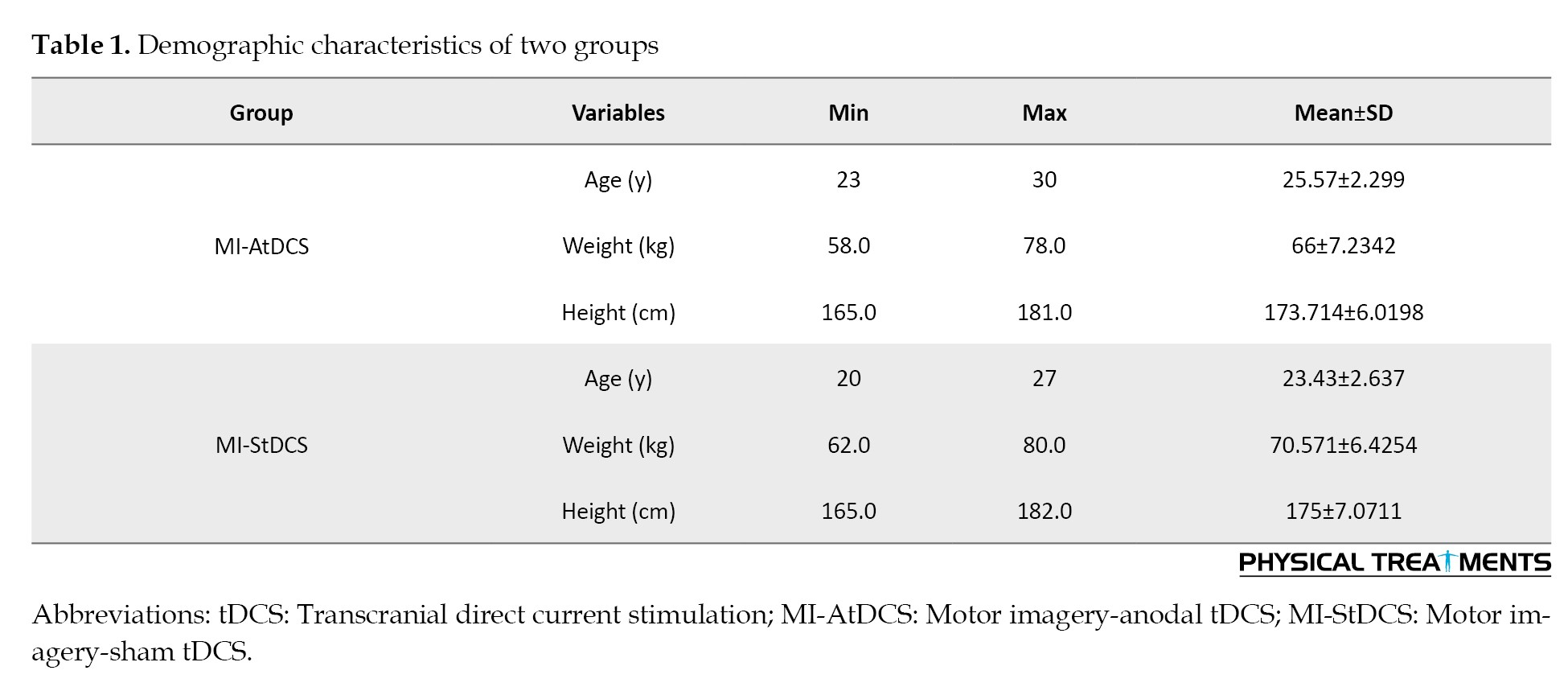
No statistically significant differences were observed between the groups regarding age, body weight, or height.
Table 2 presents the descriptive statistics for static and dynamic balance measures, reported separately for the pre-test and post-test phases.
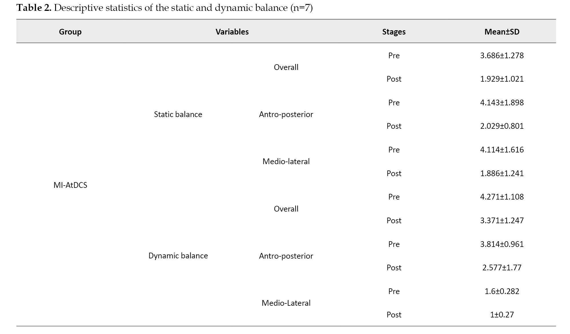
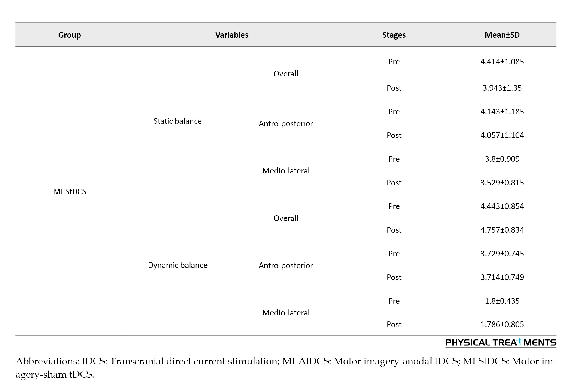
Table 2 summarizes the descriptive statistics of static and dynamic balance outcomes for each group at pre- and post-intervention stages.
Table 3 presents changes in static and dynamic balance scores across three items (two different directions: antro-posterior and medio-lateral, and overall).
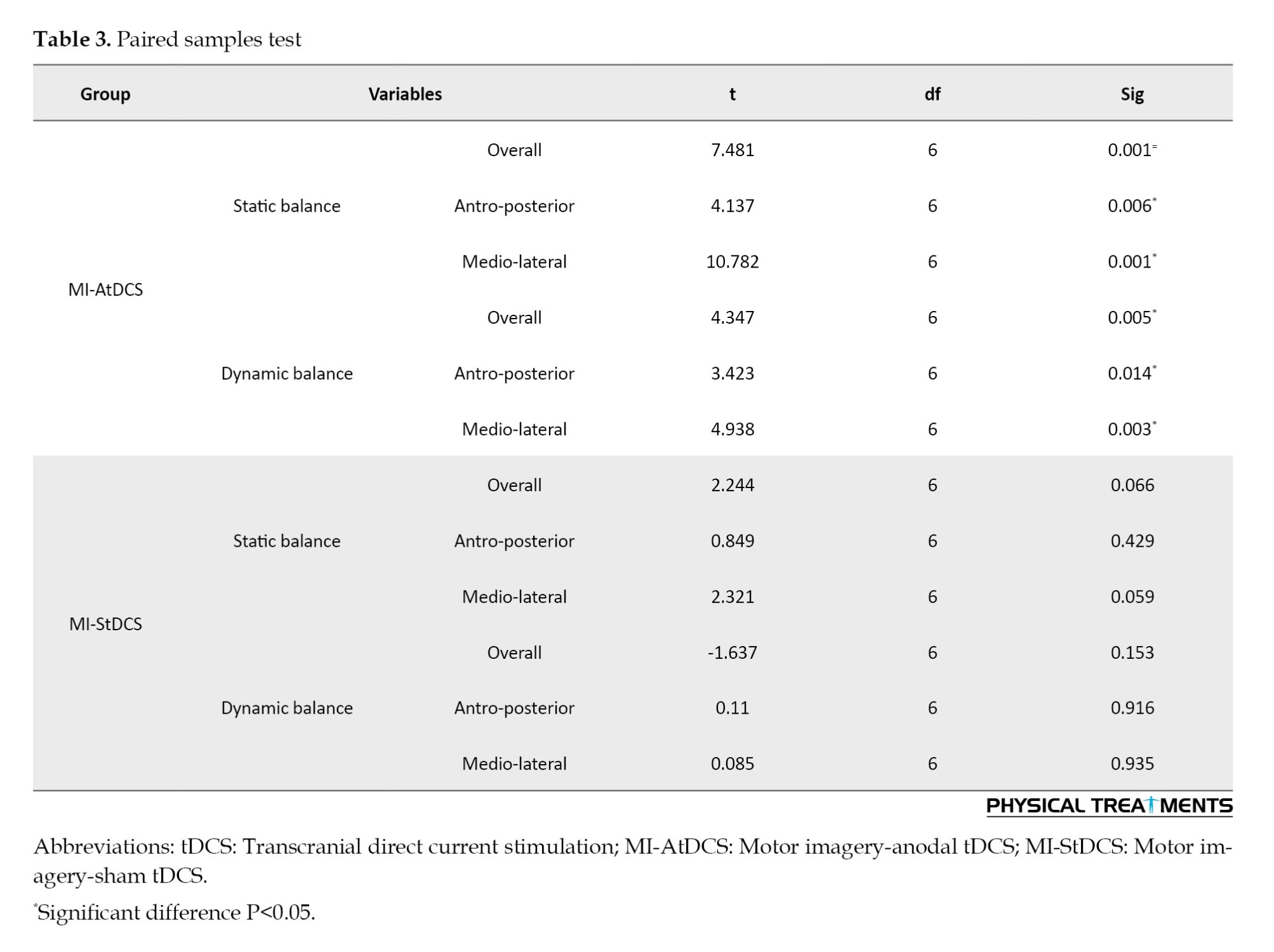
A paired t-test revealed a statistically significant change (P<0.05) in the scores on three BBT items in the active group. In contrast, no significant differences were detected in the sham group.
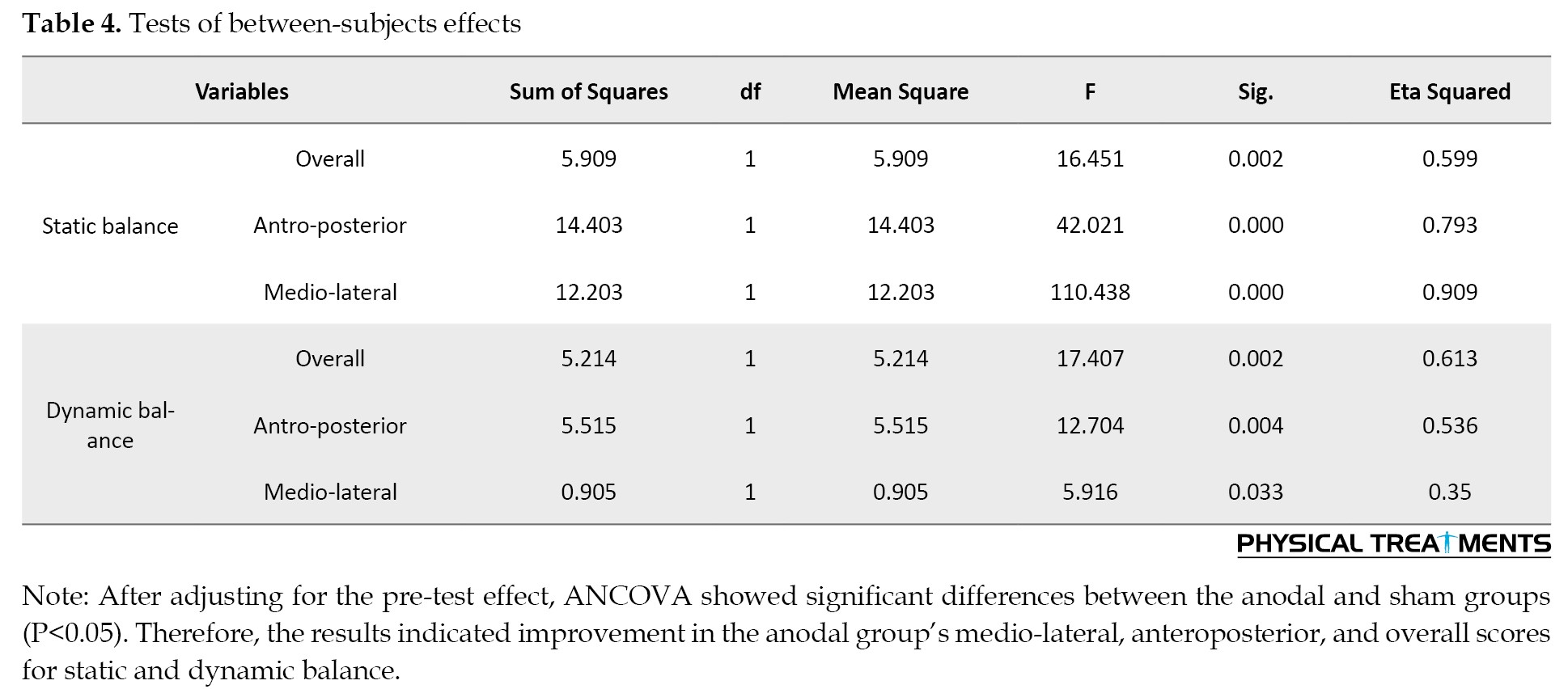
Table 4 presents a comparison of post-test scores between groups.
Discussion
This study aimed to examine the combined effects of MIP and tDCS on both dynamic and static balance in athletes diagnosed with CAI. Pre- and post-intervention assessments were conducted using the BBS. The intervention involved a single session of either anodal or sham tDCS in conjunction with MIP. Results indicated significant enhancements in both dynamic and static balance within the active stimulation group, as reflected by improvements in the anterior-posterior index, medial-lateral index, and overall stability index. Conversely, participants in the sham tDCS-MIP group exhibited no notable improvements, suggesting that MIP alone may not be sufficient to produce balance gains in athletes with CAI.
The effectiveness of tDCS in improving balance has been previously demonstrated in healthy populations [31, 32] and clinical groups [33, 34]. For example, a study combining tDCS with virtual reality training reported improved fatigue, balance, and walking ability in patients with multiple sclerosis [22]. The effects of tDCS on dynamic balance in CAI have also been investigated in non-athletic populations [21]. These studies found improvements in dynamic balance following a 4-week protocol of anodal tDCS applied to the primary motor cortex (M1). However, the consistency of these results varies across studies, with some demonstrating significant improvements and others showing minimal or no effects. Possible factors contributing to this inconsistency include differences in stimulation parameters (e.g. electrode placement, stimulation duration, and intensity), population characteristics (e.g. age, baseline motor function), and specific protocols used (e.g. exercise combinations). Similarly, significant improvements in dynamic balance and proprioception were observed when high-definition tDCS was combined with short-foot exercises, using a 4-week stimulation protocol targeting the Cz region [35].
Contrary to the aforementioned results, recent research involving a single application of cerebellar tDCS in healthy participants did not yield significant enhancements in either static or dynamic balance. The discrepancy between the results could be attributed to differences in the stimulation site and balance assessment tools (e.g. Y-balance test versus BBS). Various tDCS protocols have been employed in studies aimed at improving balance and postural control. Depending on the targeted cortical region, electrical stimulation of the motor cortex (M1), cerebellum, or frontal areas, such as the DLPFC, has shown potential for enhancing balance and postural control [36-38].
Most tDCS studies have focused on motor-specific areas, such as the motor cortex and cerebellum, to influence balance and posture. In our study, the anodal electrode was placed over F3 to stimulate the DLPFC, aiming to improve cognitive performance and postural control. However, the DLPFC has been primarily recognized for its role in cognitive functions, but it has been considered to affect motor functions in some studies. Fujita demonstrated hyperactivation of the DLPFC, indicated by increased blood flow, during dual-task posture control [39]. Similarly, an fMRI study reported increased DLPFC activity among the brain regions involved in proprioceptive stimulation, contributing to balance performance and postural control [40]. The application of tDCS to the DLPFC has been shown to improve functional mobility and balance in patients with Parkinson’s disease [41]. Researchers attributed this improvement to the DLPFC’s role in visuospatial processing, which enhances balance and mobility [42]. Another proposed mechanism is that anodal stimulation targeting F3 (DLPFC) directly and indirectly influences the premotor region through inter-neuronal connections with the prefrontal cortex. Additionally, tDCS over the DLPFC has been widely used to enhance cognitive functions, including attention and working memory [43, 44].
The DLPFC is a densely interconnected brain region believed to play a key role in integrating sensory inputs with cognitive processes [45]. Evidence from neuroimaging and repetitive transcranial magnetic stimulation studies has highlighted the DLPFC’s involvement in higher-order cognitive functions, including attention regulation and working memory [46, 47]. A recent investigation employed a combined MI and tDCS intervention to enhance mobility and reduce fall risk among older adults [48]. This protocol involved six sessions of prefrontal tDCS targeting a cortical region analogous to the one stimulated in the current study.
The current study investigated the acute effects of tDCS in athletes with CAI. Consistent with the findings, Weigie studied the effects of a one-session of anodal tDCS and reported improvements in intrinsic foot muscle activation and static balance [49]. Similarly, tDCS has been shown to enhance muscle strength, foot sensation, and static balance in other studies [50]. In contrast, the present study found that MIP alone did not significantly improve balance in athletes with CAI. This aligns with prior research reporting the limited efficacy of standalone MIP interventions [51]. It should be noted that the intervention in the present study consisted of only a single MIP session. MI activates brain regions typically involved during task performance, but its effects may require multiple sessions to elicit significant improvements [52]. The underlying mechanism of MIP is attributed to weak neuromuscular activation in the muscles involved in the imagined activity. This facilitated neural pathway improves muscle function and motor performance [53]. Neuromodulation techniques such as tDCS can enhance the effects of MIP by altering the resting membrane potential and increasing neural pathway activity [54]. The present study used tDCS and MIP to strengthen the stimulation effect. tDCS is presumed to influence the resting membrane potential during stimulation, thereby facilitating motor activity by modulating neural excitability [55].
This study is the first attempt to evaluate the impact of a single-session tDCS intervention combined with MIP in individuals with CAI. The results support the efficacy of CNS-targeted strategies in addressing balance impairments in athletes with CAI, even following a single exposure. Although the current investigation focused on immediate, short-term outcomes, prior studies have reported prolonged effects resulting from repeated tDCS sessions, including sustained increases in cortical excitability elicited by motor cortex stimulation. The present results highlight the synergistic influence of tDCS and MIP on balance regulation in this population. These findings underscore the value of examining acute responses and lay the foundation for future research to optimize CNS-based rehabilitation strategies for balance enhancement.
Conclusion
The results of this study suggest that the simultaneous use of MIP and tDCS leads to significant enhancements in both static and dynamic balance among athletes with CAI. In contrast to conventional rehabilitation approaches targeting peripheral neuromuscular mechanisms, integrating brain stimulation techniques, such as tDCS, introduces a novel strategy for engaging CNS pathways. This approach enhances motor function and postural control, addressing the neural aspects of balance deficits. Although the study has limitations, including a small sample size and the inability to isolate the effects of tDCS alone, the results suggest that CNS-based interventions may serve as valuable adjuncts to traditional rehabilitation protocols. These findings provide a foundation for future research to optimize postural control and minimize re-injury risk in athletes with CAI.
Limitations
This study has some limitations. The effects of tDCS alone were not assessed, making it impossible to isolate the individual contributions of tDCS and MIP. The limited sample size may limit the generalizability of these findings; therefore, further studies with larger participant groups are essential to confirm and extend these results. Additionally, the quality or success of MIP was not quantitatively assessed using a specific instrument, which may have influenced the intervention’s efficacy.
Recommendations for future studies
Future studies should consider employing multi-session protocols to examine the sustained effects of combining tDCS and MIP in athletes with CAI. Increasing sample sizes would also improve the generalizability of findings. Moreover, investigating the isolated effects of each intervention could help clarify their individual contributions to balance improvements.
Policy implications
Integrating tDCS-based interventions into rehabilitation protocols for athletes with CAI could improve treatment outcomes by addressing the central nervous system’s contribution to balance dysfunction. Sports organizations and rehabilitation centers may consider adopting this novel approach to reduce the risk of recurrent injuries, improve postural control, and enhance athletic performance. Policies supporting research funding for non-invasive neuromodulation techniques could accelerate advancements in rehabilitation science and promote evidence-based clinical practices. Moreover, incorporating tDCS and MIP into professional training programs for sports medicine practitioners could facilitate the broader adoption of CNS-based therapies in athletic rehabilitation.
Ethical Considerations
Compliance with ethical guidelines
All stages of the research were approved by the approval of the Ethics Committee of the Faculty of Sport Sciences and Health at the University of Tehran, Tehran, Iran (Code: IR.UT.SPORT.REC.1401.033).
Funding
This research was supported by the University of Tehran, Tehran, Iran (Grant No.: 25208/1/04).
Authors' contributions
All authors equally participated in the preparation and development of this manuscript.
Conflict of interest
The authors declared no conflict of interest.
Acknowledgments
The authors gratefully acknowledge the participation of all patients involved in this study.
An acute ankle sprain represents one of the most prevalent musculoskeletal injuries in physically active populations. Approximately one-third of those who sustain such sprains may develop chronic ankle instability (CAI), characterized by recurrent ankle sprains and a subjective sensation of the joint “giving way” [1]. This condition is commonly associated with functional deficits, including impaired balance, diminished proprioceptive acuity, and altered neuromuscular control [2]. A previous study identified static and dynamic balance as key variables commonly assessed and targeted in CAI rehabilitation [3]. The somatosensory system is critical in postural and balance control by integrating sensory input with the central nervous system (CNS). Mechanoreceptors and joint sensory information, transmitted by peripheral nerves to the CNS, are essential for maintaining balance [4]. CAI alters sensory receptors and proprioception, leading to impaired transmission of information to the cerebral cortex and resulting in balance deficits [5]. These impairments in postural control can reduce functional independence, limit daily activities, and negatively affect quality of life [6]. Therefore, restoring postural function is a central goal in rehabilitation programs for individuals with significant motor impairments [7]. Conventional rehabilitation for CAI primarily targets peripheral neuromuscular elements [8]. However, research has also linked reduced cortical activation to biomechanical impairments and a heightened likelihood of recurrent ankle sprains [9]. Integrating approaches that stimulate central neural pathways alongside peripheral neuromuscular interventions may enhance recovery outcomes in individuals with CAI. Techniques such as action observation and motor imagery (MI) have shown promise in promoting balance and improving postural control [10, 11].
MI, mental rehearsal of movements without physical execution, is an effective intervention for motor skill development and rehabilitation. Extensive research indicates repeated motor imagery practice (MIP) significantly benefits athletic performance and physical rehabilitation [12]. For instance, MIP has been shown to improve motor function in postural control tasks, such as weight shifting, by reducing the time required to execute the movement [13]. While physical training typically yields more pronounced improvements, MIP has proven effective at facilitating the acquisition of postural control skills and improving dynamic balance in unpredictable environments [14]. Additionally, MIP activates brain regions involved in gait planning, making it particularly valuable for populations with balance deficits, such as older adults and stroke survivors [15]. MIP enhances dynamic postural control and stabilizes movement by engaging neural pathways similar to those activated during physical exercise, particularly in the primary motor cortex (M1). Neuroimaging studies have demonstrated that MIP promotes activity-dependent plasticity, which is essential for motor learning, as it parallels the increases in neural excitability observed during physical training [16].
Transcranial direct current stimulation (tDCS) is another noteworthy non-invasive brain stimulation method that alters cortical excitability and facilitates motor learning. Anodal tDCS increases neural excitability and enhances interneuronal communication during motor tasks, facilitating learning. This stimulation improves performance on sequential and visuomotor tasks, reduces reaction times, and enhances functional abilities [17, 18]. Prior studies have shown that tDCS can augment motor learning, particularly in populations requiring rehabilitative care or skill acquisition. Similar improvements in balance and gait performance have been observed with tDCS interventions, comparable to those observed with physical exercises [19]. Recently, tDCS has been combined with various therapeutic modalities, such as physical therapy, virtual reality, and mindfulness, to optimize functional outcomes by facilitating neuroplasticity [20, 21].
Using anodal tDCS over the right dorsolateral prefrontal cortex (DLPFC) during MI has enhanced Mu desynchronization, a neural marker of motor execution and imagery [22]. This suggests that anodal tDCS can augment the neuroplastic effects of MIP, thereby enhancing performance. For instance, Yavari et al. reported that the simultaneous application of tDCS and mindfulness training led to significant improvements in self-control and emotion regulation among skilled badminton players, with the combined approach proving more effective than mindfulness alone [23]. Similarly, Matsumoto et al. found greater performance improvements in sequential tasks when MIP was paired with anodal tDCS than either intervention alone [24]. While prior research has explored the individual effects of MIP and tDCS on motor performance, the integrated application of these two approaches—especially with stimulation targeting the DLPFC—remains underexplored in the context of balance regulation among athletes experiencing CAI. Given the DLPFC’s involvement in executive functioning, attentional processes, and motor coordination, it is a potentially valuable neural target for enhancing MIP outcomes via tDCS-induced modulation [25, 26]. Moreover, effective balance regulation is a key component in the rehabilitation of individuals with CAI. Nonetheless, the neural mechanisms underlying cognitive-motor integration achieved through the combined use of MIP and DLPFC-targeted stimulation remain insufficiently understood. To address this, the present study investigates whether a single session of anodal tDCS over the right DLPFC can amplify the effects of MIP on both static and dynamic balance in physically active individuals with CAI.
Materials and Methods
The current study utilized a double-blind, randomized clinical trial with a pre-test-post-test design and a control (sham) group. Fourteen right-handed athletes (mean age: 24.5±2.62 years; seven females) who had sustained an ankle sprain more than a year prior and reported experiencing at least two episodes of ankle “giving way” on the same side were recruited for the study [27], and physician-diagnosed CAI were randomly selected based on predefined inclusion criteria. Participants were recruited from the Faculty of Sport Sciences and Health at the University of Tehran. Before initiating the intervention, all subjects completed written informed consent for voluntary participation and submitted demographic, sports, and medical history data. Participants were excluded if they had a history of seizures, any neurological or psychiatric conditions, previous lower limb surgery or fractures, or if they were concurrently involved in other intervention programs. The Ethics Committee of the Faculty of Sport Sciences and Health at the University of Tehran approved the study protocol. Baseline assessments were conducted using the Biodex Balance System (BBS), in which participants performed both static and dynamic balance tests to establish pre-intervention balance metrics. Participants were then randomly assigned to one of two groups: The anodal stimulation group (MI + AtDCS) or the imagery sham stimulation group (MI + StDCS), each consisting of seven individuals. The intervention included an MI session and tDCS targeting the DLPFC. All experimental procedures, including the intervention and subsequent assessments, were conducted at the laboratory of the Faculty of Sport Sciences and Health. Participants were randomly allocated to the experimental or control group using a computer-generated random number sequence. The study was double-blind, with both the participants and the assessors unaware of group assignments. After the intervention, post-test assessments were performed to evaluate dynamic and static balance using the BBS. The BBS was selected due to its high reliability and validity in assessing both static and dynamic balance. Given the study’s focus on balance performance in athletes with CAI and to ensure methodological clarity and control, a single outcome measure was used. This allowed for a focused evaluation of the intervention’s effectiveness without introducing confounding variability from multiple assessment tools.
Study design
This experiment employed a randomized, double-blind design with placebo control involving sham and anodal stimulation. Participants were randomly allocated into two groups: imagery anodal stimulation (MI-AtDCS) and imagery sham stimulation (MI-StDCS), with seven participants in each group.
tDCS
A tDCS device (ActivaDose) was used to administer the stimulation protocol. For electrode placement, a 5×5 cm saline-soaked sponge served as the anodal electrode and was positioned over the left DLPFC (F3), The cathodal electrode, identical in size, was positioned on the right forehead. The stimulation protocol involved delivering a direct current of 1.5 mA for a single 15-minute session [23]. For the sham condition, the device delivered the current for 60 seconds before turning off to mimic stimulation without maintaining active current flow, thereby ensuring participant blinding to the condition.
MI
The MI exercise program consisted of a guided imagery procedure tailored to balance tasks. Participants were first instructed to visualize, step by step, the process of maintaining static balance as assessed during the pre-test using the BBS, while focusing on executing the task positively. This visualization lasted approximately two minutes, followed by a one-minute rest period, and was repeated twice. After completing the static balance imagery phase, the participants engaged in the dynamic balance imagery procedure, which followed the same protocol as the static balance exercise. The total imaging and rest duration for static and dynamic balance exercises were 12 minutes [13]. Before the guided imagery exercises began, participants received tDCS stimulation for an initial three-minute period without performing any specific activity, allowing them to adapt to the stimulation protocol.
Assessments
The BBS is reliable for evaluating static and dynamic balance in clinical and athletic populations. The BBS measures participants’ ability to maintain stability on a fixed platform during static balance assessments. Dynamic balance assessments require participants to respond to multi-directional platform tilts, thereby evaluating functional stability and proprioception [28]. These metrics are essential for identifying balance deficits that may increase injury risk and monitoring improvements following interventions. The BBS has demonstrated high test, re-test reliability for dynamic balance measurements in injured and non-injured athletes [29]. This study used the BBS (manufactured in the USA, 2018) to measure static and dynamic balance. Postural maintenance time during each assessment was set at 30 s, followed by a 60-s rest period. These durations were selected based on previously established protocols to ensure adequate time for balance evaluation while minimizing fatigue [30]. Three indices—overall stability, anterior-posterior stability, and medial-lateral stability—were used to assess balance. Each test was repeated three times, with the average values recorded for analysis.
Statistics
Data analysis was conducted using paired-sample t-tests to evaluate within-group difference measurements. For between-group comparisons, analysis of covariance (ANCOVA) was performed to adjust for baseline differences. To summarize the data, descriptive statistics were computed for all variables, including Means±SD. Data were analyzed using SPSS software, version 26.0 with statistical significance at P<0.05.
Results
All 14 participants (n=14) completed the study protocol. Table 1 presents a summary of their demographic information.
No statistically significant differences were observed between the groups regarding age, body weight, or height.
Table 2 presents the descriptive statistics for static and dynamic balance measures, reported separately for the pre-test and post-test phases.
Table 2 summarizes the descriptive statistics of static and dynamic balance outcomes for each group at pre- and post-intervention stages.
Table 3 presents changes in static and dynamic balance scores across three items (two different directions: antro-posterior and medio-lateral, and overall).
A paired t-test revealed a statistically significant change (P<0.05) in the scores on three BBT items in the active group. In contrast, no significant differences were detected in the sham group.
Table 4 presents a comparison of post-test scores between groups.
Discussion
This study aimed to examine the combined effects of MIP and tDCS on both dynamic and static balance in athletes diagnosed with CAI. Pre- and post-intervention assessments were conducted using the BBS. The intervention involved a single session of either anodal or sham tDCS in conjunction with MIP. Results indicated significant enhancements in both dynamic and static balance within the active stimulation group, as reflected by improvements in the anterior-posterior index, medial-lateral index, and overall stability index. Conversely, participants in the sham tDCS-MIP group exhibited no notable improvements, suggesting that MIP alone may not be sufficient to produce balance gains in athletes with CAI.
The effectiveness of tDCS in improving balance has been previously demonstrated in healthy populations [31, 32] and clinical groups [33, 34]. For example, a study combining tDCS with virtual reality training reported improved fatigue, balance, and walking ability in patients with multiple sclerosis [22]. The effects of tDCS on dynamic balance in CAI have also been investigated in non-athletic populations [21]. These studies found improvements in dynamic balance following a 4-week protocol of anodal tDCS applied to the primary motor cortex (M1). However, the consistency of these results varies across studies, with some demonstrating significant improvements and others showing minimal or no effects. Possible factors contributing to this inconsistency include differences in stimulation parameters (e.g. electrode placement, stimulation duration, and intensity), population characteristics (e.g. age, baseline motor function), and specific protocols used (e.g. exercise combinations). Similarly, significant improvements in dynamic balance and proprioception were observed when high-definition tDCS was combined with short-foot exercises, using a 4-week stimulation protocol targeting the Cz region [35].
Contrary to the aforementioned results, recent research involving a single application of cerebellar tDCS in healthy participants did not yield significant enhancements in either static or dynamic balance. The discrepancy between the results could be attributed to differences in the stimulation site and balance assessment tools (e.g. Y-balance test versus BBS). Various tDCS protocols have been employed in studies aimed at improving balance and postural control. Depending on the targeted cortical region, electrical stimulation of the motor cortex (M1), cerebellum, or frontal areas, such as the DLPFC, has shown potential for enhancing balance and postural control [36-38].
Most tDCS studies have focused on motor-specific areas, such as the motor cortex and cerebellum, to influence balance and posture. In our study, the anodal electrode was placed over F3 to stimulate the DLPFC, aiming to improve cognitive performance and postural control. However, the DLPFC has been primarily recognized for its role in cognitive functions, but it has been considered to affect motor functions in some studies. Fujita demonstrated hyperactivation of the DLPFC, indicated by increased blood flow, during dual-task posture control [39]. Similarly, an fMRI study reported increased DLPFC activity among the brain regions involved in proprioceptive stimulation, contributing to balance performance and postural control [40]. The application of tDCS to the DLPFC has been shown to improve functional mobility and balance in patients with Parkinson’s disease [41]. Researchers attributed this improvement to the DLPFC’s role in visuospatial processing, which enhances balance and mobility [42]. Another proposed mechanism is that anodal stimulation targeting F3 (DLPFC) directly and indirectly influences the premotor region through inter-neuronal connections with the prefrontal cortex. Additionally, tDCS over the DLPFC has been widely used to enhance cognitive functions, including attention and working memory [43, 44].
The DLPFC is a densely interconnected brain region believed to play a key role in integrating sensory inputs with cognitive processes [45]. Evidence from neuroimaging and repetitive transcranial magnetic stimulation studies has highlighted the DLPFC’s involvement in higher-order cognitive functions, including attention regulation and working memory [46, 47]. A recent investigation employed a combined MI and tDCS intervention to enhance mobility and reduce fall risk among older adults [48]. This protocol involved six sessions of prefrontal tDCS targeting a cortical region analogous to the one stimulated in the current study.
The current study investigated the acute effects of tDCS in athletes with CAI. Consistent with the findings, Weigie studied the effects of a one-session of anodal tDCS and reported improvements in intrinsic foot muscle activation and static balance [49]. Similarly, tDCS has been shown to enhance muscle strength, foot sensation, and static balance in other studies [50]. In contrast, the present study found that MIP alone did not significantly improve balance in athletes with CAI. This aligns with prior research reporting the limited efficacy of standalone MIP interventions [51]. It should be noted that the intervention in the present study consisted of only a single MIP session. MI activates brain regions typically involved during task performance, but its effects may require multiple sessions to elicit significant improvements [52]. The underlying mechanism of MIP is attributed to weak neuromuscular activation in the muscles involved in the imagined activity. This facilitated neural pathway improves muscle function and motor performance [53]. Neuromodulation techniques such as tDCS can enhance the effects of MIP by altering the resting membrane potential and increasing neural pathway activity [54]. The present study used tDCS and MIP to strengthen the stimulation effect. tDCS is presumed to influence the resting membrane potential during stimulation, thereby facilitating motor activity by modulating neural excitability [55].
This study is the first attempt to evaluate the impact of a single-session tDCS intervention combined with MIP in individuals with CAI. The results support the efficacy of CNS-targeted strategies in addressing balance impairments in athletes with CAI, even following a single exposure. Although the current investigation focused on immediate, short-term outcomes, prior studies have reported prolonged effects resulting from repeated tDCS sessions, including sustained increases in cortical excitability elicited by motor cortex stimulation. The present results highlight the synergistic influence of tDCS and MIP on balance regulation in this population. These findings underscore the value of examining acute responses and lay the foundation for future research to optimize CNS-based rehabilitation strategies for balance enhancement.
Conclusion
The results of this study suggest that the simultaneous use of MIP and tDCS leads to significant enhancements in both static and dynamic balance among athletes with CAI. In contrast to conventional rehabilitation approaches targeting peripheral neuromuscular mechanisms, integrating brain stimulation techniques, such as tDCS, introduces a novel strategy for engaging CNS pathways. This approach enhances motor function and postural control, addressing the neural aspects of balance deficits. Although the study has limitations, including a small sample size and the inability to isolate the effects of tDCS alone, the results suggest that CNS-based interventions may serve as valuable adjuncts to traditional rehabilitation protocols. These findings provide a foundation for future research to optimize postural control and minimize re-injury risk in athletes with CAI.
Limitations
This study has some limitations. The effects of tDCS alone were not assessed, making it impossible to isolate the individual contributions of tDCS and MIP. The limited sample size may limit the generalizability of these findings; therefore, further studies with larger participant groups are essential to confirm and extend these results. Additionally, the quality or success of MIP was not quantitatively assessed using a specific instrument, which may have influenced the intervention’s efficacy.
Recommendations for future studies
Future studies should consider employing multi-session protocols to examine the sustained effects of combining tDCS and MIP in athletes with CAI. Increasing sample sizes would also improve the generalizability of findings. Moreover, investigating the isolated effects of each intervention could help clarify their individual contributions to balance improvements.
Policy implications
Integrating tDCS-based interventions into rehabilitation protocols for athletes with CAI could improve treatment outcomes by addressing the central nervous system’s contribution to balance dysfunction. Sports organizations and rehabilitation centers may consider adopting this novel approach to reduce the risk of recurrent injuries, improve postural control, and enhance athletic performance. Policies supporting research funding for non-invasive neuromodulation techniques could accelerate advancements in rehabilitation science and promote evidence-based clinical practices. Moreover, incorporating tDCS and MIP into professional training programs for sports medicine practitioners could facilitate the broader adoption of CNS-based therapies in athletic rehabilitation.
Ethical Considerations
Compliance with ethical guidelines
All stages of the research were approved by the approval of the Ethics Committee of the Faculty of Sport Sciences and Health at the University of Tehran, Tehran, Iran (Code: IR.UT.SPORT.REC.1401.033).
Funding
This research was supported by the University of Tehran, Tehran, Iran (Grant No.: 25208/1/04).
Authors' contributions
All authors equally participated in the preparation and development of this manuscript.
Conflict of interest
The authors declared no conflict of interest.
Acknowledgments
The authors gratefully acknowledge the participation of all patients involved in this study.
References
- Herzog MM, Kerr ZY, Marshall SW, Wikstrom EA. Epidemiology of ankle sprains and chronic ankle instability. Journal of Athletic Training. 2019; 54(6):603-10. [DOI:10.4085/1062-6050-447-17] [PMID]
- Akbari M, Karimi H, Farahini H, Faghihzadeh S. Balance problems after unilateral lateral ankle sprains. Journal of Rehabilitation Research and Development. 2006; 43(7):819-24. [DOI:10.1682/JRRD.2006.01.0001] [PMID]
- Nozu S, Takemura M, Sole G. Assessments of sensorimotor deficits used in randomized clinical trials with individuals with ankle sprains and chronic ankle instability: A scoping review. PM&R. 2021; 13(8):901-4. [DOI:10.1002/pmrj.12487] [PMID]
- Van der Cruyssen F, Politis C. Neurophysiological aspects of the trigeminal sensory system: An update. Reviews in the Neurosciences. 2018; 29(2):115-23. [DOI:10.1515/revneuro-2017-0044] [PMID]
- Pietrosimone BG, Gribble PA. Chronic ankle instability and corticomotor excitability of the fibularis longus muscle. Journal of Athletic Training. 2012; 47(6):621-6. [DOI:10.4085/1062-6050-47.6.11] [PMID]
- Guetiti S, Cadoret G, Chénier F, Blanchet M. Impacts of developmental coordination disorder on postural control mechanisms in children and early adolescents. Journal of Motor Learning and Development. 2024; 12(3):635-65. [DOI:10.1123/jmld.2023-0030]
- Rybski MF, Juckett L. Posture. In: Rybski MF, editor. Kinesiology for occupational therapy. Milton Park: Routledge; 2024. [DOI:10.4324/9781003524724-11]
- Alahmari KA, Kakaraparthi VN, Reddy RS, Silvian P, Tedla JS, Rengaramanujam K, et al. Combined effects of strengthening and proprioceptive training on stability, balance, and proprioception among subjects with chronic ankle instability in different age groups: Evaluation of clinical outcome measures. Indian Journal of Orthopaedics. 2021; 55(Suppl 1):199-208. [DOI:10.1007/s43465-020-00192-6] [PMID]
- Needle AR, Lepley AS, Grooms DR. Central nervous system adaptation after ligamentous injury: A summary of theories, evidence, and clinical interpretation. Sports Medicine. 2017; 47(7):1271-88. [DOI:10.1007/s40279-016-0666-y] [PMID]
- Yakovlev L, Syrov N, Kaplan A. Investigating the influence of functional electrical stimulation on motor imagery related μ-rhythm suppression. Frontiers in Neuroscience. 2023; 17:1202951. [DOI:10.3389/fnins.2023.1202951] [PMID]
- Nedelko V, Hassa T, Hamzei F, Schoenfeld MA, Dettmers C. Action imagery combined with action observation activates more corticomotor regions than action observation alone. Journal of Neurologic Physical Therapy. 2012; 36(4):182-8. [DOI:10.1097/NPT.0b013e318272cad1] [PMID]
- Choi JW, Kim BH, Huh S, Jo S. Observing actions through immersive virtual reality enhances motor imagery training. IEEE Transactions on Neural Systems and Rehabilitation Engineering. 2020; 28(7):1614-22. [DOI:10.1109/TNSRE.2020.2998123] [PMID]
- Saruco E, Di Rienzo F, Nunez-Nagy S, Rubio-Gonzalez MA, Jackson PL, Collet C, et al. Anodal tDCS over the primary motor cortex improves motor imagery benefits on postural control: A pilot study. Scientific Reports. 2017; 7(1):480. [DOI:10.1038/s41598-017-00509-w] [PMID]
- Guillot A, Collet C. Construction of the motor imagery integrative model in sport: A review and theoretical investigation of motor imagery use. International Review of Sport and Exercise Psychology. 2008; 1(1):31-44. [DOI:10.1080/17509840701823139]
- Taube W, Leukel C, Gollhofer A. Influence of enhanced visual feedback on postural control and spinal reflex modulation during stance. Experimental Brain Research. 2008; 188(3):353-61. [DOI:10.1007/s00221-008-1370-4] [PMID]
- Debarnot U, Clerget E, Olivier E. Role of the primary motor cortex in the early boost in performance following mental imagery training. Plos One. 2011; 6(10):e26717. [DOI:10.1371/journal.pone.0026717] [PMID]
- Hétu S, Grégoire M, Saimpont A, Coll MP, Eugène F, Michon PE, et al. The neural network of motor imagery: An ALE meta-analysis. Neuroscience & Biobehavioral Reviews. 2013; 37(5):930-49. [DOI:10.1016/j.neubiorev.2013.03.017] [PMID]
- Reis J, Fritsch B. Modulation of motor performance and motor learning by transcranial direct current stimulation. Current Opinion in Neurology. 2011; 24(6):590-6. [DOI:10.1097/WCO.0b013e32834c3db0] [PMID]
- Nitsche MA, Cohen LG, Wassermann EM, Priori A, Lang N, Antal A, et al. Transcranial direct current stimulation: state of the art 2008. Brain Stimulation. 2008; 1(3):206-23. [DOI:10.1016/j.brs.2008.06.004] [PMID]
- Mohammadkhanbeigi S, Moghadas Tabrizi Y, Nabavi SM, Minoonejad H. The comparable effect of tDCS and core exercises on Balance and mobility in patients with multiple sclerosis. Iranian Rehabilitation Journal. 2022; 20(4):569-78. [DOI:10.32598/irj.20.4.1699.1]
- Bruce AS, Howard JS, Van Werkhoven H, McBride JM, Needle AR. The effects of transcranial direct current stimulation on chronic ankle instability. Medicine and Science in Sports and Exercise. 2020; 52(2):335-44. [DOI:10.1249/MSS.0000000000002129] [PMID]
- Charehjou B, Moghadas Tabrizi Y, Minoonejad H. The combination of transcranial direct current stimulation and virtual reality training on fatigue, balance and walking in patients with multiple sclerosis. Physical Treatments-Specific Physical Therapy Journal. 2024; 14(3):171-82. [DOI:10.32598/ptj.14.3.578.2]
- Kateb MY, Ameri EA, Rostami R, Zandi HG, Tabrizi YM. the effect of tdcs-mindfulness program on self-control and emotion regulation in badminton players. International Journal of Sport Studies for Health. 2024; 7(1):11-6. [DOI:10.61838/kman.intjssh.7.1.2]
- Matsumoto J, Fujiwara T, Takahashi O, Liu M, Kimura A, Ushiba J. Modulation of mu rhythm desynchronization during motor imagery by transcranial direct current stimulation. Journal of NeuroEngineering and Rehabilitation. 2010; 7:27. [DOI:10.1186/1743-0003-7-27] [PMID]
- Cieslik EC, Zilles K, Caspers S, Roski C, Kellermann TS, Jakobs O, et al. Is there “one” DLPFC in cognitive action control? Evidence for heterogeneity from co-activation-based parcellation. Cerebral Cortex. 2013; 23(11):2677-89. [DOI:10.1093/cercor/bhs256] [PMID]
- Ko JH, Monchi O, Ptito A, Bloomfield P, Houle S, Strafella AP. Theta burst stimulation‐induced inhibition of dorsolateral prefrontal cortex reveals hemispheric asymmetry in striatal dopamine release during a set‐shifting task-a TMS-[11C] raclopride PET study. European Journal of Neuroscience. 2008; 28(10):2147-55. [DOI:10.1111/j.1460-9568.2008.06501.x] [PMID]
- Brinkman RE, Evans TA. History of ankle sprain as a risk factor of future lateral ankle sprain in athletes. Journal of Sport Rehabilitation. 2011; 20(3):384-8. [DOI:10.1123/jsr.20.3.384] [PMID]
- Cachupe WJ, Shifflett B, Kahanov L, Wughalter EH. Reliability of biodex balance system measures. Measurement in Physical Education and Exercise Science. 2001; 5(2):97-108. [DOI:10.1207/S15327841MPEE0502_3]
- Oliver GD, Weimar W. Hip range of motion and scapula position in youth baseball pitching pre and post simulated game. Journal of Sports Sciences. 2015; 33(14):1447-53. [DOI:10.1080/02640414.2014.990493] [PMID]
- Ahmadipoor A, Khademi-Kalantari K, Rezasoltani A, Naimi SS, Akbarzadeh-Baghban A. Effect of forward head posture on dynamic balance based on the Biodex Balance System. Journal of Biomedical Physics & Engineering. 2022; 12(5):543-8. [DOI:10.31661/jbpe.v0i0.1912-1036]
- Lee YS, Yang HS, Jeong CJ, Yoo YD, Jeong SH, Jeon OK, et al. The effects of transcranial direct current stimulation on functional movement performance and balance of the lower extremities. Journal of Physical Therapy Science. 2012; 24(12):1215-8. [DOI:10.1589/jpts.24.1215]
- Erdoğan ET, Kır C, Beycan E, Karakaya E, Altınçınar S, Bayramoğlu T, et al. Acute effect of single-session cerebellar anodal transcranial direct current stimulation on static and dynamic balance in healthy volunteers. Brain Sciences. 2023; 13(7):1107. [DOI:10.3390/brainsci13071107] [PMID]
- Marotta N, de Sire A, Marinaro C, Moggio L, Inzitari MT, Russo I, et al. Efficacy of transcranial direct current stimulation (tDCS) on balance and gait in multiple sclerosis patients: A machine learning approach. Journal of Clinical Medicine. 2022; 11(12):3505. [DOI:10.3390/jcm11123505] [PMID]
- Beretta VS, Santos PCR, Orcioli-Silva D, Zampier VC, Vitorio R, Gobbi LTB. Transcranial direct current stimulation for balance rehabilitation in neurological disorders: A systematic review and meta-analysis. Ageing Research Reviews. 2022; 81:101736. [DOI:10.1016/j.arr.2022.101736] [PMID]
- Ma Y, Yin K, Zhuang W, Zhang C, Jiang Y, Huang J, et al. Effects of combining high-definition transcranial direct current stimulation with short-foot exercise on chronic ankle instability: A pilot randomized and double-blinded study. Brain Sciences. 2020; 10(10):749. [DOI:10.3390/brainsci10100749] [PMID]
- Giancatarina M, Grandperrin Y, Nicolier M, Gimenez P, Vidal C, Tio G, et al. Acute effect of transcranial direct current stimulation (tDCS) on postural control of trained athletes: A randomized controlled trial. Plos One. 2024; 19(1):e0286443. [DOI:10.1371/journal.pone.0286443] [PMID]
- Baharlouei H, Sadeghi-Demneh E, Mehravar M, Manzari P, Yazdi MJS, Joghataei MT, et al. Comparison of transcranial direct current stimulation of the primary motor cortex and cerebellum on static balance in older adults. Iranian Red Crescent Medical Journal. 2020; 22(3):e188815. [DOI:10.5812/ircmj.96259]
- Zhou J, Hao Y, Wang Y, Jor’dan A, Pascual‐Leone A, Zhang J, et al. Transcranial direct current stimulation reduces the cost of performing a cognitive task on gait and postural control. European Journal of Neuroscience. 2014; 39(8):1343-8. [DOI:10.1111/ejn.12492] [PMID]
- Fujita N, Kunimune S, Okada S. Contribution of the dorsolateral prefrontal cortex activation, ankle muscle activities, and coactivation during dual-tasks to postural steadiness: A pilot study. Journal of Physical Therapy Science. 2020; 32(7):467-72. [DOI:10.1589/jpts.32.467] [PMID]
- Goble DJ, Coxon JP, Van Impe A, Geurts M, Doumas M, Wenderoth N, et al. Brain activity during ankle proprioceptive stimulation predicts balance performance in young and older adults. Journal of Neuroscience. 2011; 31(45):16344-52. [DOI:10.1523/JNEUROSCI.4159-11.2011] [PMID]
- Lattari E, Costa SS, Campos C, de Oliveira AJ, Machado S, Neto GA. Can transcranial direct current stimulation on the dorsolateral prefrontal cortex improves balance and functional mobility in Parkinson’s disease? Neuroscience Letters. 2017; 636:165-9. [DOI:10.1016/j.neulet.2016.11.019] [PMID]
- Salehinejad MA, Rostami R, Ghanavati E. Transcranial direct current stimulation of dorsolateral prefrontal cortex of major depression: Improving visual working memory, reducing depressive symptoms. NeuroRegulation. 2015; 2(1):37. [DOI:10.15540/nr.2.1.37]
- Teymoori H, Amiri E, Tahmasebi W, Hoseini R, Grospretre S, Machado DGdS. Effect of tDCS targeting the M1 or left DLPFC on physical performance, psychophysiological responses, and cognitive function in repeated all-out cycling: A randomized controlled trial. Journal of NeuroEngineering and Rehabilitation. 2023; 20(1):97. [DOI:10.1186/s12984-023-01221-9] [PMID]
- Dubreuil-Vall L, Chau P, Ruffini G, Widge AS, Camprodon JA. tDCS to the left DLPFC modulates cognitive and physiological correlates of executive function in a state-dependent manner. Brain stimulation. 2019; 12(6):1456-63. [DOI:10.1016/j.brs.2019.06.006] [PMID]
- Selemon LD, Goldman-Rakic PS. Common cortical and subcortical targets of the dorsolateral prefrontal and posterior parietal cortices in the rhesus monkey: Evidence for a distributed neural network subserving spatially guided behavior. Journal of Neuroscience. 1988; 8(11):4049-68. [DOI:10.1523/JNEUROSCI.08-11-04049.1988] [PMID]
- Webler RD, Fox J, McTeague LM, Burton PC, Dowdle L, Short EB, et al. DLPFC stimulation alters working memory related activations and performance: An interleaved TMS-fMRI study. Brain Stimulation. 2022; 15(3):823-32. [DOI:10.1016/j.brs.2022.05.014] [PMID]
- Wagner AD, Maril A, Bjork RA, Schacter DL. Prefrontal contributions to executive control: fMRI evidence for functional distinctions within lateral prefrontal cortex. Neuroimage. 2001; 14(6):1337-47. [DOI:10.1006/nimg.2001.0936] [PMID]
- Swanson CW, Vial SE, Manini TM, Sibille KT, Clark DJ. Protocol for a home-based self-delivered prehabilitation intervention to proactively reduce fall risk in older adults: A pilot randomized controlled trial of transcranial direct current stimulation and motor imagery. Pilot and Feasibility Studies. 2024; 10(1):89. [DOI:10.1186/s40814-024-01516-1] [PMID]
- Xiao S, Wang B, Zhang X, Zhou J, Fu W. Acute effects of high-definition transcranial direct current stimulation on foot muscle strength, passive ankle kinesthesia, and static balance: A pilot study. Brain Sciences. 2020; 10(4):246. [DOI:10.3390/brainsci10040246] [PMID]
- Xiao S, Wang B, Yu C, Shen B, Zhang X, Ye D, et al. Effects of intervention combining transcranial direct current stimulation and foot core exercise on sensorimotor function in foot and static balance. Journal of NeuroEngineering and Rehabilitation. 2022; 19(1):98. [DOI:10.1186/s12984-022-01077-5] [PMID]
- Mozafaripour E, Sadati SKM, Najafi L, Zoghi M. The effect of motor imaginary combined with transcranial direct current stimulation (tDCS) on balance in middle‐aged women with high fall risk: A double‐blind randomized controlled trial. Neural Plasticity. 2023; 2023:9680371. [DOI:10.1155/2023/9680371] [PMID]
- Grezes J, Decety J. Functional anatomy of execution, mental simulation, observation, and verb generation of actions: A meta‐analysis. Human Brain Mapping. 2001; 12(1):1-19. [DOI:10.1002/1097-0193(200101)12:13.0.CO;2-V] [PMID]
- Kearns DW, Crossman J. Effects of a cognitive intervention package on the free-throw performance of varsity basketball players during practice and competition. Perceptual and Motor Skills. 1992; 75(3 Pt 2):1243-53 [DOI:10.2466/pms.1992.75.3f.1243] [PMID]
- Xie J, Peng M, Lu J, Xiao C, Zong X, Wang M, et al. Enhancement of event-related desynchronization in motor imagery based on transcranial electrical stimulation. Frontiers in Human Neuroscience. 2021; 15:635351. [DOI:10.3389/fnhum.2021.635351] [PMID]
- Stagg CJ, Nitsche MA. Physiological basis of transcranial direct current stimulation. The Neuroscientist. 2011; 17(1):37-53. [DOI:10.1177/1073858410386614] [PMID]
Type of Study: Research |
Subject:
General
Received: 2024/11/24 | Accepted: 2025/05/31 | Published: 2026/07/1
Received: 2024/11/24 | Accepted: 2025/05/31 | Published: 2026/07/1
Send email to the article author
| Rights and permissions | |
 |
This work is licensed under a Creative Commons Attribution-NonCommercial 4.0 International License. |

This is an open access article distributed under the terms of the Creative Commons Attribution-NonCommercial 4.0 (CC-By-NC 4.0), which permits use, distribution, and reproduction in any medium, provided the original work is properly cited and is not used for commercial purposes.
Contact Information