Mon, Feb 16, 2026
Volume 12, Issue 4 (Autumn 2022)
PTJ 2022, 12(4): 261-268 |
Back to browse issues page


Download citation:
BibTeX | RIS | EndNote | Medlars | ProCite | Reference Manager | RefWorks
Send citation to:


BibTeX | RIS | EndNote | Medlars | ProCite | Reference Manager | RefWorks
Send citation to:
Seyedi M, Torkaman M, Soleiman-fallah M, Khezri D, Ashroshtaghi M. The Effect of Core Fatigue on Lower Extremity Kinematic Variables During Squats in Female Body Builders. PTJ 2022; 12 (4) :261-268
URL: http://ptj.uswr.ac.ir/article-1-560-en.html
URL: http://ptj.uswr.ac.ir/article-1-560-en.html
Mohammadreza Seyedi *1 

 , Maryam Torkaman2 , Mohammadali Soleiman-fallah2 , Davood Khezri1 , Mojtaba Ashroshtaghi1
, Maryam Torkaman2 , Mohammadali Soleiman-fallah2 , Davood Khezri1 , Mojtaba Ashroshtaghi1
1- Department of Sports Medicine, Sport Sciences Research Institute, Tehran, Iran.
2- Department of Corrective Exercises and Sport Injuries, Faculty of Sport Sciences and Physical Education, Karaj Branch, Islamic Azad University, Karaj, Iran.
2- Department of Corrective Exercises and Sport Injuries, Faculty of Sport Sciences and Physical Education, Karaj Branch, Islamic Azad University, Karaj, Iran.
Full-Text [PDF 754 kb]
(1375 Downloads)
| Abstract (HTML) (3728 Views)

Full-Text: (1888 Views)
1. Introduction
Joint stability in the lower extremity depends on neuromuscular control of other involved segments, including trunk control throughout all dynamic movements, such as squatting maneuvers [1].
Core stability and function referred to bedrock for trunk dynamic control that helps generate, control, and transfer forces, and also it can control dynamic motion in distal segments of the lower limb kinetic chain [2].
Any impairment of core muscle function can result in uncontrolled movement of the trunk and other extremities through the functional activity that can place the lower extremity in a faulty movement pattern and increase the risk of lower extremity injury [3-5]. One of the most common deficiencies in athletes is neuromuscular core muscle fatigue. Neuromuscular fatigue is defined as the reduction of the maximum force produced voluntarily by a muscle or group of muscles resulting from exercise. Research has shown that a deficit in muscle activation patterns resulting from fatigue can lead to kinematic changes in the lower extremities, resulting in an increased risk of injury [6]. It has been shown that fatigue can reduce the ability of muscles to create strength and change neuromuscular control during various activities, such as squats. On the other hand, studies have shown that lower extremity neuromuscular fatigue leads to changes in knee flexion, knee valgus angle, and internal rotation of the femur during double and single-legged squat maneuvers [6, 7].
Squats are a fundamental movement in athletic performance and are one of the most widely used exercises in bodybuilding for the development of lower extremity muscles. This exercise is used in resistance training programs as well as to assess the control of the lower extremity in transitional and dynamic movements [8]. Extensive use of squats for training and evaluation purposes has led to this movement being considered a fundamental part of athletes’ training, evaluation, screening, and diagnosis of musculoskeletal problems. Core muscle function is vital in performing squat movements. For example, the amount of trunk leaning forward is a crucial factor in increasing shear force on the spinal column [9] and it seems that the good performance of core muscles can affect this situation.
In recent years, the effect of musculoskeletal conditions and related factors such as pain, fatigue, and muscle stiffness on squat performance has been repeatedly investigated [8, 10]. In this regard, it has been reported that fatigue as one of the negative factors affecting core muscle function alters the kinematics of the lower and upper extremities in various movements. Considering the importance of squat movement in training, rehabilitation, and musculoskeletal screening protocols, examining the direct effects of fatigue in core muscles on lower and upper limb kinematics during squatting can be a critical first step in the screening for sports injuries and further guidance on future sports injury prevention studies. Moreover, some factors, such as gender-related anatomical differences or differences in lower extremity muscle activation patterns make female athletes more vulnerable to above mention injury in any movement, such as squatting [11, 12]. Therefore, it is clinically important to appraise the main effects of neuromuscular core fatigue on functional movement kinematics in female athletes. Because this can be considered in better designing of training programs to increase performance and prevent injuries, hence this study was conducted to investigate the effect of core muscles fatigue on lower extremity kinematic variables of the squatting movement in female bodybuilders.
2. Methods and Methods
The present study is a quasi-experimental pre-test-post-test study design and the study population included female bodybuilders in Alborz Province, Iran who exercised professionally and regularly. Based on the results of Weeks et al.’s study [7] and using G-Power software, version 3.1 and considering the Mean±SD of knee valgus in healthy women before and after core fatigue, assuming α=0.05 and 1-β=0.95, 16 female athletes of Alborz Province were considered as study samples. All samples were included in the research based on the entry variables according to the inclusion criteria and sampling method and took part in the study voluntarily. The Mean±SD of demographic variables, such as age, height, and mass of the participants was 23.31±1.6 years, 168.51±3.65 cm, and 62.93±3.65 kg, respectively. The inclusion criteria included being in the desired age range of motions, and a history of regular physical activity in the field of bodybuilding for at least 2 years. The exclusion criteria included an abnormal range of motion in the ankle dorsiflexion [13], history of any musculoskeletal injury in the past 6 months that makes movement restriction [14, 15], obvious postural abnormalities [16, 17], having an abnormal BMI between 18 to 24 [18]. In the briefing session before participating in the study, the informed consent form and voluntary participation were completed by the subjects.
The kinematic data of the lower extremity in frontal and sagittal planes obtain by two digital cameras (Sony cyber-shot DSC HX400V made in Japan) with 50 Hz image recording speed and 21 megapixels Kinova motion analysis software, version 8.0.15 was used to analyze the obtain date. In this regard, the reliability between the examiner of this software was reported 0.95-0.99 and the internal reliability of the examiner was reported 0.99-99.98 [19].
Squatting motion analysis
All subjects performed a squat movement in front of two professional digital video cameras before and immediately after the fatigue protocol. The video camera was placed on a fixed base at a distance of 1.5 m from the person’s frontal and lateral view and at the height of the person’s pelvis in a standing position. The captured video images were analyzed by Quinoa software to evaluate two-dimensional kinematic for frontal and sagittal plane movements. The height of the cameras was adjusted using a tripod equal to the hip joint and perpendicular to the movement plane of each subject. Reflective markers are set on the right and left large trochanter, patella center, anterior superior iliac spine (ASIS), both ends of the barbell, the lateral part of the patella, and ankle [20]. As the samples were all female professional athletes in the field of bodybuilding and fitness, they were familiar enough with how to perform the squat movement, but to get acquainted with the weight and conditions, each subject had a brief time to do some experimental tries before performing the main movements. To measure the joint angles, three squats were performed each time, and the average of different angles in performing three movements was considered joint angles.
Before measuring, subjects used standard warm-ups on a stationary bicycle for 5 minutes at a power of 50 (W) and speed of 60 (RPM) [7]. The saddle height was also adjusted for all subjects so that when the pedal was in the bottom position, the flexion angle of the knee was approximately 30 degrees [21].
The number of weights that each subject had to lift while squatting was equal to 15% of their body weight [22]. Subjects were also instructed to squat at a normal 8-s speed (8 s of the squat: 3 s of descent, 2 s of pause, and 3 s of ascent) to approximately 60 degrees of knee flexion.
In the present study, the hip and knee flexion, knee valgus, and dorsiflexion of the ankle were measured as kinematic variables. To evaluate the hip angle, the marker on the ASIS was considered as the apex, and the marker at the end of the barbell and the lateral part of the patella were considered as the angle arms. To evaluate the knee angle, the marker on the lateral part of the patella was considered as the apex, and the marker on the ASIS and the ankle were considered as the angle arms [20]. Also, to evaluate the knee dynamic valgus in the frontal plane during squat movement, the angle between the ASIS patella and the lateral part of the ankle was measured. To evaluate the ankle angle, the marker was placed on the lateral part of the ankle as the apex, and the marker located on the lateral part of the patella and the head of the fifth metatarsal was considered angular [20]. All kinematic data were measured at maximum knee flexion angle during descending.
Core muscles fatigue protocol
In the present study, to exhaust the stabilizing core muscles of the body, the modified fatigue protocol of Abt et al. was used. The validity and ability of this exercise protocol to induce core muscle fatigue were demonstrated by isokinetic strength testing of trunk muscles [23].The protocol duration was 32 minutes and consisted of four consecutive sets of seven exercises, such that the subject performed each exercise 20 times for 40 s (each repetition in two seconds) with 20 s of rest between each exercise [23].
This protocol exercise included rotation of the trunk while sitting with a physioball, extension of the trunk while prone lying with a physioball, lateral sit-ups with a physioball, sit-ups with weights, bending to the sides (lateral flexion of the trunk) while standing with dumbbells, extension of the trunk with weights while prone lying, and rotating the trunk against the pulling resistance while standing.
To ensure the fatigue of the core muscles of the body in the fourth set of the fatigue protocol, the subject was asked to perform each movement until reaching complete exhaustion, and two criteria were considered to end each exercise, the subject may not be able to continue to move in the correct form or may not be able to move at a rate of one repetition per two seconds.
Statistical methods
Excel 2016 and SPSS software, version 21 was used for statistical analysis. The significance level in the present study was considered at 95% level with an α≤0.05. The descriptive Mean±SD were used to describe respondents’ demographic characteristics and the Shapiro-Wilk test was used to assess the normality of data distribution. In addition, Levene’s test was used to examine the homogeneity of variances. Finally, a paired sample t-test was used to compare the results of the kinematic variables in pre and post-tests.
3. Results
Table 1 presents the demographic characteristics of the study subject.
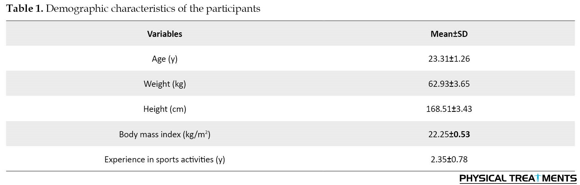
Figure 1 shows the statistical data related to the mean joint angles of the research samples in performing squats, before and after the implementation of the core muscle fatigue protocol.
Joint stability in the lower extremity depends on neuromuscular control of other involved segments, including trunk control throughout all dynamic movements, such as squatting maneuvers [1].
Core stability and function referred to bedrock for trunk dynamic control that helps generate, control, and transfer forces, and also it can control dynamic motion in distal segments of the lower limb kinetic chain [2].
Any impairment of core muscle function can result in uncontrolled movement of the trunk and other extremities through the functional activity that can place the lower extremity in a faulty movement pattern and increase the risk of lower extremity injury [3-5]. One of the most common deficiencies in athletes is neuromuscular core muscle fatigue. Neuromuscular fatigue is defined as the reduction of the maximum force produced voluntarily by a muscle or group of muscles resulting from exercise. Research has shown that a deficit in muscle activation patterns resulting from fatigue can lead to kinematic changes in the lower extremities, resulting in an increased risk of injury [6]. It has been shown that fatigue can reduce the ability of muscles to create strength and change neuromuscular control during various activities, such as squats. On the other hand, studies have shown that lower extremity neuromuscular fatigue leads to changes in knee flexion, knee valgus angle, and internal rotation of the femur during double and single-legged squat maneuvers [6, 7].
Squats are a fundamental movement in athletic performance and are one of the most widely used exercises in bodybuilding for the development of lower extremity muscles. This exercise is used in resistance training programs as well as to assess the control of the lower extremity in transitional and dynamic movements [8]. Extensive use of squats for training and evaluation purposes has led to this movement being considered a fundamental part of athletes’ training, evaluation, screening, and diagnosis of musculoskeletal problems. Core muscle function is vital in performing squat movements. For example, the amount of trunk leaning forward is a crucial factor in increasing shear force on the spinal column [9] and it seems that the good performance of core muscles can affect this situation.
In recent years, the effect of musculoskeletal conditions and related factors such as pain, fatigue, and muscle stiffness on squat performance has been repeatedly investigated [8, 10]. In this regard, it has been reported that fatigue as one of the negative factors affecting core muscle function alters the kinematics of the lower and upper extremities in various movements. Considering the importance of squat movement in training, rehabilitation, and musculoskeletal screening protocols, examining the direct effects of fatigue in core muscles on lower and upper limb kinematics during squatting can be a critical first step in the screening for sports injuries and further guidance on future sports injury prevention studies. Moreover, some factors, such as gender-related anatomical differences or differences in lower extremity muscle activation patterns make female athletes more vulnerable to above mention injury in any movement, such as squatting [11, 12]. Therefore, it is clinically important to appraise the main effects of neuromuscular core fatigue on functional movement kinematics in female athletes. Because this can be considered in better designing of training programs to increase performance and prevent injuries, hence this study was conducted to investigate the effect of core muscles fatigue on lower extremity kinematic variables of the squatting movement in female bodybuilders.
2. Methods and Methods
The present study is a quasi-experimental pre-test-post-test study design and the study population included female bodybuilders in Alborz Province, Iran who exercised professionally and regularly. Based on the results of Weeks et al.’s study [7] and using G-Power software, version 3.1 and considering the Mean±SD of knee valgus in healthy women before and after core fatigue, assuming α=0.05 and 1-β=0.95, 16 female athletes of Alborz Province were considered as study samples. All samples were included in the research based on the entry variables according to the inclusion criteria and sampling method and took part in the study voluntarily. The Mean±SD of demographic variables, such as age, height, and mass of the participants was 23.31±1.6 years, 168.51±3.65 cm, and 62.93±3.65 kg, respectively. The inclusion criteria included being in the desired age range of motions, and a history of regular physical activity in the field of bodybuilding for at least 2 years. The exclusion criteria included an abnormal range of motion in the ankle dorsiflexion [13], history of any musculoskeletal injury in the past 6 months that makes movement restriction [14, 15], obvious postural abnormalities [16, 17], having an abnormal BMI between 18 to 24 [18]. In the briefing session before participating in the study, the informed consent form and voluntary participation were completed by the subjects.
The kinematic data of the lower extremity in frontal and sagittal planes obtain by two digital cameras (Sony cyber-shot DSC HX400V made in Japan) with 50 Hz image recording speed and 21 megapixels Kinova motion analysis software, version 8.0.15 was used to analyze the obtain date. In this regard, the reliability between the examiner of this software was reported 0.95-0.99 and the internal reliability of the examiner was reported 0.99-99.98 [19].
Squatting motion analysis
All subjects performed a squat movement in front of two professional digital video cameras before and immediately after the fatigue protocol. The video camera was placed on a fixed base at a distance of 1.5 m from the person’s frontal and lateral view and at the height of the person’s pelvis in a standing position. The captured video images were analyzed by Quinoa software to evaluate two-dimensional kinematic for frontal and sagittal plane movements. The height of the cameras was adjusted using a tripod equal to the hip joint and perpendicular to the movement plane of each subject. Reflective markers are set on the right and left large trochanter, patella center, anterior superior iliac spine (ASIS), both ends of the barbell, the lateral part of the patella, and ankle [20]. As the samples were all female professional athletes in the field of bodybuilding and fitness, they were familiar enough with how to perform the squat movement, but to get acquainted with the weight and conditions, each subject had a brief time to do some experimental tries before performing the main movements. To measure the joint angles, three squats were performed each time, and the average of different angles in performing three movements was considered joint angles.
Before measuring, subjects used standard warm-ups on a stationary bicycle for 5 minutes at a power of 50 (W) and speed of 60 (RPM) [7]. The saddle height was also adjusted for all subjects so that when the pedal was in the bottom position, the flexion angle of the knee was approximately 30 degrees [21].
The number of weights that each subject had to lift while squatting was equal to 15% of their body weight [22]. Subjects were also instructed to squat at a normal 8-s speed (8 s of the squat: 3 s of descent, 2 s of pause, and 3 s of ascent) to approximately 60 degrees of knee flexion.
In the present study, the hip and knee flexion, knee valgus, and dorsiflexion of the ankle were measured as kinematic variables. To evaluate the hip angle, the marker on the ASIS was considered as the apex, and the marker at the end of the barbell and the lateral part of the patella were considered as the angle arms. To evaluate the knee angle, the marker on the lateral part of the patella was considered as the apex, and the marker on the ASIS and the ankle were considered as the angle arms [20]. Also, to evaluate the knee dynamic valgus in the frontal plane during squat movement, the angle between the ASIS patella and the lateral part of the ankle was measured. To evaluate the ankle angle, the marker was placed on the lateral part of the ankle as the apex, and the marker located on the lateral part of the patella and the head of the fifth metatarsal was considered angular [20]. All kinematic data were measured at maximum knee flexion angle during descending.
Core muscles fatigue protocol
In the present study, to exhaust the stabilizing core muscles of the body, the modified fatigue protocol of Abt et al. was used. The validity and ability of this exercise protocol to induce core muscle fatigue were demonstrated by isokinetic strength testing of trunk muscles [23].The protocol duration was 32 minutes and consisted of four consecutive sets of seven exercises, such that the subject performed each exercise 20 times for 40 s (each repetition in two seconds) with 20 s of rest between each exercise [23].
This protocol exercise included rotation of the trunk while sitting with a physioball, extension of the trunk while prone lying with a physioball, lateral sit-ups with a physioball, sit-ups with weights, bending to the sides (lateral flexion of the trunk) while standing with dumbbells, extension of the trunk with weights while prone lying, and rotating the trunk against the pulling resistance while standing.
To ensure the fatigue of the core muscles of the body in the fourth set of the fatigue protocol, the subject was asked to perform each movement until reaching complete exhaustion, and two criteria were considered to end each exercise, the subject may not be able to continue to move in the correct form or may not be able to move at a rate of one repetition per two seconds.
Statistical methods
Excel 2016 and SPSS software, version 21 was used for statistical analysis. The significance level in the present study was considered at 95% level with an α≤0.05. The descriptive Mean±SD were used to describe respondents’ demographic characteristics and the Shapiro-Wilk test was used to assess the normality of data distribution. In addition, Levene’s test was used to examine the homogeneity of variances. Finally, a paired sample t-test was used to compare the results of the kinematic variables in pre and post-tests.
3. Results
Table 1 presents the demographic characteristics of the study subject.
Figure 1 shows the statistical data related to the mean joint angles of the research samples in performing squats, before and after the implementation of the core muscle fatigue protocol.
The results of the Shapiro-Wilk test showed that the variables had a normal distribution, the paired samples t-test was then used to compare the kinematic data before and after the implementation of the fatigue protocol. The results of the statistical test showed a significant difference in the mean angle of flexion and valgus of the knee in squat movement before and after the protocol of core muscles fatigue, but no significant change was found in the mean angle of hip flexion and ankle dorsiflexion (Table 2).

4. Discussion
The present study investigated the effect of core muscle fatigue on selected lower extremity kinematic variables in the squat movement of professional female bodybuilders. The results indicated that core muscles fatigue may be related to impaired knee flexion (P=0.001) and increased knee valgus (P=0.001) during squatting, but with a difference in hip flexion (P=0.371) and ankle dorsiflexion (P=0.078) were not observed before and after the fatigue protocol.
Correct positioning of the pelvis in the coronal plane is essential to avoid excessive and uncontrolled knee valgus movement during a dynamic task [24]. Abnormal or incorrect pelvis or upper extremity movements can affect the moments acting on the knee structure [25]. During functional tasks, such as a double-leg squat, excessive trunk movements in the sagittal and frontal planes, which occurs due to insufficient core muscles function due to core muscles fatigue, can lead to produce compensatory adjustments and limitations of movement in the distal joints to compensate for the lack of pelvic control [26]. Inadequate control of trunk stability which may be due to core muscle function can lead to the increased dynamic valgus alignment of the lower extremities [27]. Previous studies have shown that core muscle underperformance results in poor control of hip adduction and hip internal rotation during functional weight-bearing activities, such as double-leg squats, and increased lower extremity valgus collapse [26, 28, 29].
Hewett et al. found that trunk movement can affect knee kinematics through mechanical and neuromuscular mechanisms [27]. Any lateral movement of the trunk during functional activities due to a deficit in the core muscle function can deflect the ground reaction force vector laterally and create a higher lever arm that is higher than the center of the knee joint [27]. This deviation has the potential to increase knee abduction loading and medial displacement of the knee joint, leading to an increase in knee valgus angle during dynamic movement [27, 30]. To counteract the excessive lateral trunk motion and maintain an upright posture and also good balance, the neuromuscular system increased hip adductor torque by activating the hip adductor muscle [31]. This can increase knee abduction moments [32]. Depending on the factors listed, they can increase the knee valgus during a dynamic task, such as the double-leg squat.
Another finding of the current study revealed that core muscle fatigue can produce less knee flexion angle during double-leg squats.
Our results are consistent with the idea of a feedforward recruitment pattern of proximal to distal musculature. Hodges et al conducted a series of studies showing that activity of the trunk muscles, particularly the transversus abdominis and multifidus muscles, precede lower and upper extremity muscle activity in healthy individuals, while this function is absent or reversed in individuals with movement disorders associated with back pain [33, 34]. This core muscle recruitment pattern is thought to provide a more stable neuromuscular basis for movement. Similarly, other authors have described increased neuromuscular control of the hip as a significant contributor to knee kinematics [26, 35, 36]. The interpretation of reduced knee flexion during the double-leg squat can be explained by this theory, the results of the current study support this theory of proximal stability promoting greater distal mobility in the lower extremities and vice versa [37]. However, the extent to which the core muscles involved prepare or influence lower extremity movement is not fully understood.
The results regarding the hip joint angle showed that the fatigue of the core muscles did not affect the amount of hip flexion before and after the fatigue protocol during squat movement. In this regard, it has been reported that muscle fatigue in the core region disrupts pelvic control at the sagittal plane and will affect the flexion angles of the hip and trunk [38], but in the present study, no significant difference was observed in hip flexion before and after fatigue protocol. The probable reason for non-compliance with this study is related to the type of evaluated task and the special subjects who always do squats in their routine exercises.
The current study had some limitations. It was initially performed on female subjects, while it has been shown that differences are observed in lower extremity kinematics between genders [39] and on specific athletes (bodybuilders) and these factors can affect the generalizability of the obtained results.
5. Conclusion:
The result of the current study showed that core muscle fatigue can negatively affect lower extremity kinematics in squatting and increase the risk of related injuries. Clinicians and coaches must consider this factor when designing the workout for bodybuilders.
Clinical implications: Many bodybuilding athletes perform their core muscle exercises at beginning of their workout and then move to other segment exercises, based on the current results, this routine can increase the risk of lower extremity injury in these athletes, especially on leg days’ exercise.
Ethical Considerations
Compliance with ethical guidelines
All ethical principles are considered in this article. The participants were informed of the purpose of the research and its implementation stages. They were also assured about the confidentiality of their information and were free to leave the study whenever they wished, and if desired, the research results would be available to them. A written consent has been obtained from the subjects. principles of the Helsinki Convention was also observed.
Funding
The paper was extracted from the MSc thesis of Maryam Torkaman, Department of Corrective Exercises and Sport Injuries, Islamic Azad University, Karaj Branch,
Authors' contributions
All authors equally contributed to preparing this article.
Conflict of interest
The authors declared no conflict of interest.
References
- Burnham JM, Yonz MC, Robertson KE, McKinley R, Wilson BR, Johnson DL, et al. Relationship of hip and trunk muscle function with single leg step-down performance: implications for return to play screening and rehabilitation. Physical Therapy in Sport. 2016; 22:66-73. [DOI:10.1016/j.ptsp.2016.05.007] [PMID]
- De Blaiser C, Roosen P, Willems T, Danneels L, Bossche LV, De Ridder R. Is core stability a risk factor for lower extremity injuries in an athletic population? A systematic review. Physical Therapy in Sport. 2018; 30:48-56. [DOI:10.1016/j.ptsp.2017.08.076] [PMID]
- Raschner C, Platzer HP, Patterson C, Werner I, Huber R, Hildebrandt C. The relationship between ACL injuries and physical fitness in young competitive ski racers: A 10-year longitudinal study. British Journal of Sports Medicine. 2012; 46(15):1065-71. [DOI:10.1136/bjsports-2012-091050] [PMID]
- Jamison ST, McNally MP, Schmitt LC, Chaudhari AM. The effects of core muscle activation on dynamic trunk position and knee abduction moments: Implications for ACL injury. Journal of Biomechanics. 2013; 46(13):2236-41.[DOI:10.1016/j.jbiomech.2013.06.021] [PMID]
- Seyedi M, Rajabi R, Shirzad E, Zareei M. [Comparison of high-risk movement patterns of ACL injury in male and female adolescent soccer players during cutting maneuver (Persian)]. Studies in Sport Medicine. 2016; 8(19):77-94. [DOI:10.22089/smj.2016.894]
- McLean SG, Fellin RE, Suedekum N, Calabrese G, Passerallo A, Joy S. Impact of fatigue on gender-based high-risk landing strategies. Medicine and Science in Sports and Exercise. 2007; 39(3):502-14. [DOI:10.1249/mss.0b013e3180d47f0] [PMID]
- Weeks BK, Carty CP, Horan SA. Effect of sex and fatigue on single leg squat kinematics in healthy young adults. BMC Musculoskeletal Disorders. 2015; 16:271. [DOI:10.1186/s12891-015-0739-3] [PMID] [PMCID]
- Cheatham SW, Stull KR, Fantigrassi M, Montel I. Hip musculoskeletal conditions and associated factors that influence squat performance: A systematic review. Journal of Sport Rehabilitation. 2018; 27(3):263-73. [DOI:10.1123/jsr.2016-0246] [PMID]
- Potvin JR, McGill SM, Norman RW. Trunk muscle and lumbar ligament contributions to dynamic lifts with varying degrees of trunk flexion. Spine. 1991; 16(9):1099-107. [DOI:10.1097/00007632-199109000-00015] [PMID]
- Bagwell JJ, Snibbe J, Gerhardt M, Powers CM. Hip kinematics and kinetics in persons with and without cam femoroacetabular impingement during a deep squat task.Clinical Biomechanics. 2016; 31:87-92. [DOI:10.1016/j.clinbiomech.2015.09.016] [PMID]
- Brophy R, Silvers HJ, Gonzales T, Mandelbaum BR. Gender influences: The role of leg dominance in ACL injury among soccer players. British Journal of Sports Medicine. 2010; 44(10):694-7. [DOI:10.1136/bjsm.2008.051243] [PMID]
- Cheung EC, Boguszewski DV, Joshi NB, Wang D, McAllister DR. Anatomic factors that may predispose female athletes to anterior cruciate ligament injury. Current Sports Medicine Reports. 2015; 14(5):368-72. [DOI:10.1249/JSR.0000000000000188] [PMID]
- Dill KE, Begalle RL, Frank BS, Zinder SM, Padua DA. Altered knee and ankle kinematics during squatting in those with limited weight-bearing-lunge ankle-dorsiflexion range of motion. Journal of Athletic Training. 2014; 49(6):723-32. [DOI:10.4085/1062-6050-49.3.29] [PMID] [PMCID]
- Orishimo KF, Kremenic IJ, Mullaney MJ, McHugh MP, Nicholas SJ. Adaptations in single-leg hop biomechanics following anterior cruciate ligament reconstruction. Knee Surgery, Sports Traumatology, Arthroscopy. 2010; 18(11):1587-93. [DOI:10.1007/s00167-010-1185-2] [PMID]
- del Pozo-Cruz B, Gusi N, Adsuar JC, del Pozo-Cruz J, Parraca JA, Hernandez-Mocholí M. Musculoskeletal fitness and health-related quality of life characteristics among sedentary office workers affected by sub-acute, non-specific low back pain: A cross-sectional study. Physiotherapy. 2013; 99(3):194-200. [DOI:10.1016/j.physio.2012.06.006] [PMID]
- Lin JJ, Chen WH, Chen PQ, Tsauo JY. Alteration in shoulder kinematics and associated muscle activity in people with idiopathic scoliosis. Spine. 2010; 35(11):1151-7. [DOI:10.1097/BRS.0b013e3181cd5923] [PMID]
- Farr S, Kranzl A, Pablik E, Kaipel M, Ganger R. Functional and radiographic consideration of lower limb malalignment in children and adolescents with idiopathic genu valgum. Journal of Orthopaedic Research. 2014; 32(10):1362-70. [DOI:10.1002/jor.22684] [PMID]
- Global BMI Mortality Collaboration, Di Angelantonio E, Bhupathiraju ShN, Wormser D, Gao P, Kaptoge S, et al. Body-mass index and all-cause mortality: individual-participant-data meta-analysis of 239 prospective studies in four continents. Lancet. 2016; 388(10046):776-86. [DOI:10.1016/S0140-6736(16)30175-1] [PMID]
- Cunha AB, Babik I, Harbourne R, Cochran NJ, Stankus J, Szucs K, et al. Assessing the validity and reliability of a new video goniometer app for measuring joint angles in adults and children. Archives of Physical Medicine and Rehabilitation. 2020; 101(2):275-82. [DOI:10.1016/j.apmr.2019.07.008] [PMID]
- Hooper DR, Szivak TK, Comstock BA, Dunn-Lewis C, Apicella JM, Kelly NA, et al. Effects of fatigue from resistance training on barbell back squat biomechanics.Journal of Strength and Conditioning Research. 2014; 28(4):1127-34. [DOI:10.1097/JSC.0000000000000237] [PMID]
- Holliday W, Theo R, Fisher J, Swart J. Cycling: Joint kinematics and muscle activity during differing intensities. Sports Biomechanics. 2019:1-15. [DOI:10.1080/14763141.2019.1640279] [PMID]
- Ninos JC, Irrgang JJ, Burdett R, Weiss JR. Electromyographic analysis of the squat performed in self-selected lower extremity neutral rotation and 30 of lower extremity turn-out from the self-selected neutral position. Journal of Orthopaedic & Sports Physical Therapy. 1997; 25(5):307-15. [DOI:10.2519/jospt.1997.25.5.307] [PMID]
- Abt JP, Smoliga JM, Brick MJ, Jolly JT, Lephart SM, Fu FH. Relationship between cycling mechanics and core stability. Journal of Strength and Conditioning Research. 2007; 21(4):1300-4. [DOI:10.1519/R-21846.1] [PMID]
- Kirtley C. Clinical gait analysis: Theory and practice in gait analysis. Amsterdam: Elsevier Health Sciences; 2006. [Link]
- Powers CM. The influence of abnormal hip mechanics on knee injury: A biomechanical perspective. The Journal of Orthopaedic and Sports Physical Therapy. 2010; 40(2):42-51. [DOI:10.2519/jospt.2010.3337] [PMID]
- Zazulak BT, Hewett TE, Reeves NP, Goldberg B, Cholewicki J. Deficits in neuromuscular control of the trunk predict knee injury risk: A prospective biomechanical-epidemiologic study. The American Journal of Sports Medicine. 2007; 35(7):1123-30. [DOI:10.1177/0363546507301585] [PMID]
- Hewett TE, Myer GD. The mechanistic connection between the trunk, hip, knee, and anterior cruciate ligament injury. Exercise and Sport Sciences Reviews. 2011; 39(4):161-6.[DOI:10.1097/JES.0b013e3182297439] [PMID] [PMCID]
- Hewett TE, Ford KR, Myer GD, Wanstrath K, Scheper M. Gender differences in hip adduction motion and torque during a single-leg agility maneuver. Journal of Orthopaedic Research. 2006; 24(3):416-21. [DOI:10.1002/jor.20056] [PMID]
- van Dieën JH, Selen LP, Cholewicki J. Trunk muscle activation in low-back pain patients, an analysis of the literature. Journal of Electromyography and Kinesiology. 2003; 13(4):333-51. [DOI:10.1016/S1050-6411(03)00041-5] [PMID]
- Seyedi M, Zarei M, Daneshjoo A, Rajabi R, Shirzad E, Mozafaripour E, et al. Effects of FIFA 11+ warm-up program on kinematics and proprioception in adolescent soccer players: A parallelgroup randomized control trial. Scientific Reports. 2023; 13(1):5527. [DOI:10.1038/s41598-023-32774-3] [PMID] [PMCID]
- Hewett TE, Stroupe AL, Nance TA, Noyes FR. Plyometric training in female athletes. Decreased impact forces and increased hamstring torques. The American Journal of Sports Medicine. 1996; 24(6):765-73. [DOI:10.1177/036354659602400611] [PMID]
- Mozafaripour E, Seidi F, Minoonejad H, Mousavi SH, Bayattork M. Can lower extremity anatomical measures and core stability predict dynamic knee valgus in young men? Journal of Bodywork and Movement Therapies. 2021; 27:358-63.[DOI:10.1016/j.jbmt.2021.02.004] [PMID]
- Hodges PW, Richardson CA. Delayed postural contraction of transversus abdominis in low back pain associated with movement of the lower limb. Journal of Spinal Disorders. 1998; 11(1):46-56. [DOI:10.1097/00002517-199802000-00008] [PMID]
- Hodges PW, Moseley GL, Gabrielsson A, Gandevia SC. Experimental muscle pain changes feedforward postural responses of the trunk muscles. Experimental Brain Research. 2003; 151(2):262-71. [DOI:10.1007/s00221-003-1457-x] [PMID]
- Myer GD, Ford KR, Palumbo JP, Hewett TE. Neuromuscular training improves performance and lower-extremity biomechanics in female athletes. Journal of Strength and Conditioning Research. 2005; 19(1):51-60. [DOI:10.1519/00124278-200502000-00010] [PMID]
- Hollman JH, Ginos BE, Kozuchowski J, Vaughn AS, Krause DA, Youdas JW. Relationships between knee valgus, hip-muscle strength, and hip-muscle recruitment during a single-limb step-down. Journal of Sport Rehabilitation. 2009; 18(1):104-17. [DOI:10.1123/jsr.18.1.104] [PMID]
- Shirey M, Hurlbutt M, Johansen N, King GW, Wilkinson SG, Hoover DL. The influence of core musculature engagement on hip and knee kinematics in women during a single leg squat. International Journal of Sports Physical Therapy. 2012; 7(1):1. [PMID] [PMCID]
- Chuter VH, Janse de Jonge XA. Proximal and distal contributions to lower extremity injury: A review of the literature. Gait & Posture. 2012; 36(1):7-15. [DOI:10.1016/j.gaitpost.2012.02.001] [PMID]
- Decker MJ, Torry MR, Wyland DJ, Sterett WI, Richard Steadman J. Gender differences in lower extremity kinematics, kinetics and energy absorption during landing. Clinical Biomechanics. 2003; 18(7):662-9. [DOI:10.1016/S0268-0033(03)00090-1] [PMID]
Type of Study: Research |
Subject:
General
Received: 2023/02/14 | Accepted: 2023/02/26 | Published: 2022/10/20
Received: 2023/02/14 | Accepted: 2023/02/26 | Published: 2022/10/20
Send email to the article author
| Rights and permissions | |
 |
This work is licensed under a Creative Commons Attribution-NonCommercial 4.0 International License. |
This is an open access article distributed under the terms of the Creative Commons Attribution License (CC-By-NC), which permits use, distribution, and reproduction in any medium, provided the original work is properly cited and is not used for commercial purposes.
Contact Information
