Tue, Jul 23, 2024
Volume 9, Issue 2 (Spring 2019)
PTJ 2019, 9(2): 117-124 |
Back to browse issues page


Download citation:
BibTeX | RIS | EndNote | Medlars | ProCite | Reference Manager | RefWorks
Send citation to:


BibTeX | RIS | EndNote | Medlars | ProCite | Reference Manager | RefWorks
Send citation to:
Abdoli A, Nakhostin Roohi B. Effect of Extracorporeal Shockwave Therapy Versus Stretching in the Treatment of Athletes With Chronic Plantar Fasciitis. PTJ 2019; 9 (2) :117-124
URL: http://ptj.uswr.ac.ir/article-1-401-en.html
URL: http://ptj.uswr.ac.ir/article-1-401-en.html


1- Department of Physical Education and Sport Sciences, Faculty of Human Sciences, Shams Education Institute of Science and Technology, Tabriz, Iran.
2- Department of Physical Education and Sport Sciences, Faculty of Human Sciences, Ardabil Branch, Islamic Azad University, Ardabil, Iran.
2- Department of Physical Education and Sport Sciences, Faculty of Human Sciences, Ardabil Branch, Islamic Azad University, Ardabil, Iran.
Keywords: Chronic plantar fasciitis, Extracorporeal shockwave therapy, Passive Stretching, Visual analog scale
Full-Text [PDF 606 kb]
(1749 Downloads)
| Abstract (HTML) (3099 Views)

Before starting the treatment program, the patients were invited to participate in a briefing session for the treatment process. They were willing to participate in the research, and they were asked to sign a consent form. The inclusion criteria included regular participation in a field of sports for at least two years, age over 18 years, heel pain for at least four weeks, which did not respond to conservative treatments (non-steroid anti-inflammatory drug and ice therapies), no evidence in favor of the collapse of calcaneus in the radiography, pain intensity greater than 2 based on the visual analog scale (VAS), and tenderness at the site of plantar fascia attachment to calcaneus.
The exclusion criteria included heel surgery, Achilles nerve or tendon injury, rheumatoid arthritis, both types of diabetes mellitus, local or systemic infection, peripheral vascular disorders, gout, coagulation disorders, and corticosteroid injection in the last six months or non-steroid anti-inflammatory drug use in the previous seven days. VAS was used to evaluate pain. On this scale, the patient rates his status from zero (without disturbance) to ten (completely disturbed). Goniometer (Yagamy: Japan) was used to measure the active flexion-extension range of movement (ROM) of the ankle. A dynamometer (Tiger R-372: Germany) was also used to measure the strength of the plantar flexors of ankle muscles.
Interventions
The ESWT (enPlusPro Zimmer: Germany) device was set on 12 Hz to 15 Hz frequency and 2500 pulses at 2 to 3 bar. The therapy (ESWT) was performed once a week for five weeks [26]. During the ESWT sessions, the patients were in a prone position with their feet extending beyond the examination table, and their knee and hip joints in a neutral position. Using ultrasound gel as a coupling medium, the head of the ESWT device was applied to the inferior aspect of the calcaneus. The region of maximum tenderness in the medial calcaneus was the target area. No local anesthetic was applied. All patients were allowed for weight-bearing [27].
In the STCH group, the stretching of the plantar fascia, Achilles tendon, and calf muscles were performed three times a day for five weeks. Each time, ten stretches (each taking 20 seconds) were performed to each area.
Data analysis
The data were analyzed, using paired and independent t-tests for within- and between-groups differences in SPSS V. 22. A tow-way repeated measures analysis of variance (ANOVA) was performed to estimate the differences between (group effect) and within (time and time × group effects) treatment groups for each studied outcome.
3. Results
Table 1 presents the demographic characteristics of the subjects of both groups. The Shapiro-Wilk test was used to check the normality of the data. There were no significant differences in terms of these variables between the two groups (P>0.05).

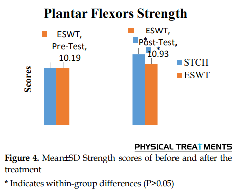
Figures 2, 3, and 4 show the results of the VAS, ROM of the ankle, and the strength of plantar flexors. According to Figures 2, VAS scores significantly decreased after the treatment in both groups (P<0.001). The VAS scores showed no significant between-group differences at any time (P>0.05). Moreover, as seen in Figures 2, in both groups after the treatment, the ROM of ankle significantly increased (P<0.001). There were no significant between-groups differences at any time (P>0.05). Finally, as observed in Figures 3, after the treatment, the strength of plantar flexors in both groups significantly increased (P<0.001). There was no significant difference between the two groups before and after the treatment (P>0.05).

4. Discussion
The results of this research showed that ESWT and STCH reduced the ankle pain, increased the ROM of ankle joint, and improved the strength of the plantar flexors in the athletes with chronic PF. However, there were no significant differences between the two groups in any variable (P>0.05).
Regarding the pain, the results of this study are consistent with the studies of Akinoglu et al. Maffulli et al. Ordahan et al. and Lizis et al. [14, 27-29]. ESWT is a treatment, in which waves are produced by electromagnetic, piezoelectric, and electrohydrolysis techniques. According to studies, shockwaves cause an inflammatory-like response in the damaged tissue and cause the body to respond to this condition that as a result, blood flow and damaged tissue metabolism increase [30, 31]. Two mechanisms have been proposed for the effectiveness of ESWT. Firstly, transmitted waves are thought to affect the physiology of nociceptors. Secondly, the transmitted waves are responsible for tissue regeneration through micro-trauma, the release of growth factors, and molecular factors [9, 31]. Increasing the ROM of the ankle joint in both groups is a remarkable result of this study. Improvement in the ROM in the ESWT group seems to result from a decrease of pain than other factors. Probably reducing pain has made it easier for the ankle joint to move. Moreover, there is a consensus among the researchers on the positive effects of STCH on increasing the ROM of joints [32].
Finally, increasing the strength of plantar flexors affecting feet in both the STCH and ESWT groups can also be as a result of pain relief. Pain relief seems to have made plantar flexor muscles work better. According to the results, both short-term methods have been effective in reducing chronic PF pain in athletes. Studies have shown that stretching is a therapeutic approach that is more effective in the short term. Stretching is also an easy and inexpensive method that can be applied by the patient. The effect of this method on improving the ROM and the strength of plantar flexor muscles are also well documented in this study. On the other hand, ESWT is a relatively expensive method, which causes pain and discomfort of the patient during the treatment. The use of costly methods in sports medicine is often applied for the faster conclusion and quicker return of athletes to sports fields.
Since the stretching method is cheaper and non-invasive, and since there is no significant difference in the effectiveness of the two methods in the short term, according to our data, using aggressive ESWT method in athletes is not much recommended. However, there are doubts about the long-term effects of STCH, and some researchers believe that the effect of STCH is temporary [20]. Therefore, more studies are required to achieve more accurate results. Of course, the limitations of this research should also be considered. The number of subjects in this research is low, and the probability of changing the results is likely with the increase in the number of subjects for both methods.
This study suggests that both methods of stretching and ESWT are suitable methods for improving the symptoms in chronic PF. There was no significant difference in the effect of both types of treatments. According to the results, stretching is easier, non-invasive, and less costly method. Thus, so, stretching seems to be preferable method to ESWT. However, more studies are required because of the limitations of this research.
Ethical Considerations
Compliance with ethical guidelines
All ethical principles were considered in this article. The participants were informed about the aim of the research and its procedure. They were also assured about the confidentiality of their information. Also, they were allowed to leave the study whenever they wish, and if desired, the results of the research would be available to them.
Funding
This research did not receive any specific grant from funding agencies in the public, commercial, or not-for-profit sectors.
Authors' contributions
All authors contributed to designing, running, and writing all parts of the research.
Conflict of interest
The authors declared no conflict of interest.
Acknowledgments
We thank Dr Rouhani, the technical director of Nikan Physical Therapy Center for his sincere cooperation.
References
Full-Text: (1643 Views)
1. Introduction
Plantar fasciitis is a musculoskeletal disorder in the lower extremities that affects the plantar fascia [1, 2]. In the past, Plantar Fasciitis (PF) was known as a chronic inflammatory disease, but today, it is considered a degenerative pathological condition like tendinopathy [3]. Plantar fasciitis occurs at the site of the attachment of the plantar fascia to the medial tubercle of calcaneus [4]. PF rapidly is converted from acute to chronic, a symptom of which is the reduction of pain and the progression of the degenerative process [5]. It has been reported in many athletes, especially soccer players and runners [3].
This damage occurs both in recreational and professional sports, and female athletes suffer from this injury slightly more than male athletes [6, 7]. In addition to the external factors such as inappropriate shoes and prolonged walking on rugged surfaces, the practical factors in the occurrence of plantar fasciitis are a number of internal factors such as obesity, flat foot , pes cavus, the difference in the size of the two limbs, the increase in external rotation of tibia, and increased femoral anteversion [8]. The patients typically suffer from a sharp pain in the medial heel of the calcaneus. Pain is often more severe when standing or in the first steps after waking up or after long periods of sitting. Severe cases limit the ability to move a patient during daily activities and climb stairs [9].
Pediatrics, orthopedic surgeons, physiotherapists, and chiropractors are involved in the treatment of this injury [3]. In 2007, the cost of PF in the United States was estimated between 192 and 376 million dollars [10]. The treatment of chronic plantar fasciitis is mainly conservative (90%), including the use of non-steroidal anti-inflammatory drugs, topical steroid injections, orthotic devices, Stretching Exercises (STCH), as well as electrotherapies such as laser therapy, ultrasound, iontophoresis, and extracorporeal shockwave therapy (ESWT) [3, 11-14].
ESWT is a relatively new method in the treatment of musculoskeletal disorders, which attracts a lot of attention and is rapidly expanding [15]. Shockwave means transferring a great deal of energy over brief periods at a rate exceeding the speed of sound. Thus, shockwave therapy uses the characteristics of these waves. Since the source of such high energy is human production, and this energy is transmitted from the outside of the body to the target tissue, the term ESWT is used for this therapeutic approach [16]. Radial shockwave therapy is a type of low-to-moderate ESWT produced pneumatically [17]. The success rate of this method for plantar fasciitis in various studies is 50%-80% [18].
Compared to ESWT, which is a relatively expensive treatment, the stretching of soft tissues is one of the very low-cost methods used to treat plantar fasciitis. Some studies suggest that Achilles tendon and plantar fascia stretching can reduce the symptoms of PF [19]. Some investigators believe that stretching is one of the easiest and most effective treatments for plantar fasciitis [20-25]. Regarding the relative effect of both types of treatment of ESWT and stretching on chronic PF and because of the significant difference in terms of the cost of treatment, the high prevalence of this injury in athletes, and the lack of research in this area, we aimed to compare the effect of these two therapies in the treatment of chronic PF in athletes.
2. Materials and Methods
The setting, participants, and randomization
This study was designed as a randomized clinical trial. Of the 61 consecutive male athletes with chronic PF, who attended the Nikan physiotherapy clinic in Tabriz, Iran, 35 patients were eligible to participate in the study. Five of these participants refused to participate in the project, and the rest were randomly assigned into two groups of ESWT (n=15) and STCH (n=15) (Figure 1).
Before starting the treatment program, the patients were invited to participate in a briefing session for the treatment process. They were willing to participate in the research, and they were asked to sign a consent form. The inclusion criteria included regular participation in a field of sports for at least two years, age over 18 years, heel pain for at least four weeks, which did not respond to conservative treatments (non-steroid anti-inflammatory drug and ice therapies), no evidence in favor of the collapse of calcaneus in the radiography, pain intensity greater than 2 based on the visual analog scale (VAS), and tenderness at the site of plantar fascia attachment to calcaneus.
The exclusion criteria included heel surgery, Achilles nerve or tendon injury, rheumatoid arthritis, both types of diabetes mellitus, local or systemic infection, peripheral vascular disorders, gout, coagulation disorders, and corticosteroid injection in the last six months or non-steroid anti-inflammatory drug use in the previous seven days. VAS was used to evaluate pain. On this scale, the patient rates his status from zero (without disturbance) to ten (completely disturbed). Goniometer (Yagamy: Japan) was used to measure the active flexion-extension range of movement (ROM) of the ankle. A dynamometer (Tiger R-372: Germany) was also used to measure the strength of the plantar flexors of ankle muscles.
Interventions
The ESWT (enPlusPro Zimmer: Germany) device was set on 12 Hz to 15 Hz frequency and 2500 pulses at 2 to 3 bar. The therapy (ESWT) was performed once a week for five weeks [26]. During the ESWT sessions, the patients were in a prone position with their feet extending beyond the examination table, and their knee and hip joints in a neutral position. Using ultrasound gel as a coupling medium, the head of the ESWT device was applied to the inferior aspect of the calcaneus. The region of maximum tenderness in the medial calcaneus was the target area. No local anesthetic was applied. All patients were allowed for weight-bearing [27].
In the STCH group, the stretching of the plantar fascia, Achilles tendon, and calf muscles were performed three times a day for five weeks. Each time, ten stretches (each taking 20 seconds) were performed to each area.
Data analysis
The data were analyzed, using paired and independent t-tests for within- and between-groups differences in SPSS V. 22. A tow-way repeated measures analysis of variance (ANOVA) was performed to estimate the differences between (group effect) and within (time and time × group effects) treatment groups for each studied outcome.
3. Results
Table 1 presents the demographic characteristics of the subjects of both groups. The Shapiro-Wilk test was used to check the normality of the data. There were no significant differences in terms of these variables between the two groups (P>0.05).
Figures 2, 3, and 4 show the results of the VAS, ROM of the ankle, and the strength of plantar flexors. According to Figures 2, VAS scores significantly decreased after the treatment in both groups (P<0.001). The VAS scores showed no significant between-group differences at any time (P>0.05). Moreover, as seen in Figures 2, in both groups after the treatment, the ROM of ankle significantly increased (P<0.001). There were no significant between-groups differences at any time (P>0.05). Finally, as observed in Figures 3, after the treatment, the strength of plantar flexors in both groups significantly increased (P<0.001). There was no significant difference between the two groups before and after the treatment (P>0.05).
4. Discussion
The results of this research showed that ESWT and STCH reduced the ankle pain, increased the ROM of ankle joint, and improved the strength of the plantar flexors in the athletes with chronic PF. However, there were no significant differences between the two groups in any variable (P>0.05).
Regarding the pain, the results of this study are consistent with the studies of Akinoglu et al. Maffulli et al. Ordahan et al. and Lizis et al. [14, 27-29]. ESWT is a treatment, in which waves are produced by electromagnetic, piezoelectric, and electrohydrolysis techniques. According to studies, shockwaves cause an inflammatory-like response in the damaged tissue and cause the body to respond to this condition that as a result, blood flow and damaged tissue metabolism increase [30, 31]. Two mechanisms have been proposed for the effectiveness of ESWT. Firstly, transmitted waves are thought to affect the physiology of nociceptors. Secondly, the transmitted waves are responsible for tissue regeneration through micro-trauma, the release of growth factors, and molecular factors [9, 31]. Increasing the ROM of the ankle joint in both groups is a remarkable result of this study. Improvement in the ROM in the ESWT group seems to result from a decrease of pain than other factors. Probably reducing pain has made it easier for the ankle joint to move. Moreover, there is a consensus among the researchers on the positive effects of STCH on increasing the ROM of joints [32].
Finally, increasing the strength of plantar flexors affecting feet in both the STCH and ESWT groups can also be as a result of pain relief. Pain relief seems to have made plantar flexor muscles work better. According to the results, both short-term methods have been effective in reducing chronic PF pain in athletes. Studies have shown that stretching is a therapeutic approach that is more effective in the short term. Stretching is also an easy and inexpensive method that can be applied by the patient. The effect of this method on improving the ROM and the strength of plantar flexor muscles are also well documented in this study. On the other hand, ESWT is a relatively expensive method, which causes pain and discomfort of the patient during the treatment. The use of costly methods in sports medicine is often applied for the faster conclusion and quicker return of athletes to sports fields.
Since the stretching method is cheaper and non-invasive, and since there is no significant difference in the effectiveness of the two methods in the short term, according to our data, using aggressive ESWT method in athletes is not much recommended. However, there are doubts about the long-term effects of STCH, and some researchers believe that the effect of STCH is temporary [20]. Therefore, more studies are required to achieve more accurate results. Of course, the limitations of this research should also be considered. The number of subjects in this research is low, and the probability of changing the results is likely with the increase in the number of subjects for both methods.
This study suggests that both methods of stretching and ESWT are suitable methods for improving the symptoms in chronic PF. There was no significant difference in the effect of both types of treatments. According to the results, stretching is easier, non-invasive, and less costly method. Thus, so, stretching seems to be preferable method to ESWT. However, more studies are required because of the limitations of this research.
Ethical Considerations
Compliance with ethical guidelines
All ethical principles were considered in this article. The participants were informed about the aim of the research and its procedure. They were also assured about the confidentiality of their information. Also, they were allowed to leave the study whenever they wish, and if desired, the results of the research would be available to them.
Funding
This research did not receive any specific grant from funding agencies in the public, commercial, or not-for-profit sectors.
Authors' contributions
All authors contributed to designing, running, and writing all parts of the research.
Conflict of interest
The authors declared no conflict of interest.
Acknowledgments
We thank Dr Rouhani, the technical director of Nikan Physical Therapy Center for his sincere cooperation.
References
- Huang YC, Wei SH, Wang HK, Lieu FK. Ultrasonographic guided botulinum toxin type a for plantar fasciitis: An outcome-based investigation for treating pain and gait changes. Journal of Rehabilitation Medicine. 2010; 42:136-40. [DOI:10.2340/16501977-0491] [PMID]
- Sahin N, Oztürk A, Atıcı T. Foot mobility and plantar fascia elasticity in patients with plantar fasciitis. Acta Orthopaedica et Traumatologica Turcica. 2010; 44:385-91. [DOI:10.3944/AOTT.2010.2348] [PMID]
- Petraglia F, Ramazzina I, Costantino C. Plantar fasciitis in athletes: Diagnostic and treatment strategies. A systematic review. Muscles, Ligaments and Tendons Journal. 2017; 7:107. [DOI:10.32098/mltj.01.2017.14] [PMID] [PMCID]
- Stuber K, Kristmanson K. Conservative therapy for plantar fasciitis: A narrative review of randomized controlled trials. The Journal of the Canadian Chiropractic Association. 2006; 50:118.
- League AC. Current concepts review: Plantar fasciitis. Foot & Ankle International. 2008; 29:358-66. [DOI:10.3113/FAI.2008.0358] [PMID]
- Orchard J. Plantar fasciitis. BMJ. 2012; 345:e6603. [DOI:10.1136/bmj.e6603] [PMID]
- Taunton JE, Ryan MB, Clement D, McKenzie DC, Lloyd-Smith D, Zumbo B. A retrospective case-control analysis of 2002 running injuries. British Journal of Sports Medicine. 2002; 36:95-101. [DOI:10.1136/bjsm.36.2.95] [PMID] [PMCID]
- Gibbon W, Long G. Ultrasound of the plantar aponeurosis (fascia). Skeletal Radiology. 1999; 28:21-6. [DOI:10.1007/s002560050467] [PMID]
- Muhammad M, Tulaar AB, Pradanasari R, Bardosono S. Effectiveness of taping for the short-term treatment of pain and walking speed in patients with plantar fasciitis. Indonesian Journal of Physical Medicine & Rehabilitation. 2016; 5(1):33-42.
- Tong KB, Furia J. Economic burden of plantar fasciitis treatment in the United States. American Journal of Orthopedics. 2010; 39:227-31.
- Crawford R. Diagnosis and treatment of plantar fasciitis. American Family Physician. 2011; 84:676-82.
- Radford JA, Landorf KB, Buchbinder R, Cook C. Effectiveness of calf muscle stretching for the short-term treatment of plantar heel pain: A randomised trial. BMC Musculoskeletal Disorders. 2007; 8:36. [DOI:10.1186/1471-2474-8-36] [PMID] [PMCID]
- Costantino C, Vulpiani M, Romiti D, Vetrano M, Saraceni V. Cryoultrasound therapy in the treatment of chronic plantar fasciitis with heel spurs. A randomized controlled clinical study. European Journal of Physical and Rehabilitation Medicine. 2014; 50:39-47.
- Maffulli G, Padulo J, Iuliano E, Furia J, Rompe J, Maffulli N. Extracorporeal shock wave therapy in the treatment of midsubstance plantar fasciitis: The ASSERT database. Muscles, Ligaments & Tendons Journal (MLTJ). 2018; 8(3):430-6. [DOI:10.32098/mltj.03.2018.10]
- Romeo P, Lavanga V, Pagani D, Sansone V. Extracorporeal shock wave therapy in musculoskeletal disorders: A review. Medical Principles and Practice. 2014; 23:7-13. [DOI:10.1159/000355472] [PMID] [PMCID]
- Maffulli G, Hemmings S, Maffulli N. Assessment of the effectiveness of extracorporeal shock wave therapy (ESWT) for soft tissue injuries (ASSERT): An online database protocol. Translational Medicine @ UniSa. 2014; 10:46.
- Cacchio A, Paoloni M, Barile A, Don R, de Paulis F, Calvisi V, et al. Effectiveness of radial shock-wave therapy for calcific tendinitis of the shoulder: Single-blind, randomized clinical study. Physical Therapy. 2006; 86:672-82.
- Rompe J, Hopf C, Nafe B, Bürger R. Low-energy extracorporeal shock wave therapy for painful heel: A prospective controlled single-blind study. Archives of Orthopaedic and Trauma Surgery. 1996; 115:75-9. [DOI:10.1007/BF00573445] [PMID]
- Digiovanni BF, Nawoczenski DA, Malay DP, Graci PA, Williams TT, Wilding GE, et al. Plantar fascia-specific stretching exercise improves outcomes in patients with chronic plantar fasciitis: A prospective clinical trial with two-year follow-up. Journal of Bone and Joint Surgery. 2006; 88:1775-81. [DOI:10.2106/00004623-200608000-00013] [PMID]
- Hyland MR, Webber-Gaffney A, Cohen L, Lichtman SW. Randomized controlled trial of calcaneal taping, sham taping, and plantar fascia stretching for the short-term management of plantar heel pain. Journal of Orthopaedic & Sports Physical Therapy. 2006; 36:364-71. [DOI:10.2519/jospt.2006.2078] [PMID]
- DiGiovanni BF, Nawoczenski DA, Lintal ME, Moore EA, Murray JC, Wilding GE, et al. Tissue-specific plantar fascia-stretching exercise enhances outcomes in patients with chronic heel pain: A prospective, randomized study. Journal of Bone and Joint Surgery. 2003; 85:1270-7. [DOI:10.2106/00004623-200307000-00013] [PMID]
- Porter D, Barrill E, Oneacre K, May BD. The effects of duration and frequency of achilles tendon stretching on dorsiflexion and outcome in painful heel syndrome: A randomized, blinded, control study. Foot & Ankle International. 2002; 23:619-24. [DOI:10.1177/107110070202300706] [PMID]
- Quaschnick MS. The diagnosis and management of plantar fasciitis. The Nurse Practitioner. 1996; 21:50-4. [DOI:10.1097/00006205-199604000-00005]
- Shrier I, Gossal K. Myths and truths of stretching: Individualized recommendations for healthy muscles. The Physician and Sportsmedicine. 2000; 28:57-63. [DOI:10.3810/psm.2000.08.1159] [PMID]
- Wapner KL, Sharkey PF. The use of night splints for treatment of recalcitrant plantar fasciitis. Foot & Ankle. 1991; 12:135-7. [DOI:10.1177/107110079101200301] [PMID]
- Buchbinder R, Ptasznik R, Gordon J, Buchanan J, Prabaharan V, Forbes A. Ultrasound-guided extracorporeal shock wave therapy for plantar fasciitis: A randomized controlled trial. JAMA. 2002; 288:1364-72. [DOI:10.1001/jama.288.11.1364] [PMID]
- Ordahan B, Türkoğlu G, Karahan AY, Akkurt HE. Extracorporeal shockwave therapy versus kinesiology taping in the management of plantar fasciitis: A randomized clinical trial. Archives of Rheumatology. 2017; 32:227. [DOI:10.5606/ArchRheumatol.2017.6059] [PMID] [PMCID]
- LIZIS P. Comparison between real and placebo extracorporeal shockwave therapy for the treatment of chronic plantar fasciitis pain in the males. Iranian Journal of Public Health. 2015; 44:1150.
- Akınoğlu B, Köse N. A comparison of the acute effects of radial extracorporeal shockwave therapy, ultrasound therapy, and exercise therapy in plantar fasciitis. Journal of Exercise Rehabilitation. 2018; 14(2):306-12. [DOI:10.12965/jer.1836048.024] [PMID] [PMCID]
- Frontera WR, Silver JK, Rizzo TD. Essentials of physical medicine and rehabilitation: musculoskeletal disorders, pain, and rehabilitation. Edinburgh: Elsevier Health Sciences; 2018.
- Maier M, Dürr H, Köhler S, Staupendahl D, Pfahler M, Refior H. Analgesic effect of low energy extracorporeal shock waves in tendinosis calcarea, epicondylitis humeri radialis and plantar fasciitis. Zeitschrift fur Orthopadie und ihre Grenzgebiete. 2000; 138(1):34-8. [DOI:10.1055/s-2000-10110] [PMID]
- Page P. Current concepts in muscle stretching for exercise and rehabilitation. International Journal of Sports Physical Therapy. 2012; 7(1):109-19. [PMID] [PMCID]
Type of Study: Research |
Subject:
Special
Received: 2019/06/17 | Accepted: 2019/08/28 | Published: 2019/11/12
Received: 2019/06/17 | Accepted: 2019/08/28 | Published: 2019/11/12
Send email to the article author
| Rights and permissions | |
 |
This work is licensed under a Creative Commons Attribution-NonCommercial 4.0 International License. |
This is an open access article distributed under the terms of the Creative Commons Attribution License (CC-By-NC), which permits use, distribution, and reproduction in any medium, provided the original work is properly cited and is not used for commercial purposes.
Contact Information
